The Erisocrinoidea: Shallow Crinoids

Articulated calyx of Erisocrinus typus, copyright Richard Paselk.


The close of the Permian period saw the largest mass extinction ever recorded. It has been estimated that about 95% of all marine species were wiped out. Many prominent Palaeozoic lineages disappeared entirely; others were reduced to a mere remnant of their former selves.

One of the casualties of the end-Permian extinction was the crinoid group known as the Erisocrinoidea (or Erisocrinacea in older texts). These were a diverse group of crinoids divided between several families, recorded from the Carboniferous and Permian periods. One species, Erisocrinus typus, is known from a large number of well-preserved, articulated specimens from the mid-Late Carboniferous of the United States and is one of the best representatives of the Palaeozoic cladid crinoids. Erisocrinoids are characterised by a low cup, dominated by the ring of radial plates. The base of cup was often recessed, meaning that the basal and infrabasal plate rings were often partially or entirely obscured in outer view. Most significantly, the array of anal plates found in other crinoids was reduced to a single plate or even lost. The insertion points of the arms bear signs of strong muscular articulation, indicating that these were animals of higher-energy environments requiring more exertion to maintain an ideal feeding position. The anal sac, where it is preserved, was only weakly plated and would have been reasonably soft in life (Moore et al. 1978).

In other respects, though, the erisocrinoids could be somewhat disparate. Many, such as the type family Erisocrinidae and the families Protencrinidae and Catacrinidae, have biserial arms in which the arm's skeleton is comprised of paired rows of plates. In other families, such as the Graphiocrinidae and Diphuicrinidae, the arms were uniserial, with only a single row of plates. Webster & Maples (2006) noted that, even though all erisocrinoids shared the character of a reduced anal plate array, the exact position in the cup of the anal plate or its remnant differed between families. They therefore suggested that the erisocrinoids might not be a monophyletic group, but members of a number of different lineages that had converged on a similar morphology and presumably lifestyle.

This was not an entirely novel suggestion. Even while recognising a single superfamily Erisocrinacea, Moore et al. (1978) had suggested connections between individual erisocrinoid families and families placed in other superfamilies. The integrity of the Erisocrinoidea had also been questioned in relation to Encrinus, a genus from the Middle Triassic that had been included with the erisocrinoids on the basis of its combination of biserial arms and lack of an anal plate. If this assignment was correct, erisocrinoids would have survived the end-Permian extinction: the only crinoid lineage to do so other than the Articulata, the clade including the living sea lilies and feather stars. Articulates retain uniserial arms, a more plesiomorphic characteristic. However, while investigating the evolutionary origins of the articulates, Simms & Sevastopulo (1993) pointed out that Encrinus shared derived features with articulates that were absent in erisocrinoids. For instance, while Encrinus and the erisocrinoids both had each of the basic five echinoderm arms branching to form a total array of ten arms, in Encrinus they branched from the second primibrachial plate as in articulates, instead of from the first as in erisocrinoids. Rather than being a late-surviving erisocrinoid, Encrinus was an early side-branch of the articulates, and as far as is known only a single crinoid lineage survived the Permian.

REFERENCES

Moore, R. C., N. G. Lane, H. L. Strimple, J. Sprinkle & R. O. Fay. 1978. Inadunata. In: Moore, R. C., & C. Teichert (eds.) Treatise on Invertebrate Paleontology pt T. Echinodermata 2. Crinoidea vol. 2 pp. T520–T759. The Geological Society of America, Inc.: Boulder (Colorado), and The University of Kansas: Lawrence (Kansas).

Simms, M. J., & G. D. Sevastopulo. 1993. The origin of articulate crinoids. Palaeontology 36 (1): 91–109.

Webster, G. D., & C. G. Maples. 2006. Cladid crinoid (Echinodermata) anal conditions: a terminology problem and proposed solution. Palaeontology 49 (1): 187–212.

A Crab Out of Water

Crabs are, of course, one of the most instantly recognisable groups of crustaceans. We all know what they look like, and we all know where can find them: under rocks at the beach, among seaweed,... climbing trees?

The Sri Lankan climbing crab Ceylonthelphusa scansor, copyright Harsha Meemaduma.


Though most of us probably think of crabs as animals of the seaside, there are several crab lineages that are found further inland, either in bodies of fresh water or among damp forests. One such group is the Parathelphusinae, an assemblage of freshwater crabs found in south-east Asia and the Indian subcontinent. A single genus, Somanniathelphusa, is found in southern China as far north as Taiwan and the adjacent mainland. Another, Austrothelphusa, is found in Australia. The group is diverse and new species continue to be described at a fair rate of knots. Most are found in swamps or on the banks of water bodies, in which they dig burrows up to a metre in depth (Davie 2002). They often emerge from the water to forage terrestrially, and at least one species, the Sri Lankan Ceylonthelphusa scansor, has been found in association with phytotelmata (water-filled hollows) in trees (Ng 2005). Parathelphusines are distinguished from the other subfamily of the Asian freshwater crab family Gecarcinucidae, the Gecarcinucinae, by the presence of a strong lateral groove on the male's second gonopods (Klaus et al. 2006). Until recently, most sources have treated these two groups as distinct families, but phylogenetic studies have suggested the Gecarcinucidae in the restricted sense to be non-monophyletic. The situation is further complicated by the diagnostic gonopod groove becoming reduced in some genera, so their gonopods look superficially more like gecarcinucines'.

Paddyfield crab Parathelphusa convexa in west Java, copyright Wibowo Djatmiko.


The Gecarcinucidae differ from the grapsoid terrestrial crabs referred to in earlier posts in that they do not need to return to the sea to release their eggs to hatch into larvae. Instead, gecarcinucids produce relatively large eggs that hatch directly into miniature crabs, that are brooded for a short period by the females before being released to face the world. Because of the lack of a planktonic stage, some parathelphusines have quite restricted ranges, and many are threatened by human developments.

REFERENCES

Davie, P. J. F. 2002. Zoological Catalogue of Australia vol. 19.3B. Crustacea: Malacostraca: Eucarida (part 2): Decapoda—Anomura, Brachyura. CSIRO Publishing: Collingwood (Australia).

Klaus, S., C. D. Schubart & D. Brandis. 2006. Phylogeny, biogeography and a new taxonomy for the Gecarcinucoidea Rathbun, 1904 (Decapoda: Brachyura). Organisms, Diversity and Evolution 6: 199–217.

Ng, P. K. L. 1995. Ceylonthelphusa scansor, a new species of tree-climbing crab from Sinharaja Forest in Sri Lanka (Crustacea: Decapoda: Brachyura: Parathelphusidae). J. South Asian nat. Hist. 1 (2): 175–184.

The Polyctenidae: Blood-sucking Bugs on Bats

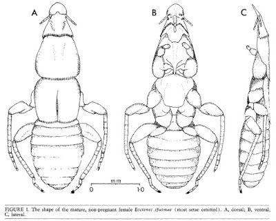
Dorsal, ventral and lateral views of Eoctenes spasmae, from Marshall (1982).


If you ever feel inclined to scan through host records for ectoparasites (and really, why wouldn't you?), you may be struck by the impression that bats seem to be peculiarly lousy animals. There seems to be an unexpected number of groups of ectoparasites that have their highest number of species on bats. One possible reason for this is that, with over 900 potential host species, bat-parasite diversity is high simply because bat diversity is high. Nevertheless, there are other features peculiar to bats that make them excellent parasite hosts. The modification of their fore-legs into wings means that their ability to groom themselves is curtailed. Because many bat species roost in dense colonies, transmission of parasites from one bat to another may happen freely. And because most bats will consistently return to the same roost, speciation is promoted by each colony becoming like an isolated island.

At the same time, referring to bats as 'lousy' is misleading because one ectoparasite group that is curiously absent from bats is the true lice (why this should be I have no idea). Instead, bats are often host to a number of parasite groups all of their own. One such group is the Polyctenidae, flightless true bugs that are found only on bats in tropical and subtropical parts of the world. Polyctenids are closely related to the bed bugs of the Cimicidae and are not dissimilar in appearance. Noticeable differences are their relatively shorter antennae and absence of eyes. They also possess a number of bristle combs at various places on the body, roughly similar in appearance to those on fleas. Their front legs are short and have sucker-like structures on the tarsi instead of claws; the hind two pairs of legs are longer and clawed. The manner of movement of the legs is specialised for crawling among the hair of their host; if removed from the host, the bug is unable to move on a flat surface. Transmission of bugs from one host to another presumably happens only through direct physical contact. Polyctenids share with bed bugs the notorious practice of traumatic insemination with each male injecting sperm directly into the female's body cavity via sharpened genitalia. However, unlike bed bugs they are viviparous, producing live nymphs instead of eggs. The developing embryos are nourished by a 'pseudoplacenta' with a single female potentially containing several developing embryos in a conveyor arrangement at different stages of development. The most mature of these embryos protrudes from the female's genital opening for some time prior to birth and may be a third of its mother's size when born (Marshall 1982).

Type specimen of Hesperoctenes giganteus, from here.


Five genera of polyctenids are generally recognised, with four genera found in the Old World and only a single genus, Hesperoctenes, in the New World (Maa 1964; Ueshima 1972). A second New World genus, Parahesperoctenes, was described in 1947 from a single female, but as the features supposedly distinguishing it from Hesperoctenes related to the consistent duplication of combs, etc., it is thought likely that this was an ordinary individual of Hesperoctenes on the cusp of moulting from a nymph to an adult (so the features of the adult cuticle were visible through the translucent nymphal cuticle). Most of the polyctenid species have a restricted host range, being found on only a single bat species or a small number of closely related species. Some species of Hesperoctenes are more flexible, being found on a range of host species. Hesperoctenes and the Old World genus Hypoctenes are found on free-tailed bats of the Molossidae. Of the other Old World genera, Adroctenes is found on horseshoe bats and leaf-nosed bats of the Rhinolophidae and Hipposideridae, Polyctenes is found on ghost bats of the Megadermatidae, and Eoctenes is found on Megadermatidae, Nycterididae and Emballonuridae. Records of polyctenids from other bat families are currently regarded as suspicious, due to either mislabelling or cross-contamination. Ueshima (1972) suggested that records of Hesperoctenes fumarius from the bulldog bat Noctilio labialis might result from bugs being transferred while the bulldog bats were sharing a roost with their more usual molossid hosts.

Relationships between the genera were discussed by Maa (1964) who divided the family between two subfamilies on the basis of comparative features; a formal phylogenetic analysis of the family appears to still be wanting. On the basis of Hesperoctenes being the 'most specialised' genus and its shared host family with Adroctenes, Maa suggested an Old World origin for Polyctenidae. Eoctenes, with its broad host family range, was regarded as 'least specialised' and likely to be evolutionarily older than other genera. Many of the features distinguishing the polyctenid genera relate to the arrangement of combs: which combs are present where and how they are developed. Prior to Maa's revision, Hesperoctenes had been regarded as likely to be primitive within the Polyctenidae due to its relatively low number of combs. The mid- and hind legs of Adroctenes are fairly short compared to those of other genera.

REFERENCES

Maa, T. C. 1964. A review of the Old World Polyctenidae (Hemiptera: Cimicoidea). Pacific Insects 6 (3): 494–516.

Marshall, A. G. 1982. The ecology of the bat ectoparasite Eoctenes spasmae (Hemiptera: Polyctenidae) in Malaysia. Biotropica 14 (1): 50–55.

Ueshima, N. 1972. New World Polyctenidae (Hemiptera), with special reference to Venezuelan species. Brigham Young University Science Bulletin, Biological Series 17 (1): 13–21.

In a Pichia

Culture of Pichia membranifaciens, from Tomas Linder.


In my previous post, I alluded to the revolutionary effect that DNA analysis had on the classification of bacteria. A similar thing happened for the study of yeasts. Previously, the taxonomy of yeasts (i.e. unicellular fungi) had suffered for the same reasons as bacterial taxonomy: a dearth of usable morphological features combined with uncertainty about the significance or otherwise of metabolic variations. With the availability of genetic information, the relations between yeast taxa became far easier to ascertain.

Needless to say, this lead to a significant shake-up in our understanding of individual yeast taxa. One of the harder-hit taxa was the genus Pichia, previously recognised as a large genus of close to 100 species. Molecular phylogenetic analyses showed that the various species of Pichia were widely scattered within the Saccharomycotina, a fungal clade that includes a large number of yeast species (including such familiar taxa as the brewer's or baker's yeast Saccharomyces cerevisiae). This probably did not come as a huge shock: part of the reason for Pichia's size was that it had not been very stringently defined. Members of this genus were characterised by multilateral budding (that is, buds could develop anywhere along the side of the yeast cell) on a narrow base. They could produce hyphae and/or pseudohyphae (except when they didn't), they might ferment sugars (except when they couldn't), and nitrate might be used as their sole source of nitrogen (except when it wasn't). Pichia spores might be hat-shaped, hemispheroidal or spherical, and they might or might not have a ledge or rim around the equator (Kurtzman 2011).

All of which adds up to a genus that probably tended to be defined as 'the genus that includes any yeast not belonging to these other genera'. In other words, the classic concept of a wastebasket taxon. As a result, the genus has been progressively pared down to a smaller array of species concentrated around the type, Pichia membranifaciens. This is a yeast commonly found as a spoilage organism on foods such as fruit or cheese. Among its other sins, it may grow as a film in the surface of wine, giving the wine an off taste. However, it's not all bad news: recently, P. membranifaciens has been studied as a potential biocontrol agent as it may produce a toxin that has an inhibitory effect on other contaminating fungi (Santos et al. 2009).

A growing culture of Komagataella pastoris, from here.


Somewhat unfortunately, one of the species to be expelled from Pichia is perhaps the best-studied: the yeast formerly known as Pichia pastoris (now supposed to be referred to as Komagataella pastoris though a quick Google Scholar search suggests that a great many authors are pretending that hasn't happened). This species can be grown using methanol as a sole carbon source, and protocols were developed in the 1970s for growing it in high densities at an industrial scale. The original plan was for it to be used for high-protein stock-feed using methanol produced as a by-product of oil refining (the modern agricultural industry has been described as the process of turning oil into food; this would have been a somewhat literal example). Rising oil prices rendered this proposal economically inviable but the P. pastoris industry was to have a reprieve, as the culture method was adopted as a means of producing active proteins (Cereghino & Cregg 2000). Procedures were developed for inserting foreign genes into the yeast, with the resulting pure methanol-based culture allowing the target protein to be generated at a greater rate and higher purity than might be possibly with a culture of the original source organism. Enzymes for laboratory studies, vaccines, medical products such as insulin: whatsitsname pastoris has been used in the production of them all.

REFERENCES

Cereghino, J. L., & J. M. Cregg. 2000. Heterologous protein expression in the methylotrophic yeast Pichia pastoris. FEMS Microbiology Reviews 24: 45–66.

Kurtzman, C. P. 2011. Phylogeny of the ascomycetous yeasts and the renaming of Pichia anomala to Wickerhamomyces anomalus. Antonie van Leeuwenhoek 99: 13–23.

Santos, A., M. San Mauro, E. Bravo & D. Marquina. 2009. PMKT2, a new killer toxin from Pichia membranifaciens, and its promising biotechnological properties for control of the spoilage yeast Brettanomyces bruxellensis. Microbiology 155: 624–634.

Alpha Bacteria

Two budding individuals of Caulobacter crescentus, from the US Dept of Energy.


From about the 1980s onwards, the increasing application of molecular data (particularly the sequences of ribosomal RNA genes) to bacterial phylogeny meant that what had previously been an intractable mass of diversity began to emerge into some sort of order. One of the major new groups of bacteria to be recognised in this way was the Proteobacteria, a hyperdiverse array that includes many of those bacteria of direct significance to ourselves. Within this bacterial supergroup, the phylogeneticists also resolved five major subgroups that, in the absence of any more obvious markers, they labelled alphabetically: the alpha, beta, gamma, delta and epsilon Proteobacteria. Eventually these convenient labels would become formalised, and it is with the group known as the Alphaproteobacteria that I am concerned today.

Like the other proteobacterial lineages, the Alphaproteobacteria are diverse in features and habits. To the best of my knowledge, no uniting characteristic has yet been identified for members of this group other than their shared ribosomal heritage. Many of the Alphaproteobacteria are associated with anoxic habitats. Many are at least facultatively photosynthetic, obtaining energy from sunlight by means of bacteriochlorophyll a and/or carotenoids; these factors give such bacteria a purple coloration. Other Alphaproteobacteria are intracellular parasites of eukaryotes, including a number that are of medical significance to humans. Earlier posts on this site have covered particular subgroups of the Alphaproteobacteria: the nitrogen fixers and plant pathogens of the Rhizobiales, and the diverse order Rhodospirillales. Another group of Proteobacteria, the Epsilonproteobacteria, was the subject of another post.

Culture of vinegar bacteria Acetobacter aceti, copyright Эрг.


A recent study of Alphaproteobacteria ribosomal phylogeny by Ferla et al. (2013) recognised three major lineages within the group which they dubbed the Magnetococcidae, Rickettsidae and Caulobacteridae. The Caulobacteridae include the greater number of the named free-living Alphaproteobacteria: both of the orders covered in earlier posts, for instance, belong to this lineage. Detailed coverage of the various members of Caulobacteridae would fill a book, so I'll just mention some highlights. The earlier post on Rhodospirillales mentioned the family Acetobacteraceae, but one important detail I neglected to mention was that many members of this family obtain their energy by oxidising ethanol to acetic acid: these are the bacteria responsible for producing ethanol. Also potentially belonging to the Rhodospirillales is Sporospirillum, a candidate genus of enormous bacteria that have been found in the intestines of tadpoles. Individuals of Sporospirillum reach up to one-tenth of a millimetre in length, potentially large enough to be observed with a standard dissecting microscope, though they are only up to 5 µm in width. Because Sporospirillum have never been cultured or studied from a molecular perspective, their relationships remain uncertain: they may alternatively belong to the Spirillaceae, a family of the Betaproteobacteria (Brenner et al. 2005).

Also belonging to the Caulobacteridae are the Caulobacterales. As recognised by Ferla et al. (2013), this order contains two families, the Caulobacteraceae and Hyphomonadaceae. Many (but not all) of the members of these families have a distinctive life cycle, in which a previously motile individual loses its flagellum and grows an elongate stalk. This now-immotile individual then produces a motile offspring by budding at one end. The manner of budding differs between the two families: in the Caulobacteraceae, the stalk functions as an attachment to the substrate and the offspring buds from the unattached end of the cell, but in the Hyphomonadaceae the stalk is not an attachment organ and the offspring buds from the end of the stalk. Similar modes of growth and budding are found in other families of the Caulobacteridae, such as the Hyphomicrobiaceae in the Rhizobiales.

TEM view of Magnetococcus marinus, showing the line of magnetic particles (magnetosomes).


The other subclasses of the Alphaproteobacteria are smaller than the Caulobacteridae in terms of numbers of named species, but this may reflect our low appreciation of bacterial diversity more than environmental reality. The Magnetococcidae are represented by only a single named species, Magnetococcus marinus. This is an aquatic chemolithoautotroph, obtaining energy from sulphur compounds. Cells of Magnetococcus contain a row of magnetic particles that the bacterium uses to orient itself. Though only one species of magnetococcid has been named to date, environmental DNA samples indicate that many more await description (Bazylinski et al. 2013).

The ciliate Paramecium, infected with Holospora (in the swollen nucleus in the lower part of the photo). Photo from here, by this network's own Psi Wavefunction (wherever she may be...)


The named members of the Rickettsidae are mostly placed in the order Rickettsiales, an assemblage of intracellular parasites of eukaryotes. This order contains two families, the Rickettsiaceae and Anaplasmataceae; a third family, the Holosporaceae, that contains intracellular endosymbionts of large protozoans such as Paramecium and Acanthamoeba, was found by Ferla et al. (2013) to be potentially closer to the Caulobacteridae than the Rickettsidae. Members of the Rickettsiales of significance to humans include those causing such diseases as typhus or spotted fever. The Anaplasmataceae also includes the genus Wolbachia which has come under the spotlight in recent years for the significance that its effects on reproductive compatibility may have for the evolution of insects.

The only free-living bacterium associated with the Rickettsidae to date is the marine Pelagibacter ubique but, again, environmental DNA samples suggest that this is merely a representative of a larger undescribed group, commonly referred to as the 'SAR11' clade. Indeed, Pelagibacter and its relatives may be the most numerous organisms on the entire planet, making up about a third of the planktonic cells in the surface layers of the world's oceans (Morris et al. 2002). Even by bacterial standards, Pelagibacter cells are small, and it has one of the smallest known genomes for any free-living organisms.

Stained sample of Pelagibacter ubique, copyright Thomas Lankiewicz & Matthew Cottrell.


There is one final important subgroup of the alphaproteobacterial lineage that I haven't mentioned yet: us. At some point in the distant past, a member of the Alphaproteobacteria developed a close and personal relationship with another micro-organism, either a member of the Archaea or a close relative thereof. Phylogenetic studies indicate that this early alphaproteobacterium was probably a close relative of the Rickettsiales. Over time, this relationship became ever closer, until the one became inseparable from the other. Together, these two microbes were to give rise to the eukaryotes, with the alphaproteobacteria becoming transformed into the mitochondria of a eukaryote cell. From the perspective of descent, then, we are all Alphaproteobacteria.

REFERENCES

Bazylinski, D. A., T. J. Williams, C. T. Lefèvre, R. J. Berg, C. L. Zhang, S. S. Bowser, A. J. Dean & T. J. Beveridge. 2013. Magnetococcus marinus gen. nov., sp. nov., a marine, magnetotactic bacterium that represents a novel lineage (Magnetococcaceae fam. nov., Magnetococcales ord. nov.) at the base of the Alphaproteobacteria. International Journal of Systematic and Evolutionary Microbiology 63: 801–808.

Brenner, D. J., N. R. Krieg & J. T. Staley. 2005. Bergey's Manual of Systematic Bacteriology 2nd ed. vol. 2 pt C. The Alpha-, Beta-, Delta-, and Epsilonproteobacteria. Springer.

Ferla, M. P., J. C. Thrash, S. J. Giovannoni & W. M. Patrick. 2013. New rRNA gene-based phylogenies of the Alphaproteobacteria provide perspective on major groups, mitochondrial ancestry and phylogenetic instability. PLoS One 8 (12): e83383. doi:10.1371/journal.pone.0083383.

Morris, R. M., M. S. Rappé, S. A. Connon, K. L. Vergin, W. A. Siebold, C. A. Carlson & S. J. Giovannoni. 2002. SAR11 clade dominates ocean surface bacterioplankton communities. Nature 420: 806–810.