Holding Forams Together

Nouria polymorphinoides, from Foraminifera.eu.


In past posts relating to the Foraminifera, I've made reference to the changes in classification undergone by this group over the years. Forams are unusual among unicellular organisms in producing a hard, often complex test that means they have both left an extensive fossil record and provided a number of characters on which to base a classification. However, there has been much disagreement over the relative attention due to particular features of the test. The classification used for forams in the Treatise on Invertebrate Paleontology by Loeblich & Tappan (1964), one of most influential sources in recent decades, made its primary divisions on the basis of the structure and chemistry of the test itself. Forams that produce a test by gluing together (agglutinating) sand particles and other foreign objects were treated as fundamentally distinct from those that secreted calcareous tests. Because the foram cell itself is amoeboid, there was an underlying assumption that the test architecture was too mutable to indicate anything more than low-level relationships.

However, there were some prominent inconsistencies with this assumption (Mikhalevich 2013). One is that the division between agglutinated and calcareous tests is not always perfect. Agglutinated forams might not secrete the bulk of the test themselves but they do secrete the cement used to hold the sand grains together, and there is a definite spectrum in the proportion of sand to cement used by a given foram. In some agglutinated forms, a distinct calcareous layer may underlie the agglutinated section of the test, and it is easy to envision how a progressive reduction in the proportion of agglutinated material could lead to the evolution of an entirely secreted test. This was not in itself fatal to the earlier system as it had generally been assumed that agglutinated forams were likely to represent a paraphyletic group. More problematic was the common appearance of foram species that were extremely similar in test architecture with the only really significant difference being that one was agglutinated and the other calcareous. This lead some authors to argue that whereas a small number of such cases might be accepted as the result of convergence, the abundance of such cases suggested that changes in test composition were more common than previously recognised. Molecular studies of forams are still in their infancy but have offered some support for the significance of test architecture, such as the division between globular and tubular forams (Pawlowski et al. 2013) that I referred to in an earlier post.

Liebusella goesi, from Foram Barcoding.


One effect of this change in focus is that the Mikhalevich (2013) classification divides the agglutinated forams between a number of groups that are not recognised in alternative systems. One such group is the Nouriida, known from the Cretaceous to the present day. Mikhalevich included the Nouriida in a larger group called the Hormosinana; at least one hormosinanan was placed by Pawlowski et al. (2013) at the base of the globular foram lineage. In contrast, Loeblich & Tappan (1964) included most of the nouriidans in the family Ataxophragmiidae, other members of which belong to the tubular forams. Nouriida and other Hormosinana are united by having the aperture of the test in a terminal position; in some nouriidans, it may be raised on a short neck. Nouriida differ from other hormosinanans in the arrangement of chambers in the test. In early stages they tend to be more or less trochospiral; with maturity, the number of chambers to a whorl decreases and the test may become biserial or uniserial. The two subfamilies recognised within the Nouriida by Mikhalevich differ in the internal structure of their chambers: Nourioidea have internally simple chambers but Liebuselloidea have the lumen of the chambers complexly subdivided.

I haven't found much about their ecological role; at least one modern species, Nouria polymorphinoides, seems to be not uncommon in shallower continental shelf waters worldwide. My general impression (just confirmed by asking a colleague who actually works on forams) is that agglutinated forams receive far less attention than calcareous ones. A big part of this is simply that they're harder to find: it takes a lot of practice to be able to pick out an actual agglutinated foram test from any other conglomeration of sand, and if they break apart during sample prep (which they often do) then there is little sign they were ever there to begin with.

REFERENCES

Loeblich, A. R., Jr, & H. Tappan. 1964. Treatise on Invertebrate Paleontology pt C. Protista 2. Sarcodina: chiefly "thecamoebians" and Foraminiferida vol. 1. The Geological Society of America, and The University of Kansas Press.

Mikhalevich, V. I. 2013. New insight into the systematics and evolution of the Foraminifera. Micropaleontology 59 (6): 493–527.

Pawlowski, J., M. Holzmann & J. Tyszka. 2013. New supraordinal classification of Foraminifera: molecules meet morphology. Marine Micropalaeontology 100: 1–10.

Walruses, Sea Lions and Fur Seals

Adaptation to a primarily aquatic lifestyle has happened numerous times within mammals, but some groups have radiated more in this environment than others. One particularly well-known group of marine mammals is the pinnipeds, the seals and sea lions.

Australian sea lions Neophoca cinerea on a beach on Kangaroo Island, copyright Diver Dave.


Pinnipeds are highly modified for life in the water, with streamlined bodies and all four limbs modified into flippers. When I was young, many of the animal books that I read referred to pinnipeds as their own distinct order within the mammals. However, it has long been recognised that pinnipeds are derived from within the Carnivora and these days they are almost universally treated as a subgroup of the latter. Modern pinnipeds are divided between three families: the Phocidae ('true' seals), Otariidae (fur seals and sea lions) and Odobenidae (which has only one living species, the walrus Odobenus rosmarus). While some morphological analyses have argued for a relationship between the walrus and the Phocidae, the majority view treats the walrus and the Otariidae as together forming a clade Otarioidea, commonly referred to as the eared seals. There has historically also been some argument about whether the pinnipeds represent a single clade; some have argued for two separate origins, Otarioidea being related to bears whereas Phocidae were supposed to be closer to otters and weasels. However, the current majority supports a single origin for the group.

Northern fur seals Callorhinus ursinus, photographed by M. Boylan.


Eared seals differ from true seals in the possession of small external ears, and the ability to turn the hind flippers back under the body so that they can still function (if somewhat awkwardly) as feet when moving on land. I have seen Australian sea lions on coastal islands near Perth (there are boat tours that will take you to see them) and I can confirm that they can run along the beach at a surprising speed when they wish to. True seals have the hind flippers permanently directed behind them and so are forced to awkwardly belly-flop along when not swimming (doubtless as a result of this, true seals also differ from eared seals in that males lack an external scrotum). In the water, the hind flippers provide the main source of propulsion in true seals whereas eared seals get more of their thrust from the fore flippers (sea lions have been said to swim like penguins). As an aside, eared seals are also apparently unusual among mammals in that their milk completely lacks lactose. The lactose intolerant among you need not be denied dairy, you need only milk a walrus.

Mounted skeleton of Allodesmus sp., copyright Momotarou2012.


The earliest eared seals are known from the Miocene when they appear to have originated in the northern Pacific. Two extinct families from this place and period, the Enaliarctidae and Desmatophocidae, are commonly included in the Otarioidea, though it remains possible that either of these families should be placed outside the pinniped crown group, or closer to the true seals. The early Miocene Enaliarctidae differ from other otarioids in retaining differentiated premolars and molars (later forms have the cheek teeth uniform in appearance) and may well represent the ancestral form of the group. The mid- to late Miocene Desmatophocidae combined a rather Phocidae-like skull with a more Otarioidea-like post-cranium; the best-known genus Allodesmus had larger eyes than other otarioids and may have hunted in deep waters. One species of desmatophocid, Allodesmus sinanoensis, may have reached a length approaching five metres, making it larger than a modern walrus and rivalling the elephant seals in size. I highly recommend a series of posts on Allodesmus written a few years back by Robert Boessenecker (1, 2, 3, 4) that cover just about everything you might want to know about this animal.

Skull of Gomphotaria pugnax, from Robert Boessenecker.


Though only one walrus species is generally recognised in the modern fauna, the family was much more diverse in the past. However, most fossil Odobenidae lacked the tusks of a modern walrus and would have been more similar at a glance to sea lions. These early odobenids would have probably been generalist fish-feeders (Boessenecker & Churchill 2013). The modern walrus, in contrast, feeds primarily on bivalves. They don't crush the clam's shell but grab it with their lips and then suck powerfully enough that the meat is ripped out. Other than the tusks, the teeth of a modern walrus are small and weak; one close fossil relative, the Pliocene Valenictus chulavistensis, went so far as to lose the non-tusk teeth entirely. The tusks themselves are usually thought to function in display and the like rather than having any prominent role in feeding. However, it is an intriguing detail that the fossil whale Odobenocetops that converged in its feeding biology with walruses also possessed a large tusk. The non-tusk teeth were still used in feeding in the fossil clam-feeding walrus genera Dusignathus and Gomphotaria, which had a pair of large forward-directed tusks in both the upper and lower jaws.

Suckling South African fur seals Arctocephalus pusillus, copyright Robur.q.


The majority of living eared seals belong to the Otariidae, which have been divided in the past between the fur seals and sea lions. Fur seals tend to be smaller than sea lions and possess a dense layer of underfur. However, more recent phylogenetic studies (particularly molecular ones) have thrown this distinction out the window (e.g. Higdon et al. 2007). Instead, the northern fur seal Callorhinus ursinus of the north Pacific is probably the sister species to all other living otariids. Even the southern fur seals, generally placed in a single genus Arctocephalus, may not be monophyletic relative to the New Zealand sea lion Phocarctos hookeri (as a result, some authors have suggested resurrecting the genus Arctophoca for all southern fur seals other than the South African fur seal Arctocephalus pusillus). The South American fur seal Otaria flavescens may also be associated with this latter group. The two north Pacific sea lions, Steller's sea lion Eumetopias jubatus and the Californian sea lion Zalophus californianus, form a clade outside the southern otariids. The remaining species is the Australian sea lion Neophoca cinerea whose position has been harder to pin down: some analyses place it close to the New Zealand sea lion but others position it well away from all other southern otariids, possibly even outside all other otariids except the northern fur seal.

Walruses Odobenus rosmarus crowded on shore, from here.


Fur seals and sea lions were heavily hunted in the past for pelts and oil and some species remain endangered. Climate change poses a particular threat to cold-water species; for instance, recent years have seen significant contractions in walrus ranges, leading to dramatic crowding in the locations remaining. Conversely, the Antarctic fur seal Arctocephalus gazella, once feared extinct, has apparently exhibited a population explosion in recent decades, perhaps because lowered whale populations have led to more food being available for seals.

REFERENCES

Boessenecker, R. W., & M. Churchill. 2013. A reevaluation of the morphology, paleoecology, and phylogenetic relationships of the enigmatic walrus Pelagiarctos. PLoS One 8 (1): e54311.

Higdon, J. W., O. R. P. Bininda-Emonds, R. M. D. Beck & S. H. Ferguson. 2007. Phylogeny and divergence of the pinnipeds (Carnivora: Mammalia) assessed using a multigene dataset. BMC Evolutionary Biology 7: 216.

Repenning, C. A., & R. H. Tedford. 1977. Otarioid seals of the Neogene. Geological Society Professional Paper 992: i–vi, 1–93, 24 pls.

Turridae

Shell of Turris crispa crispa, copyright H. Zell.


At this point, I've made numerous references on this site to the gastropod family Turridae, discussing its members and non-members and alluding to its sordid history. So maybe I should set out the basics of the story properly.

The Conoidea are a diverse group of marine predatory gastropods with over 4000 known living species. They are best known for the production by many species of venom used to paralyse their prey, in some species being potent enought to threaten humans. In the majority of conoideans, this venom is delivered via a tooth that becomes detached from the radula and is held at the end of the retractable proboscis. Until relatively recently, Conoidea were commonly divided between three families. Two of these families, the Conidae (cone shells) and Terebridae (awl shells) were well defined and constrained. The third family was the Turridae, including by far the greater number of species but not really defined within Conoidea beyond 'the rest'. Many of 'the rest' were small, many were restricted to deep water, many were poorly known. Different systems were proposed over the years in an attempt to break the turrid mass into more manageable units but each system differed significantly from the next and no one system became universally accepted. Some authors would focus on the protoconch as their guide to classification, others would focus on the radula, others might call out features of the operculum. One author commented in 1922 that turrids were "considered by those who meddle with them to be more perplexing than any other molluscan family", and this complaint was still being upheld by Kilburn (1983) over sixty years later.

Though it had long been accepted that the 'turrids' probably did not represent an evolutionarily coherent group, it wasn't really until the advent of molecular phylogenies that things started falling into place. Puillandre et al. (2011) identified two main lineages within the Conoidea, leading to the dissolution of the original Turridae into no less than 13 families in order to maintain the already-established Conidae and Terebridae. Turridae in the strict sense was restricted to a much smaller clade of a bit over a dozen genera, sister to the Terebridae (Bouchet et al. 2011).

In contrast to the bewilderment of the original turrid array, Turridae sensu Bouchet et al. is a morphologically quite coherent group. They are more or less fusiform (spindle-shaped) shells, often with a narrow, high spire and relatively weak sculpture. Indeed, but for the fact that most tend to have a long siphonal canal at the base of the shell, they often bear a distinct resemblance to their sister group, the terebrids. The majority of turrids have a multispiral protoconch, indicating an extended, planktonic-feeding larval stage in development, but there are some species with a paucispiral protoconch indicative of direct development.

Radula of Xenuroturris legitima, from Kantor & Puillandre (2012); ct = central tooth.


The radula of turrids usually comprises three apparent teeth in each row. The central tooth is actually formed from three teeth (the original pointed central tooth and two plate-like lateral teeth) fused together; in some species the division between these teeth remains visible whereas in others the central tooth disappears entirely. The main business part of the radula is the single pair of marginal teeth which, as in other conoideans, are enlarged and modified for venom delivery. They have a distinctive 'duplex' form; in older publications, this was referred to as a 'wishbone' form because the tooth appears under light microscopy to be divided between two branches. After the advent of electron microscopy, it was discovered that these two 'branches' in fact represent the thickened margins of an undivided tooth. The larger of the two margins is mostly attached to the radular membrane with only the tip of the tooth being free; the smaller margin is held free of the radula. The thinner part of the tooth between the two margins forms a gutter along which venom can flow. However, the radula is placed in such a position that it cannot be protruded through the mouth in the manner of grazing gastropods. As with other conoideans, prey (in this case probably worms) is despatched through the use of a detached marginal tooth transferred to the end of the proboscis. However, whereas other conoideans such as cone shells may have the tooth functioning like a hypodermic syringe for delivering prey, turrids use their tooth to slash at the prey like a switchblade, with venom passively entering through the resulting cuts. The proboscis is then used to draw the prey back into the mouth, where the radula is used to grasp and swallow it, sucking the unlucky worm down the gullet like spaghetti.

REFERENCES

Bouchet, P., Y. I. Kantor, A. Sysoev & N. Puillandre. 2011. A new operational classification of the Conoidea (Gastropoda). Journal of Molluscan Studies 77: 273–308.

Kantor, Y. I., & N. Puillandre. 2012. Evolution of the radular apparatus in Conoidea (Gastropoda: Neogastropoda) as inferred from a molecular phylogeny. Malacologia 55 (1): 55–90.

Kilburn, R. N. 1983. Turridae (Mollusca: Gastropoda) of southern Africa and Mozambique. Part 1. Subfamily Turrinae. Annals of the Natal Museum 25 (2): 549–585.

Puillandre, N., Y. I. Kantor, A. Sysoev, A. Couloux, C. Meyer, T. Rawlings, J. A. Todd & P. Bouchet. 2011. The dragon tamed? A molecular phylogeny of the Conoidea (Gastropoda). Journal of Molluscan Studies 77: 259–272.

Blister Beetles

Zonitis sayi, copyright Carol Davis.


This is a blister beetle of the genus Zonitis. Blister beetles, the family Meloidae, get their name from their production of cantharidin, a defensive chemical that can burn the skin of would-be predators. Zonitis is a widespread genus of blister beetles with over 100 species described from around the world. However, it should be noted that its wide distribution may relate to the genus being poorly defined and future revisions may divide its members between other genera (as I believe has already happened for the Australasian 'Zonitis'). As it is, Zonitis species are characterised by fully developed elytra and functional wings, and cleft tarsal claws with two rows of teeth on the upper section (Enns 1956). Adult Zonitis are flower-feeders, visiting composite plants (i.e. daisies and similar plants), and some species have the mouthparts modified into a tube for sucking nectar.

Most blister beetles exhibit what is known as hypermetamorphism or hypermetaboly, where the larvae pass through morphologically differentiated stages before reaching pupation. Zonitis species develop as parasitoids or kleptoparasites of bees. Females lay large numbers of eggs (up to and exceeding 500 in a batch) on their host plant, most commonly on the flowers though sometimes on the undersides of leaves. The eggs hatch into active, long-legged larvae that attach to bees visiting the flowers and so get carried to the bee's nest. Once there, they moult into a less mobile stage and feed on the food stores laid aside for the bee's larva, and potentially on the larva itself. In the North American species Zonitis atripennis flavida, the beetle larva completes its development in a single cell but European species consume the contents of two bee cells before reaching maturity. Following the initial active instar, meloid larvae pass through four feeding instars before entering a quiescent, immobile stage called the hypnotheca or prepupa. The hypnotheca moults into another feeding instar before the larva finally enters the pupal stage (Bologna et al. 2008). What the point (if anything) of the hypnotheca is, I have no idea. However, it is worth noting that hypnothecae of another meloid species, Hornia boharti, have been recorded surviving for multiple years without feeding before moulting to the next instar.

Once adults emerge from the host cell, they of course disperse to conduct their own affairs. Natural history data is patchy but indications are that many species are picky in their choice of host plant. From an economic perspective, their damaging role as a parasite of pollinating bees may be partially counterbalanced by their potential role as pollinators in their own right, but who can say which way the scales lean?

REFERENCES

Bologna, M. A., M. Oliverio, M. Pitzalis & P. Mariottini. 2008. Phylogeny and evolutionary history of the blister beetles (Coleoptera, Meloidae). Molecular Phylogenetics and Evolution 48: 679–693.

Enns, W. R. 1956. A revision of the genera Nemognatha, Zonitis, and Pseudozonitis (Coleoptera, Meloidae) in America north of Mexico, with a proposed new genus. University of Kansas Science Bulletin 37 (2): 685–909.

Cicadomorpha

Textbooks will tell you that the term 'bug' should be restricted to insects of the order Hemiptera though, as I've noted before, I don't know if I've ever met anyone who actually used the word that way. For many people, one of the groups of actual bugs that they are most likely to be aware of are members of the Cicadomorpha.

Tasmanian hairy cicada Tettigarcta tomentosa, copyright Simon Grove.


Cicadomorphs include the cicadas (Cicadoidea), leafhoppers (Membracoidea) and spittlebugs (Cercopoidea). As a group, they are distinguished by an enlarged postclypeus (the upper part of the front of the head below the antennae), simple antennae with a whip-like flagellum, and small and narrowly placed mid-coxae (Dietrich 2005). The enlarged postclypeus is associated with adaptations for feeding on xylem, deeper in the plant stem than many other plant-sucking bugs prefer, though derived subgroups of the leafhoppers have changed back to phloem or parenchyma. Well over 30,000 species of cicadomorph are known from around the world. Cicadas can be readily distinguished from other cicadomorphs by their possession of three ocelli in a triangle on the top of the head whereas leafhoppers and spittlebugs have only two or no ocelli.

Male bladder cicada Cystosoma saundersii, one of the world's more ridiculous animals, from Brisbane Insects.


Cicadas are best known, of course, for their singing. The songs are produced by a pair of membranous 'drums', the tymbals, at the base of the abdomen; muscular vibration of the membranes produces the sound. In most cicadas, only the male possesses these tymbals. However, both sexes possess tymbals in the hairy cicadas Tettigarcta, two species found in alpine regions in south-eastern Australia. Hairy cicadas also differ from the remaining cicadas in other ways, most notably in lacking the well-developed tympana on the underside of the abdomen that typical cicadas hear with (hairy cicadas have simpler hearing organs in their place). As a result, Tettigarcta is placed in its own distinct family, sister group to the remaining cicadas in the Cicadidae. Though now restricted to Australia, fossil species from the Mesozoic and Palaeogene of other parts of the world have also been placed in the Tettigarctidae (Shcherbakov 2008); however, they are mostly so placed on the basis of shared primitive rather than derived features and may well represent stem taxa for Cicadoidea as a whole. Other derived features of the cicadas proper in the Cicadidae include gas-filled chambers in the abdomen that resonate the calls produced by the tymbals. In males of another Australian species, the bladder cicada Cystosoma saundersii, these resonating chambers reach a remarkable size and the entire abdomen looks to have been blown up like a beach ball.

Froghopper Cercopis vulnerata, copyright Richard Bartz.


The spittlebugs or froghoppers of the Cercopoidea are smaller cicadomorphs, distinguished from species of the Membracoidea by their short and cylindrical (rather than long and quadrate) hind tibiae. The name 'spittlebug' refers to the nymphs of these bugs living covered with a protective covering of foam. In one family, the Machaerotidae, the nymph produces a calcareous tube around itself that it fills with fluid. The foam or fluid used for protection by cercopoids is primarily composed of the nymph's own excrement: the xylem fluids that they feed on are mostly water, after all, so they produce a large quantity of watery excreta.

Mango leafhopper Idioscopus nagpurensis, one of the world's many, many species of Cicadellidae, copyright Arian Suresh.


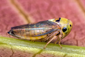
The third main subgroup of the cicadomorphs, the Membracoidea, is by far the most diverse, particularly the largest family Cicadellidae (leafhoppers). My own impression from my experience of collecting insects in various locations is that cicadellids are just everywhere. Over 20,000 species of this family have been described to date, and it has been estimated that the true number may be much higher. For instance, at one location in North America close to 100 species of a single genus Erythroneura have been recorded from a single plant (Dietrich 2002). Just how such a high diversity of closely related species can live in such close proximity remains a largely unanswered question, though some studies have apparently suggested the possibility of very fine micro-habitat partitions (making sense of the great mass of cicadellid diversity is not helped by many species exhibiting dimorphism between flying and flightless forms, similar to that I recently described for delphacids). Another notable feature of cicadellids is the protection of brochosomes, tiny, hollow, soccerball-like granules constructed of protein nets with which the leafhopper coats itself after moulting. The hydrophobic brochosomes help to keep the hopper free of water droplets and its own wet, sticky excreta. They may also serve other protective functions: females will coat newly laid eggs with a layer of brochosomes that may serve to prevent egg parasitoids such as micro-wasps from attacking the eggs.

Membracid leafhopper Cladonota benitzei, copyright P. Lahmann.


The membracoids also include the Membracidae, renowned for the remarkable appearance of the pronotal shield (the top and front of the thorax) in many species. In more humble membracids, the pronotum may form a high mound or pillar, but in others it may extend into bizarre arrangements of globules and branched spines hanging above the leafhopper like a baroque chandelier. Again, just what the purpose of this extravagant morphology is remains unknown but many authors have proposed some sort of protective function. It has been suggested that pronotal projections may help membracids mimic part of their host plant, or potential predators such as parasitic wasps. Alternatively, they mean that potential predators such as birds find the hopper just too hard to swallow.

REFERENCES

Dietrich, C. H. 2002. Evolution of Cicadomorpha (Insecta, Hemiptera). Denisia, Neue Folge 4 (176): 155–170.

Dietrich, C. H. 2005. Keys to the families of Cicadomorpha and subfamilies and tribes of Cicadellidae (Hemiptera: Auchenorrhyncha). Florida Entomologist 88 (4): 502–517.

Shcherbakov, D. E. 2008. Review of the fossil and extant genera of the cicada family Tettigarctidae (Hemiptera: Cicadoidea). Russian Entomological Journal 17 (4): 343–348.