Field of Science

Showing posts with label Parasitiformes. Show all posts

In Honour of Amblyseius

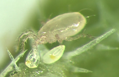
At this point in time, the Phytoseiidae are one of the most intensely studied families of mites. They are the only group of mesostigmatan mites to have significantly diversified among the foliar environment (on and around plant leaves) where they are mostly predators on other small invertebrates. The taxonomic history of phytoseiids is storied and complex but one taxon that has been consistently recognised as a major part of the family is the genus Amblyseius.

Swirski mite Amblyseius swirskii, from here.


When reviewed by Chant & McMurtry in 2004, Amblyseius was a sizeable assemblage of close to 350 known species (I quite expect that number to have expanded by now). Species of Amblyseius are lightly sclerotised, mostly pale in colour, and usually have a smooth shield covering most of the dorsum. The genus is characterised by the presence of eighteen or nineteen pairs of setae on the dorsum of the idiosoma (the central body) with three sublateral pairs being particularly long: one about the level of the third pair of legs (referred to as the s4 pair) and the other two towards the rear of the body. Except for a few pairs forward of the s4 setae, the remaining dorsal setae are all minute.

The primary focus of human interest in phytoseiids has been their role as predators of crop pests. I described some of the ways in which phytoseiids have been commercially utilised in an earlier post. Species used in this way include several Amblyseius though matters are complicated slightly by changes in taxonomy (for instance, one species which has been widely traded as Amblyseius cucumeris is now placed in the genus Neoseiulus). One of the most widely used of the commercial phytoseiids in recent years has been Amblyseius swirskii, commonly known as the Swirski mite (E. Swirski being an acarologist after whom the species was named). This species was first described in 1962 from almond trees in Israel and subsequently identified from a wide range of plant and crop species. Its history in pest control has been described in detail by Calvo et al. (2015).

The Swirski mite feeds on a range of prey, including mite, thrips and whitefly species, as well as on pollen and micro-fungi. It was first promoted as a commercial control for silverleaf whitefly Bemisia tabaci in the early 2000s. However, it did not get taken up in a big way until media publicity about pesticide residues on capsicum crops in Spain led to a crash in demand. Farmers in that country were forced to look for alternative means of pest control and found great success with A. swirskii (previous attempts to use the cooler-clime preferring Neoseiulus cucumeris in Spain had not been promising). Since then, the Swirski mite has been adopted in numerous countries for use on a range of crops to control various pests such as western flower thrips Frankliniella occidentalis. Because of its ability to grow and thrive on non-insect foods, including artificial diets, this mite is easily cultured commercially. It may also be released on crops before pest infestations develop, building up numbers on a diet of pollen until suitable prey presents itself. For the same reason, Swirski mite populations do not crash before pest control is complete. Overall, a remarkable success and a prime example of the value of Amblyseius species to mankind.

REFERENCES

Calvo, F. J., M. Knapp, Y. M. van Houten, H. Hoogerbrugge & J. E. Belda. 2015. Amblyseius swirskii: what made this predatory mite such a successful biocontrol agent? Experimental and Applied Acarology 65: 419–433.

Chant, D. A., & J. A. McMurtry. 2004. A review of the subfamily Amblyseiinae Muma (Acari: Phytoseiidae): part III. The tribe Amblyseiini Wainstein, subtribe Amblyseiina n. subtribe. International Journal of Acarology 30 (3): 171–228.

The Dermacentor Ticks

Pacific Coast tick Dermacentor occidentalis, copyright Jerry Kirkhart.


Among the ticks of most concern to humans are species of the genus Dermacentor. This genus of about forty known species is widely distributed in Africa, Eurasia and the Americas. Examples include the meadow tick D. reticulatus in Europe, and the wood tick D. variabilis and Rocky Mountain wood tick D. andersoni in North America. They are parasites of mammals, including both generalist and more host-specific species; records of Dermacentor individuals from reptiles and even carpenter bees (Goddard & Bircham 2010) presumably represent incidental and/or accidental associations. Species of Dermacentor are responsible for the spread of bacteria causing diseases such as Rocky Mountain spotted fever (which, despite sounding like a 1950s dance craze, is presumably not much fun), Q fever and tularemia. The ticks can also be more directly hazardous, as their bites inject a toxin that can cause tick paralysis.

Distinguishing features of Dermacentor species relative to other ticks include a rectangular base to the capitulum, relatively short, broad palps, well-developed eyes and the presence of festoons (impressed divisions of the posterior margin of the body) (Keirans 2009). Most are ornate—that is, marked on the dorsum with contrasting pale patterns—with the notable exception of the tropical horse tick D. nitens of the Americas (until recently, often treated as forming its own genus Anocentor). The function of such markings is unknown though suggestions include environmental protection, warning predators of distastefulness, or sexual signalling.

Meadow tick Dermacentor reticulatus, copyright Ferran Turmo Gort.


The majority of Dermacentor species have a three-host life cycle, dropping off the host between each of the life stages of larva, nymph and adult, and seeking out a new host after moulting. However, at least two New World species, the aforementioned D. nitens and the winter tick D. albipictus (a parasite of deer), are one-host ticks that remain on their original host between instars. In general, Dermacentor species are more resilient to dry climates than many other tick species. Individual species can differ in their climate tolerance, however. In North America, the geographical divide between D. variabilis in the east of the continent and D. andersoni in the west seems to be driven by the need for the latter of drier conditions (Yoder et al. 2007). Older instars also tend to be hardier than younger. Females of the ornate sheep tick D. marginatus, a European species, leave their host after gorging at the beginning of winter and then wait for more amoenable spring conditions before laying their delicate eggs (Dörr & Gothe 2001).

Higher relationships within the genus do not appear to have been extensively studied. A preliminary molecular phylogeny of hard ticks has suggested the possibility of a basal division between Afrotropical, Eurasian and New World lineages (Barker & Murrell 2004). Comparison with related tick genera raises the possibility of an Afrotropical origin for Dermacentor, though the genus has only a relictual presence in that continent now. However, with only a handful of species subjected to broad phylogenetic analysis to date, further testing is demanded. Does the continental divide hold true? Do the one-host species form a single clade within the genus? Inquiring minds wish to know.

REFERENCES

Barker, S. C., & A. Murrell. 2004. Systematics and evolution of ticks with a list of valid genus and species names. Parasitology 129: S15–S36.

Dörr, B., & R. Gothe. 2001. Cold-hardiness of Dermacentor marginatus (Acari: Ixodidae). Experimental and Applied Acarology 25: 151–169.

Goddard, J., & L. Bircham. 2010. Parasitism of the carpenter bee, Xylocopa virginica (L.) (Hymenoptera: Apidae), by larval Dermacentor variabilis (Say) (Acari: Ixodidae). Systematic and Applied Acarology 15: 195–196.

Keirans, J. E. 2009. Order Ixodida. In: Krantz, G. W., & D. E. Walter (eds) A Manual of Acarology 3rd ed. pp. 111–123. Texas Tech University Press.

Yoder, J. A., D. R. Buchan, N. F. Ferrari & J. L. Tank. 2007. Dehydration tolerance of the Rocky Mountain wood tick, Dermacentor andersoni Stiles (Acari: Ixodidae), matches preference for a dry environment. International Journal of Acarology 33 (2): 173–180.

Ferreting up a Bird's Nose

Mites, as I may have commented before, seem to have an almost fractal level of diversity: the closer you look, the more there is of it. This is nowhere more apparent than when it comes to parasitic mites which infest almost any host in any way that you can imagine. For the subject of this post, I drew one such mite: the honeyeater nasal mite Ptilonyssus myzanthae.

Venter (left) and dorsum of female Ptilonyssus myzanthae, from Domrow (1964). The scale bar equals 500 µm.


Bird nasal mites of the family Rhinonyssidae are, as their name indicates, inhabitants of the nasal passages of birds. General adaptations of the family for their parasitic lifestyle include tendencies towards reduction of the body sclerotisation and reduction in the length and number of setae. They use the claws on their front legs to tear openings in the host's mucous membranes and then feed on its blood. Transmission of nasal mites seems to happen during bill-to-bill contact such as when parents are feeding their young or during mating activities, or indirectly through water or on the surface of perches or the like. Rhinonyssid nasal mites are not known to transmit any actual diseases between hosts but they can cause the formation of lesions or inflammation or the like. All in all, probably not very pleasant for the bird (see here for some more details).

Whole-body illustration of a different rhinonyssid species, from Greg Spicer.


Nevertheless, infection rates in bird populations can be very high and most (if not all) bird species will be host to some nasal mite species. Most species of nasal mite are very host specific, known on only one or a few bird species (it must be noted, though, that the question of just how many researchers choose to look up a bird's schnozz in search of mites may not be irrelevant here). Ptilonyssus myzanthae was described by Domrow (1964) from two species of honeyeater in Queensland, Australia: the noisy miner Manorina melanocephala and the little wattlebird Anthochaera chrysoptera. Distinctive features of this species compared to others in the genus include a subhexagonal anterior dorsal shield on the body, a narrow genital shield, and a divided pygidial shield (the small pair of shields near the rear of the dorsum). Both of the known hosts are widespread and common in eastern Australia and it is likely that this mite is similarly ubiquitous. Studies of honeyeater phylogeny tend to place the genera Manorina and Anthochaera as close relatives, so it is possible that P. myzanthae has been infesting them since before their lineages diverged. It would be worth looking for the species in other related honeyeaters to see if we find any further clues.

REFERENCE

Domrow, R. 1964. Fourteen species of Ptilonyssus from Australian birds (Acarina, Laelapidae). Acarologia 6 (4): 595–623.

Zerconids

Slide-mounted male of Zercon gurensis, copyright Holger Müller.


The animal depicted above is a mite of the Zerconidae, one of the numerous families in the major mite clade known as the Mesostigmata. This family is mostly found in soil habitats such as leaf litter, mosses, decaying vegetation, or occasionally in animal nests (Lindquist et al. 2009). The zerconids are restricted to the Northern Hemisphere and are most diverse in temperate to Arctic regions; those species found in tropical parts of the world are restricted to high altitudes away from the hot lowlands (Ujvári 2012). Like many other Mesostigmata, they have the dorsal surface of the body mostly covered by shields of hardened cuticle. In most zerconids, separate shields cover the front (podonotal) and rear (opisthonotal) sections of the dorsum; the opisthonotal shield wraps around the rear margin of the mite and forms a continuous unit with the ventrianal shield that usually protects most of the underside of the mite behind the legs. Among the most noticeable features of the zerconids are two pairs of large openings near the rear of the opisthonotal shield (the four orange-segment-like structures in the photo above). These represent the openings of secretory glands, but I don't know if it has been established just what they're secreting; comparable structures in other mites may secrete pheromones, or defensive chemicals, or oils that prevent debris from sticking to the body. Other features of the zerconids include slender, relatively simple chelicerae that lack the modifications seen in the males of some other Mesostigmata, and peritremes (grooves on the underside of the body that channel air to the openings of the respiratory stigmata) that are relatively short. These peritremes are longer in zerconid nymphs, but become shortened when the mite moults to maturity.

Zerconids are another of those mite groups where the vast majority of what has been written about them relates to their basic taxonomy, with little yet known about their natural history. Several genera of zerconids are recognised, distinguished by features such as the shape of the body's various shields and the appearance of various setae. The form of their chelicerae indicates that zerconids are predatory like many other Mesostigmata. Because they are mostly found at ground level rather than on vegetation they have not attracted the economic interest of other predatory mites, but those few species that have been observed feeding were chowing down on nematodes. Mating does not appear to have been directly observed in zerconids, but again their anatomy and comparison with other mesostigs allows us to infer that the male fertilises the female by using his chelicerae to pass a spermatophore from his own genital opening on the underside of the body between the legs to hers. Where she then lays her eggs, and how her offspring spend their time to maturity, seem to be questions still awaiting an answer.

REFERENCES

Lindquist, E. E., G. W. Krantz & D. E. Walter. 2009. Order Mesostigmata. In: Krantz, G. W., & D. E. Walter (eds) A Manual of Acarology 3rd ed. pp. 124–232. Texas Tech University Press.

Ujvári, Z. 2012. Draconizercon punctatus gen. et sp. nov., a peculiar zerconid mite (Acari: Mesostigmata: Zerconidae) from Taiwan. Opusc. Zool. Budapest 43 (1): 79–87.

Phytoseiids and the Importance of Taxonomy

Gupta's (1975) original figures for Amblyseius syzygii.


For this week's semi-random post, I drew the mite species Amblyseius syzygii. Or perhaps that should be Typhlodromips syzygii, as it's more likely to be designated now. Typhlodromips syzygii is a member of the Phytoseiidae, a diverse family of about 1600 known species of predatory mite. Despite, or perhaps because of, their being an economically significant group (more on that in a bit), phytoseiids have been somewhat plagued by competing nomenclatural systems. Until relatively recently, they were predominately classified into just a few, very large genera. In the last couple of decades, however, there has been a move towards a much more finely divided classification, but you will still find many sources that will continue to use the more conservative system, particularly among those with more of an economic interest in the group than a taxonomic one.

It is as a result of this taxonomic changing of the guard that syzygii, once nestled in the broad genus Amblyseius, has been separated as part of a genus Typhlodromips that was placed by Chant & McMurtry (2005) in an entirely separate tribe from its former host. The genera are separated by features such as the number, shape and proportions of the dorsal setae, and the arrangement of macrosetae on the legs. In fact, counting setae seems to be a major part of taxonomy in the Mesostigmata (the major mite group to which the phyoseiids belong) as a whole, which is part of why this is one group of mites I've so far had difficulty in coming to terms with. Counting setae sounds like it should be easy, but in my experience it's usually not. Especially when the animal is slide-mounted, requiring you to move to focus on the microscope up and down in order to see all the setae, leading to confusion about whether a given visible setae is one you've already counted or not.

Anywho, even after the split, T. syzygii is one of about sixty species in its genus. It was first described in 1975 from West Bengal, from a specimen collected on a jambul tree Syzygium cumini (Gupta 1975; hence the species name). Since then, it has been recorded all around southern and eastern Asia, and from a wide variety of different plants. Distinguishing features of the species include (again) proportions of the dorsal setae, with T. syzygii possessing features such as a pair of large posterolateral serrate setae, as well as details such as the shape of the ventral plates on the body.

A related amblyseiid, Amblyseius swirskii, attacking a thrips. Copyright Steven Arthurs.


I referred before to the economic significance of phytoseiids. This is because a number of species in this family have been utilised as biocontrol agents for plant-feeding mites and other minute pests such as thrips. In some places, you can even buy commercially-produced satchels containing colonies of phytoseiids that can be hung in an orchard and allow the mites to disperse among your trees (arachnids in a bag, people, arachnids in a bag). Alternatively, industrial-size blowers may be used to fire clouds of mites across a crop. I have come across reference to Amblyseius syzygii as a predator of the tea red spider mite Oligonychus coffeae, a significant crop pest. The use of phytoseiids in pest control has, in turn, lead to a massive amount of research on phytoseiid distribution, prey preferences, and pesticide resistances (some phytoseiids can be applied in combination with pesticides that affect the target pest but not the phytoseiids). However, this has also required a lot of attention to phytoseiid taxonomy. For all that phytoseiid species may be obscenely difficult to distinguish, even closely related species may vary significantly in each of the aforementioned factors. For instance, Beard (1999) refers to a number of morphologically all-but-indistinguishable but behaviourally distinct species/populations/whatever that have been identified as the biocontrol 'species' Neoseiulus cucumeris. Some strains might be found only on low-growing plants and never on trees, others may be quite high above the ground. Some may cluster around the flowers of their host plants, others may prefer the fruits and/or young leaves. And they might differ in prey preferences: of two strains found in Britain, one would munch quite happily on the broad mite Polyphagotarsonemus latus, the other would refuse to touch it. Obviously, introducing the wrong strain in a pest control effort could lead to a lot of money being spent on a futile attempt.

REFERENCES

Beard, J. J. 1999. Taxonomy and biological control: Neoseiulus cucumeris (Acari: Phytoseiidae), a case study. Australian Journal of Entomology 38: 51–59.

Chant, D. A., &. J. A. McMurtry. 2005. A review of the subfamily Amblyseiinae Muma (Acari: Phytoseiidae): part VII. Typhlodromipsini n. tribe. International Journal of Acarology 31 (4): 315–340.

Gupta, S. K. 1975. Mites of the genus Amblyseius (Acarina: Phytoseiidae) from India with descriptions of eight new species. International Journal of Acarology 1 (2): 26–45.

Stygophalangium: Harvestman or Mite?

The original illustration of Stygophalangium karamani, from Oudemans (1933).


In 1933, the Dutch zoologist Anthonie Oudemans described what he believed to be a remarkable new species of harvestman. Based on two specimens collected from an underground spring in modern-day Macedonia and dubbed Stygophalangium karamani, Oudemans regarded this as a highly degenerate form as a result of its habitat: small, soft-bodied, and eyeless. It exhibited some significant differences to other harvestmen: in particular, the body lacked obvious signs of external segmentation. Also, its apparent aquatic collection point stood in direct contrast to the otherwise terrestrial habitats of other species. Nevertheless, Oudemans placed this unusual animal in a new family, the Stygophalangiidae, and suggested that its reduced morphology compared to other harvestmen might be compared to the position of Eriophyes (a plant-feeding, four-legged genus) among the mites. However, due to its anomalous character, subsequent authors have not paid much attention to little Stygophalangium. Mello-Leitão (1944) briefly suggested that it might represent a primitive form, placing it at the base of a branch of the phylogenetic tree leading to the Cyphophthalmi (mite-like harvestmen) and Palpatores (long-legged harvestmen). A number of online sources, such as Wikipedia, refer to Stygophalangium as being classified with the Eupnoi (a subgroup of the Palpatores), but this claim seems to be baseless. It seems to be derived from Joel Hallan's online list of harvestman species (which no longer appears to be available) but while Oudemans did compare Stygophalangium to the eupnoin Phalangium opilio (the common field harvestman) in his original description, he did not actually classify his new species with any particular subgroup of harvestmen. Eventually, Kury (2011) dismissed Stygophalangium from consideration in his summary of harvestman classification, stating that it 'is probably a member of the Acari'.

Unfortunately, as much as Stygophalangium might not be a convincing harvestman, it is also not a very convincing mite. One of the primary features that lead Oudemans to see Stygophalangium as a harvestman was its possession of three-segmented chelicerae. Most arachnids have chelicerae with only two segments (the basal segment and an opposing mobile claw or fang); three-segmented chelicerae are only found in two groups, the harvestmen and the mite group Parasitiformes. Of the four main groups (Opilioacarida, Holothyrida, ticks and Mesostigmata) within the Parasitiformes, none are similar to Stygophalangium. The ticks have distinctly modified (and kind of terrifying) blood-sucking mouthparts. The Holothyrida and Mesostigmata are both armoured to varying degrees, and mesostigs also bear a branched structure called the tritosternum underneath the mouthparts that is not described for Stygophalangium. The Opilioacarida are large, superficially harvestman-like mites that also have visible indications of external segmentation. And while there are a number of known lineages of aquatic mites, none of them really looks anything like Stygophalangium. It would be surprising if Oudemans, one of the leading mite researchers of his time, failed to recognise a mite when he had one in front of him! It is true that Oudemans' work underwent a precipitous decline in his last years as a result of problems with his mental health (Southcott 1961), but at the time of Stygophalangium's publication Oudemans remained alert and well.

Ventral view of Stygophalangium, with close-ups of chelicera, terminal pedipalp segments, and leg claw, from Oudemans (1933).


So if Stygophalangium was not a harvestman, and not a mite, then what was it? It is possible, of course, that it represented some taxon that has never been recorded since, but such an agnostic interpretation simply leaves the question of its affinities open. We can still at least try and compare it to other animals as best we can. One quite important point that I have avoided mentioning so far is that Oudemans' specimens were apparently not mature: Oudemans was unable to find indications of either a genital or anal opening. Though he described the body as unsegmented, it should be noted that his illustration is a reconstruction of what was apparently a not so smoothly mounted animal. Oudemans did note that a number of creases were visible on the bodies of his specimens, though he interpreted these as artefacts of slide-mountaing rather than segment boundaries because they did not appear to be placed evenly (with some creases even crossing over each other). Also, the supposed aquatic habitat may be a red herring. Subterranean samples are commonly collected by lowering sampling devices down a borehole, and it is not unknown for surface-dwelling organisms to fall in the borehole or be picked up when the traps are raised or lowered. So is Stygophalangium a larval harvestman or mite?

Again, we can rule out any arachnid except harvestmen or parasitiform mites due to the three-segmented chelicerae. The objections given above to adult ticks or Mesostigmata apply equally well to their juveniles, so they're also out. Larval Holothyrida lack the heavy armour of the adults, but these large litter-dwelling mites are not found anywhere near Europe. On the harvestman side of things, most harvestmen as both adults and nymphs have the second pair of legs particularly long and filamentous, functioning in a similar manner to the antennae of insects. The only harvestmen to lack this feature are the Cyphophthalmi, and together with the Opilioacarida they are the only real candidates for comparison with Stygophalangium. Both are soil-dwelling animals, and both are known from the Balkan region.

Larva of Opilioacarus texanus, from Klompen (2000).


One point in favour of an opilioacarid identity is that Oudemans described the chelicerae of Stygophalangium as inserted more dorsally than in other harvestmen. Opilioacarids have similarly inserted chelicerae, with a hypostome extending underneath the chelicerae. Oudemans also described Stygophalangium as lacking setae dorsally (instead having a somewhat scaly texture); opilioacarids have dorsal setae on the prosoma only. The opiliacarid prelarva (the earliest stage of its life cycle) has a scaly texture very similar to Stygophalangium (Klompen 2000), but mite larvae and prelarvae have only three pairs of legs. If Stygophalangium is an opilioacarid, it would have to be one of the later nymphal instars in which the fourth pair of legs has developed. Other features of opilioacarid juveniles conflict with Stygophalangium, such as the two pairs of large eyes on the opilioacarid prosoma. Also, Oudemans illustrated the venter of Stygophalangium with the coxae (the basalmost leg segment) integrated with the underside of the body, whereas opilioacarids (like other Parasitiformes) have the coxae free from the venter and attached by sockets. As Oudemans indicated the coxae of Stygophalangium with dotted lines only, it is possible that he inferred their position under the assumption of harvestman affinities. However, even if we assume this to be the case and that what Oudemans took to be the trochanters (the second leg segment) were actually the coxae, then Stygophalangium is left with one leg segment too few.

Larva of Siro rubens, from Juberthie (1964).


The only information on the juvenile stages of Cyphophthalmi is a brief description of the larva of Siro rubens by Juberthie (1964). Cyphophthalmi lack obvious eyes, and their legs do have the right number of segments for Stygophalangium. Juberthie described the cyphophthalmid larva as lacking a developed anus, which correlates with Oudeman's description of Stygophalangium (opilioacarid nymphs, in contrast, have a well-developed anal cone). He also recorded the presence of a pair of egg-teeth in the midline of the prosoma near the front of the body, in the same position where Oudemans described a distinctive pigmented spot on Stygophalangium. Points against a cyphophthalmid identification include the non-dorsal insertion of the chelicerae (though, again, one can't help wondering about the possibility of distortion through slide-mounting) and the presence of sparse but distinct dorsal setae. Especially difficult are the pairs of large setae marking the positions of the repugnatorial tubercles on either side of the prosoma. Unfortunately, Juberthie did not describe the venter of the cyphophthalmid larva, or comment on the degree of sclerotisation (mature cyphophthalmids are heavily sclerotised, whereas Stygophalangium is explicitly soft-bodied).

And that is about as far as we can go without looking at the original specimens. Personally, I suspect the issues with a cyphophthalmid identification are easier to overcome than those with an opilioacarid one (perhaps Oudemans did indeed mistake segment boundaries for mounting artefacts, and perhaps the dorsal setae had been lost post-mortem and Oudemans overlooked their sockets) but any such judgement requires the original description to be at least partially erroneous. Oudemans said that his type specimens were deposited in the Rijksmuseum van Natuurlijke Historie in Leiden; I wonder if they're still there?

REFERENCES

Juberthie, C. 1964. Recherches sur la biologie des opilions. Annales de Spéléologie 19 (1): 5–244.

Klompen, J. S. H. 2000. Prelarva and larva of Opilioacarus (Neocarus) texanus (Chamberlin and Mulaik) (Acari: Opilioacarida) with notes on the patterns of setae and lyrifissures. Journal of Natural History 34 (10): 1977–1992.

Oudemans, A. C. 1933. Ein neuer Stygobiont, Stygophalangium karamani Oudms. Zoologischer Anzeiger 103: 193–198.

Southcott, R. V. 1961. Studies on the systematics and biology of the Erythraeoidea (Acarina), with a critical revision of the genera and subfamilies. Australian Journal of Zoology 9: 367–610.