Field of Science

Leandra

I'm sure I've noted before that there are a number of plant families that form significant components of the world's flora but tend to glide under the radar of popular representation owing to their largely tropical distributions. One of the prime examples is the Melastomataceae, an assemblage of over 5000 known species that represents one of the ten largest recognised plant families. Melastomes often stand out from other tropical plants by their distinctive leaves, which are opposite with acrodromous venation (several strong longitudinal veins arch outwards from the base to converge near the tip) and flowers that often bear large, colourful anthers (New York Botanical Garden). They are most diverse in the Neotropics with one of the significant genera found in this region being Leandra.

Leandra subseriata, copyright James Gaither.


As currently recognised, Leandra includes over two hundred species with the highest diversity centred in southeastern Brazil. Leandra forms part of the tribe Miconieae, distinguished by flowers with more or less inferior ovaries and fleshy berry fruits. Genera within the Miconieae have historically been difficult to define; as early as 1891, the Belgian botanist Alfred Cogniaux declared that they were essentially arbitrary. Leandra was supposed to be defined by its acute petals and terminal inflorescences but it has not always been clear whether a given species can be said to possess these features or not. It should therefore come as no surprise that the genus Leandra proved to be polyphyletic with the advent of molecular analysis (Martin et al. 2008). Nevertheless, a large clade centered on southern Brazil has continued to be referred to as Leandra sensu stricto.

There appear to be few if any direct observations of pollination in Leandra but flower morphology and comparison with related genera suggests that they are buzz-pollinated with pollinators taking pollen as a reward (Reginato & Michelangeli 2016b; buzz-pollination referring to pollination by bees where the bee's buzzing induces the flower to release pollen). Apomixis, with seeds being produced directly from ovule tissue without pollination, is not uncommon and may even be the majority condition (Reginato & Michelangeli 2016a). Seeds are dispersed by birds feeding on the berries. Many Leandra species appear very localised in distribution and they are particularly diverse in a number of high altitude areas. Species vary in their preferred habitat from disturbed to undisturbed; those species found in undisturbed locations are rare components of the understory, but those found in disturbed habitats may be among the most abundant shrubs in the area.

REFERENCES

Martin, C. V., D. P. Little, R. Goldenberg & F. A. Michelangeli. 2008. A phylogenetic evaluation of Leandra (Miconieae, Melastomataceae): a polyphyletic genus where the seeds tell the story, not the petals. Cladistics 24: 315–327.

Reginato, M., & F. A. Michelangeli. 2016. Diversity and constraints in the floral morphological evolution of Leandra s.str. (Melastomataceae). Annals of Botany 118: 445–458.

Reginato, M., & F. A. Michelangeli. 2016. Untangling the phylogeny of Leandra s.str. (Melastomataceae, Miconieae). Molecular Phylogenetics and Evolution 96: 17–32.

The Sordariales: In the Soil and Under the Skin

Microfungi are a very important factor in our lives. They play a key role in assuring that we are not literally up to our armpits in shit. Their hungry little hyphae break down ordure, cleaning up the planet and unlocking nutrients that will then be made available to other organisms. And among the most significant lineages of these largely unseen decomposers are the members of the order Sordariales.

Lab culture of Sordaria fimicola, copyright BlueRidgeKitties.


Members of the Sordariales are, without exception, minute. Many species are coprophilous, growing on dung. Others may be found on rotting wood, or other decaying plant matter or soil. Fruiting bodies, when they appear, are flask-shaped perithecia protruding to a greater or lesser degree from the surface of their substrate. The walls of the perithecia are made up of large cells and have a membranous or coriaceous (leathery) texture. Within the fruiting body, the asci are single-walled and contain one- or two-celled ascospores that are often surrounded by a gelatinous sheath or bear various appendages. If the ascospores are two-celled, the cells are typically differentiated into an apical head and a basal tail (Kruys et al. 2015; Marin-Felix et al. 2020). Genera of Sordariales have historically been recognised on the basis of ascospore morphology but the advent of molecular data has indicated that such genera are highly polyphyletic. As a result, the Sordariales have seen (and are still seeing) a great deal of taxonomic reassessment. Miller & Huhndorf (2005) suggested that the structure of the fruiting body walls are more consistent with molecular phylogenies than ascospore morphology.

Cake of oncom-fermented beans, copyright Hariadhi.


Apart from their significant role as decomposers, most Sordariales have little direct impact on human economics. The mould Neurospora intermedia is used to make oncom, a fermented food similar to tempeh. A number of species of Sordariales such as Neurospora crassa and Sordaria fimicola have been widely used in genetic research, to the extent that they have been labelled the 'fruit flies of the fungal world'. Seriously, it's one of those expressions almost every publication seems obliged to crow-bar in somewhere. The analogy is made even more apropos by the fact that one of the most widely used species, Triangularia née Podospora anserina, has been made the subject of debate whether taxonomic considerations should be allowed to shake up the name of a popular model organism.

Molecular studies have also shown that the Sordariales encompass Madurella mycetomatis, a fungus causing subcutaneous inflammation in humans (van de Sande 2012). Seeing as sexual fruiting bodies are unknown in this species, and even asexual spore-producing structures are exceedingly rare, this organism would have previously been all but impossible to classify. Infection by M. mycetomatis is characterised by the production of granular swellings. It is most significant in central Africa but is also known from other tropical regions of the world. Madurella mycetomatis infects people via trauma such as animal bites and other wounds, and it has been isolated from soil and ant nests. In its normal state, M. mycetomatis is probably a quite innocent soil fungus. The trouble comes when it finds itself somewhere it shouldn't be.

REFERENCES

Kruys, Ã…., S. M. Huhndorf & A. N. Miller. 2015. Coprophilous contributions to the phylogeny of Lasiosphaeriaceae and allied taxa within Sordariales (Ascomycota, Fungi). Fungal Diversity 70: 101–113.

Marin-Felix, Y., A. N. Miller, J. F. Cano-Lira, J. Guarro, D. García, M. Stadler, S. M. Huhndorf & A. M. Stchigel. 2020. Re-evaluation of the order Sordariales: delimitation of Lasiosphaeriaceae s. str., and introduction of the new families Diplogelasinosporaceae, Naviculisporaceae, and Schizotheciaceae. Microorganisms 8: 1430.

Miller, A. N., & S. M. Huhndorf. 2005. Multi-gene phylogenies indicate ascomal wall morphology is a better predictor of phylogenetic relationships than ascospore morphology in the Sordariales (Ascomycota, Fungi). Molecular Phylogenetics and Evolution 35: 60–75.

van de Sande, W. W. J. 2012. Phylogenetic analysis of the complete mitochondrial genome of Madurella mycetomatis confirms its taxonomic position within the order Sordariales. PLoS One 7 (6): e38654.

The Age of the Perisphinctoid

During the Mesozoic era, the world's oceans were dominated by the ammonites. The coiled shells of these extinct cephalopods can be found preserved in rocks of this era around the planet, encompassing a bewildering array of species. During the latter half of the Jurassic, the most diverse ammonites were members of the superfamily Perisphinctoidea.

Likely Perisphinctes, copyright Spacebirdy.


Perisphinctoids first appear around the mid-point of the Jurassic, during what is known as the Bajocian epoch (Énay & Howarth 2019). As with other major ammonite groups, perisphinctoids are characterised by features of the folding around the edges of the septa that separate chambers of the shell. Perisphinctoids have basally five-lobed septa that differ from their ancestors in the Stephanoceratoidea in the loss of the UII lobe towards the outer edge of the whorl. The earliest perisphinctoids had more or less evolute shells (that is, later whorls did not significantly overlap the predecessors) with a rounded venter. Some later lineages would become more involute, with older whorls becoming partially hidden, and the venter might get sharper or flatter. Others would pretty much retain the original conformation to the end. The majority of perisphinctoids exhibited strong ribs on the outside of the shell, these ribs usually branching towards the outer rim of the whorl. Some forms developed further elaborations of the shells such as prominent nodules or spines.

Dimorphism was widespread in the perisphinctoids, if not universal. As with other dimorphic ammonites, populations included distinct microconches and macroconches (the majority interpretation is that macroconches were female and microconches male, but of course this is speculative). Macroconches usually had simple peristomes whereas microconches commonly had the mature shell aperture flanked by elongate lappets. The early Late Jurassic (Bathonian and Callovian) Tulitidae had a tendency in macroconches for the shell coiling to become eccentric in the outermost whorls, the peristome being distinctly skewed from the main plane of the shell.

Aspidoceras hirsutum, copyright Daderot.


Perisphinctoid faunas were often markedly provincial with many lineages being restricted to particular regions (such as the bipolar Perisphinctes or the western Eurasian Parkinsoniinae). They were mostly animals of shallower waters, perhaps foraging close to the bottom. This may go some way to explaining their high diversity but it can provide a challenge to their use in stratigraphy. Ammonites of the 'perisphinctoid' type would survive the end of the Jurassic but would fade from the fossil record not too long afterwards. Nevertheless, that would not be the end of their lineage: at the beginning of the Cretaceous, they would also spawn two derived descendants (Besnosov & Michailova 1991), the largely smooth-shelled Desmoceratoidea and the Ancyloceratoidea with four-lobed septa, that would continue to dominate the Mesozoic seas.

And while I'm on the subject of ammonites, I have another correction to make to an earlier post. However, while I was able to shift some of the blame for the correction in my last post onto my original source, in this case the blame is entirely mine. In a prior discussion of the live anatomy of ammonites, I discussed the evidence that the aptychus (a pair of calcified plates that probably functioned as an operculum) originated as a modification of the lower jaw. As such, I criticised reconstructions of ammonites that showed the aptychus articulating with the shell in the manner of a nautilus' hood. Unfortunately, I had overlooked a significant difference between ammonites and nautiluses. The coiled shell of a nautilus is exogastric—that is, when they evolved from their straight-shelled ancestors, the shell coiled upwards so the original lower edge corresponded to the outside of the whorl. However, the shell of ammonites was endogastric, with the shell coiled downwards so the original venter was on the inside (in the absence of preserved soft anatomy, we can infer this from the position of the siphuncle within the shell). This means that, even though the lower ammonite aptychus was anatomically on the opposite side of the animal from the upper nautilus hood, functionally they would have appeared in life to occupy much the same position. Entirely my mistake, and a reminder to me that describing orientation in coiled animals can be confusing.

REFERENCES

Besnosov, N. V., & I. A. Michailova. 1991. Higher taxa of Jurassic and Cretaceous Ammonitida. Paleontological Journal 25 (4): 1–19.

Énay, R., & M. K. Howarth. 2019. Part L, revised, volume 3B, chapter 7: Systematic descriptions of the Perisphinctoidea. Treatise Online 120: 1–184.

Ophiusini Corrections

Earlier this year, I presented a post on the noctuoid moth tribe Ophiusini. As it turns out, that post includes some notable errors. One of the main sources I used, Zahiri et al. (2012), stated that Ophiusini "have a strongly modified apex to the proboscis, with strong and enlarged spines and erectile, reversed hooks that are used in fruit-piercing or lachrymal-feeding behaviour". As reviewed by Zilli (2021), such hooks on the proboscis are unique to a separate subgroup of the family Erebidae, the Calpinae. Ophiusini have thin, nail-like spines on the proboscis but no erectile hooks. They are still fruit-piercers but no ophiusins have been observed to date engaging in lachrymal feeding.

Artena dotata, copyright Shipher Wu.


Zilli (2021) had further comments on the historically fraught concept of Ophiusini. As noted in my earlier post, 'Ophiusini' has historically been recognised as a cosmopolitan group of moths but molecular studies have lead to its restriction to the Old World, North American exemplars being transferred to the related tribe Poaphilini. However, though the two groups are each supported as monophyletic by molecular data, they are not well defined morphologically. Characters previously thought distinct to one or the other do not always hold true. Ophiusini have been described as having reduced coremata but some ophiusins have coremata larger than those of some poaphilins. Ophiusins have been supposed to lack the waxy bloom on the pupa found in other noctuoids but some species do indeed have such a bloom. Some have pointed to the use of Euphorbiaceae as host plants by Poaphilini but not Ophiusini, but not all poaphilins feed on Euphorbiaceae and their use of this plant family is generally correlated with species being more generalist feeders overall.

One character that may yet distinguish the two tribes is the location of the androteca, a groove along the top of one of the leg segments in the male that contains a long brush of dense hairs (I'm not sure just what the function of this structure is meant to be but I would suspect something to do with dispersing pheromones). In Ophiusini, this structure is found on the femur of the fore leg. In Poaphilini, it is on the tibia of the mid leg. Nevertheless, Zilli (2021) questions the reliability of this feature: both arrangments are found in other tribes and neither alone is diagnostic.

Conversely, molecular phylogenies support the two tribes as sister taxa, and they share a number of distinctive features of the terminalia. While he does not formalise the suggestion, Zilli (2021) seems to feel that we might be better served by a return to a broader Ophiusini uniting the two tribes as one. I commented in my previous post that noctuoid classification has been in a continuous flux for as long as it has been a thing. It would be presumptuous to believe that it has finally been settled.

REFERENCES

Zahiri, R., J. D. Holloway, I. J. Kitching, J. D. Lafontaine, M. Mutanen & N. Wahlberg. 2012. Molecular phylogenetics of Erebidae (Lepidoptera, Noctuoidea). Systematic Entomology 37: 102–124.

Zilli, A. 2021. Tabwecala robinsoni gen. nov., sp. nov., from Vanuatu and its systematic postion in the 'Ophiusini-Poaphilini' clade (Lepidoptera, Erebidae). Nota Lepidopterologica 44: 193–211.

Rasahus albomaculatus, the White-Spotted Corsair

Though the Hemiptera began their long evolutionary history as plant-feeders, many of their subgroups later switched to a predatory lifestyle, their suctorial mouthparts being just as suited for stabbing flesh as vegetation. Among the most successful of the predatory bugs where the assassin bugs of the family Reduviidae.

Image copyright Jacob Gorneau.


This is Rasahus albomaculatus, a widespread assassin of the Neotropical region, found from Mexico to Argentina (Coscaron 1983). Though not one of the largest members of its genus, R. albomaculatus is a decent-sized bug, growing close to an inch in length. Rasahus is a genus of the reduviid subfamily Peiratinae, commonly known as corsairs for their fearsome aspect. Features distinguishing Rasahus from other genera of corsairs include their large eyes, a deep grove across the head in front of the ocelli, long procoxae, and well-developed spongy pads on the fore- and mid-tibiae. Rasahus albomaculatus is distinguished from other species of the genus by its colour pattern. The body is mostly black with white patterning on the wings. Stripes along the top of the wing and across the mid-length form a crude H-shape when the wings are closed, with separate spots towards the base of the wing and towards the tip. Other noteworthy features include a lack of granulation on the pronotum, and a rounded apex to the scutellum (Swanson 2018).

Corsairs are mostly predators of other insects and not often dangerous to humans (though their bite is supposed to be very painful). Indeed, they may be beneficial to humans as among their prey are believed to be other reduviids of the subfamily Triatominae, the blood-sucking "kissing bugs" that spread Chagas disease (contrary to the Wikipedia page on the western corsair R. thoracicus, corsairs do not spread Chagas themselves). Rasahus albomaculatus may provide its vertebrate co-habitants with far more comfortable living conditions.

REFERENCES

Coscarón, M. del C. 1983. Revision del genero Rasahus (Insecta, Heteroptera, Reduviidae). Revista del Museo de La Plata (nueva serie) (Zoologia) 13: 75–138.

Swanson, D. R. 2018. Three new species of Rasahus, with clarification on the identities of three other Neotropical corsairs (Heteroptera: Reduviidae: Peiratinae). Zootaxa 4471 (3): 446–472.

The Dermacentor Ticks

Pacific Coast tick Dermacentor occidentalis, copyright Jerry Kirkhart.


Among the ticks of most concern to humans are species of the genus Dermacentor. This genus of about forty known species is widely distributed in Africa, Eurasia and the Americas. Examples include the meadow tick D. reticulatus in Europe, and the wood tick D. variabilis and Rocky Mountain wood tick D. andersoni in North America. They are parasites of mammals, including both generalist and more host-specific species; records of Dermacentor individuals from reptiles and even carpenter bees (Goddard & Bircham 2010) presumably represent incidental and/or accidental associations. Species of Dermacentor are responsible for the spread of bacteria causing diseases such as Rocky Mountain spotted fever (which, despite sounding like a 1950s dance craze, is presumably not much fun), Q fever and tularemia. The ticks can also be more directly hazardous, as their bites inject a toxin that can cause tick paralysis.

Distinguishing features of Dermacentor species relative to other ticks include a rectangular base to the capitulum, relatively short, broad palps, well-developed eyes and the presence of festoons (impressed divisions of the posterior margin of the body) (Keirans 2009). Most are ornate—that is, marked on the dorsum with contrasting pale patterns—with the notable exception of the tropical horse tick D. nitens of the Americas (until recently, often treated as forming its own genus Anocentor). The function of such markings is unknown though suggestions include environmental protection, warning predators of distastefulness, or sexual signalling.

Meadow tick Dermacentor reticulatus, copyright Ferran Turmo Gort.


The majority of Dermacentor species have a three-host life cycle, dropping off the host between each of the life stages of larva, nymph and adult, and seeking out a new host after moulting. However, at least two New World species, the aforementioned D. nitens and the winter tick D. albipictus (a parasite of deer), are one-host ticks that remain on their original host between instars. In general, Dermacentor species are more resilient to dry climates than many other tick species. Individual species can differ in their climate tolerance, however. In North America, the geographical divide between D. variabilis in the east of the continent and D. andersoni in the west seems to be driven by the need for the latter of drier conditions (Yoder et al. 2007). Older instars also tend to be hardier than younger. Females of the ornate sheep tick D. marginatus, a European species, leave their host after gorging at the beginning of winter and then wait for more amoenable spring conditions before laying their delicate eggs (Dörr & Gothe 2001).

Higher relationships within the genus do not appear to have been extensively studied. A preliminary molecular phylogeny of hard ticks has suggested the possibility of a basal division between Afrotropical, Eurasian and New World lineages (Barker & Murrell 2004). Comparison with related tick genera raises the possibility of an Afrotropical origin for Dermacentor, though the genus has only a relictual presence in that continent now. However, with only a handful of species subjected to broad phylogenetic analysis to date, further testing is demanded. Does the continental divide hold true? Do the one-host species form a single clade within the genus? Inquiring minds wish to know.

REFERENCES

Barker, S. C., & A. Murrell. 2004. Systematics and evolution of ticks with a list of valid genus and species names. Parasitology 129: S15–S36.

Dörr, B., & R. Gothe. 2001. Cold-hardiness of Dermacentor marginatus (Acari: Ixodidae). Experimental and Applied Acarology 25: 151–169.

Goddard, J., & L. Bircham. 2010. Parasitism of the carpenter bee, Xylocopa virginica (L.) (Hymenoptera: Apidae), by larval Dermacentor variabilis (Say) (Acari: Ixodidae). Systematic and Applied Acarology 15: 195–196.

Keirans, J. E. 2009. Order Ixodida. In: Krantz, G. W., & D. E. Walter (eds) A Manual of Acarology 3rd ed. pp. 111–123. Texas Tech University Press.

Yoder, J. A., D. R. Buchan, N. F. Ferrari & J. L. Tank. 2007. Dehydration tolerance of the Rocky Mountain wood tick, Dermacentor andersoni Stiles (Acari: Ixodidae), matches preference for a dry environment. International Journal of Acarology 33 (2): 173–180.

Shadow of the Palaeoniscoids

Palaeoniscum freieslebeni, copyright James St. John.


Depending how you cut it, the ray-finned fishes (Actinopterygii) are arguably the most diverse group of vertebrates in the modern fauna. They are the dominant vertebrates in all aquatic environments, they encompass an enormous array of species, and they have evolved a bewildering assemblage of morphologies. But despite their current pre-eminence, the early evolution of actinopterygians remains rather understudied. The earliest actinopterygians appear in the fossil record in the Late Silurian/Early Devonian but, until fairly recently, the majority of Palaeozoic ray-finned fishes have often been lumped into a catch-all holding tank, the 'Palaeonisciformes'. This was a vague assemblage of fishes united by plesiomorphic features such as ganoid scales (heavy, bony scales with an outer layer of enamel, also found in modern gars and sturgeons), a single dorsal fin and a heterocercal tail (with the upper arm of the tail fin longer than the lower). The key genus of the group, the Permian Palaeoniscum, had a fusiform (or torpedo-shaped) body; at first glance, it would not have looked dissimilar to a modern herring. However, it lacked the mobile jaw structure of modern teleost fishes, with the maxilla and preopercular bones being fixed together. As such, it would have lacked the modern fish's capacity for suction feeding (Lauder 1980). Prey capture by Palaeoniscum would have been a simple smash-and-grab affair. Palaeoniscoid fishes remained a component of both marine and freshwater faunas until the end of the Cretaceous before being entirely supplanted by modern teleost radiations such as the ostariophysans and percomorphs.

Reconstruction of Acrolepis gigas, copyright DiBgd.


The core concept of 'Palaeonisciformes' has united fishes with a fusiform body shape like Palaeoniscum; depending on the author, more divergent contemporary fishes such as the deep-body platysomoids might be combined in the same order or treated separately. By modern standards, former 'Palaeonisciformes' probably combine stem-actinopterygians, stem-chondrosteans, stem-holosteans and possibly even stem-teleosts. As such, the term Palaeonisciformes has tended to fall out of favour, though the less formal 'palaeoniscoid' remains a useful descriptor. Nevertheless, the exact phylogenetic position of many palaeoniscoid taxa remains unestablished. Part of this is due to a lack of observable detail: though those heavy ganoid scales preserve well, they effectively cover up internal skeletal features. Many palaeoniscoids are preserved as compression fossils, effectively not much more than intriguing silhouettes. However, part of the problem is simple neglect. Palaeoniscoids are not rare fossils; in some formations, they may be the dominant part of the fauna by a large margin. They certainly deserve a closer look.

REFERENCE

Lauder, G. V., Jr. 1980. Evolution of the feeding mechanism in primitive actinopterygian fishes: a functional anatomical analysis of Polypterus, Lepisosteus, and Amia. Journal of Morphology 163: 283–317.

Herbs of Dragons and Worms

Preparing for this post has inspired me to some low-key experimentation. When it came time to assign myself its topic, I landed on the plant genus Artemisia. This is the genus that, among others, includes the culinary herb tarragon, Artemisia dracunculus. Which got me thinking that I wasn't sure if I'd ever actually eaten tarragon. I asked Christopher if he was familiar with it; he responded that all he knew about tarragon was that you had to consume it in the 1970s. Without access to a functioning Delorean, I did the next best thing and prepared a dish of tarragon chicken myself. The verdict: very tasty, though I could appreciate why tarragon might have a reputation for being somewhat difficult as it had a light flavour that I could imagine being easily overwhelmed.

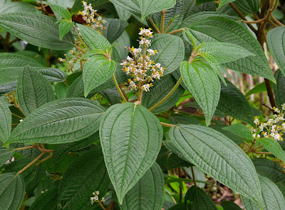
Tarragon Artemisia dracunculus, copyright Cillas.


Tarragon is not the only species of Artemisia of significance to humans. This genus of composite-flowered plants comprsises over five hundred species and subspecies of herbs and small shrubs. The greatest diversity is found in arid and semi-arid regions of the Northern Hemisphere temperate zone (Sanz et al. 2008). The genus is characterised by its distinctive pollen with surface spinules reduced or absent. This pollen type is associated with the wind pollination typical of the genus, though some species do exhibit features such as sticky pollen and colourful flower-heads associated with insect visitation (Hayat et al. 2009). The flower-heads or capitula (a reminder that the 'flowers' of composite plants such as daisies and thistles actually represent a fusion of multiple flowers) of Artemisia are either disciform, with an outer circle of reduced ray florets surrounding the inner disc florets, or discoid, with disc florets only. In disciform capitula, the outer limb of the ray florets is reduced to a membranous vestige, not readily visible without minute examination. The ray florets are female whereas the disc florets are ancestrally hermaphroditic (more on that shortly). In discoid capitula, where the ray florets have been lost, all florets are uniformly hermaphroditic.

Mugwort Artemisia vulgaris, copyright Christian Fischer.


Historically, there has been some variation in the classification of Artemisia but a popular system divides the genus between five subgenera. A phylogenetic analysis of Artemisia and related genera by Sanz et al. (2008) found that the genus as currently recognised is not monophyletic, with a handful of small related genera being embedded within the clade. Time will tell whether this inconsistency is resolved by subdividing Artemisia or simply rolling in these smaller segregates, but for the purposes of this post they can be simply set aside. The subgenus Dracunculus, including tarragon and related species, falls in the sister clade to all other Artemisia. As well as being united by molecular data, members of this clade are distinguished by disciform capitula in which the central disc florets have become functionally male (female organs have been rendered sterile).

Wormwoood Artemisia absinthium, copyright AfroBrazilian.


The second clade encompasses the subgenera Artemisia and Absinthium, with disciform capitula, and Seriphidium and Tridentatae, with discoid capitula. Not all authors have supported the distinction of Artemisia and Absinthium, and Sanz et al. identify both as non-monophyletic, both to each other and to the discoid subgenera. Because of their similar flower-heads, most authors have presumed a close relationship between the Eurasian Seriphidium and the North American Tridentatae (commonly known as sagebrushes). Some have even suggested the former to be ancestral to the latter. However, Sanz et al.'s results questioned such a relationship, instead placing the Tridentatae species in a clade that encompassed all the North American representatives of the Artemisia group.

As well as the aforementioned tarragon, economically significant representatives of Artemisia include wormwood A. absinthium, best known these days as the flavouring agent of absinthe (though historically it has also been used for more innocuous concoctions). Mugworts (A. vulgaris and related species) have also been used for culinary and medicinal purposes. Sagebrushes are a dominant component of the vegetation in much of the Great Basin region of North America, providing crucial habitat for much of the region's wildlife. Artemisia species have shaped the lives of many of their co-habitants, both animal and human.

REFERENCES

Hayat, M. Q., M. Ashraf, M. A. Khan, T. Mahmood, M. Ahmad & S. Jabeen. 2009. Phylogeny of Artemisia L.: recent developments. African Journal of Biotechnology 8 (11): 2423–2428.

Sanz, M., R. Vilatersana, O. Hidalgo, N. Garcia-Jacas, A. Susanna, G. M. Schneeweiss & J. Vallès. 2008. Molecular phylogeny and evolution of floral characters of Artemisia and allies (Anthemideae, Asteraceae): evidence from nrDNA ETS and ITS sequences. Taxon 57 (1): 66–78.

The Bolivinitids

The Cretaceous was a period of significant innovation in the evolution of Foraminifera with a number of distinct new lineages making their appearance during this period. Among those, appearing in the latter part of the Cretaceous, were the first members of the modern family Bolivinitidae.

Bolivinita costifera, from the Smithsonian National Museum of Natural History.


The Bolivinitidae are free-living benthic forams with a calcareous, hyaline (glassy) test. The overall shape of the test is elongate with chambers arranged in biserial coils (that is, there are two chambers per loop). The terminal aperture is usually loop-shaped with a surrounding lip. Inside the chamber, a tooth plate (an inner protrusion of the test) runs from the aperture to the opening of the previous chamber and may protrude through the aperture (Revets 1996).

Representatives of the Bolivinitidae are found in a wide range of depths, from the shallow waters of the ocean to the bathyal zone. They may be among the most abundant forams in areas of low oxygen concentrations and are commonly associated with sustained organic matter input (Erdem & Schönfeld 2017). In other words, these are muck-lovers. Individuals growing in low oxygen conditions tend to show less pronounced surface sculpture on the test than those where the oxygen levels are higher. Conversely, individuals at deeper levels tend to be larger overall than those in shallower waters (Brun et al. 1984). As such, bolivinitids have received their fair share of attention as potential indicators of changes in environmental condition over time.

REFERENCES

Brun, L., M. A. Chierici & M. Meijer. 1984. Evolution and morphological variations of the principal species of Bolivinitidae in the Tertiary of the Gulf of Guinea. Géologie Méditerranéenne 11 (1): 13–57.

Erdem, Z., & J. Schönfeld. 2017. Pleistocene to Holocene benthic foraminiferal assemblages from the Peruvian continental margin. Palaeontologica Electronica 20.2.35A: 1–32.

Revets, S. A. 1996. The generic revision of the Bolivinitidae Cushman, 1927. Cushman Foundation for Foraminiferal Research Special Publication 34: 1–55.

Crossing the Busycon

I must admit that when I think about the biodiversity hotspots of the world, the eastern seabord of the United States would not be among the first regions to come to mind. But for this post, I'm looking at a dramatic and eye-catching radiation of molluscs for which this is their centre of distribution. I speak of the giant whelks of the Busyconidae.

Left-handed whelk or lightning whelk Sinistrofulgur sinistrum, copyright Andrea Westmoreland.


Busyconid whelks first appeared in the waters of eastern North America during the early Oligocene, about 32 million years ago, in what was then the Mississippi Sea and is now the Mississippi River Basin. As the oceans receded from the Mississippi, they spread into the Gulf of Mexico and are now found between Massachusetts in the north and the Yucatan Peninsula in the south. Except for an introduced population of the channeled whelk Busycotypus canaliculatus that has become established in San Francisco Bay in California since the 1930s, the family has never been found elsewhere. These are remarkably large snails: smaller examples are still more than five centimetres in length, and the largest of all get close to a foot (Petuch et al. 2015). Mature shells have a large body whorl, generally higher than the visible spire, with a long siphonal canal. SCulpture of the shell, if present, is dominated by spiral elements, and the shoulder of the whorls may be marked by prominent carinae and/or spines. As is standard for neogastropods, the classification of this group has shifted around a bit over the years, whether treated as their own family or as a subfamily Busyconinae of the related families Buccinidae or Melongenidae. In a recent review of the busyconids, Petuch et al. (2015) recognised fifteen living species in six genera. The number of fossil species that has been described is significantly larger (over one hundred); not surprisingly, these large solid shells have an excellent fossil record. However, it is worth noting that some of the living species may be remarkably variable in shell morphology and I don't know whether fossil representatives have been subject to the same systematic scrutiny.

Knobbed whelk Busycon carica, copyright Matt Tillett.


All busyconids are predators on bivalves, particularly on burrowing clams. In general, the whelk envelops its victim in its muscular foot and then uses the edge of the shell lip to open the clam's shell, allowing the whelk to insert its radula and rasp out the clam's flesh. The preferred method of opening the shell depends on the species of whelk and may be classed as 'wedging' and 'chipping'. 'Wedging' is the most straightforward method and believed to be the more primitive; wedgers insert the shell lip into the gap between valves and directly force them apart and/or prevent the clam shell from closing. 'Chipping' is more involved and performed by members of the genera Busycon and Sinistrofulgur. In this method, the edge of the whelk shell is rhythmically pounded against the commissure between the clam shell valves, progressively wearing at the valve margins until enough of an opening has been made to insert the radula. The process may take multiple hours of patient hammering. Chipping requires more power and a heavier shell than wedging (chipping whelks may damage their own shell as well as the prey's) but also allows the whelk to attack thicker-shelled clams.

Though each species of busyconid will generally use one or the other method of opening prey, there are borderline examples. Larger individuals of Busycotypus canaliculatus, usually a wedger, may adopt a process like chipping though their attacks on the prey shell are usually less systematic than true chippers. And while I haven't found anywhere that says as much, I suspect that young chippers may spend the earlier parts of their life as wedgers untill they have developed the shell strength for chipping. Dietl (2004) suggested that chipping behaviour may have originated twice among busyconids, based on the fossil evidence of its traces left on clam shells. The modern chippers appear to derive from a single origin in the later Pliocene. However, evidence of an earlier and now seemingly extinct chipping lineage was also found in shells from the late Miocene. These earlier chippers seemingly did not belong to any of the modern chipping genera which are not known from the Miocene deposits in which chipped clams were found. Instead, Dietl proposed that the culprit was a large Busycotypus.

Channeled whelk Busycotypus canaliculatus laying a string of egg cases, copyright Eric Heupel.


Busyconid whelks have long been of significance to people living in areas where they are found. Not only are the shells eye-catching and ornamental objects in themselves, the animals are also harvested for food (though their meat is often sold under misleading names such as 'conch' or 'clam strips'). Archaeological examples have been found of busycon shells being used for tools; Petuch et al. (2015) illustrate an example of a left-handed whelk Sinistrofulgur sinistrum shell with holes drilled into it that would have allowed it to be attached to a stick and used as a shovel. These animals are truly an icon of North America's eastern seaboard.

REFERENCES

Dietl, G. P. 2004. Origins and circumstances of adaptive divergence in whelk feeding behavior. Palaeogeography, Palaeoclimatology, Palaeoecology 208: 279–291.

Petuch, E. J., R. F. Myers & D. P. Berschauer. 2015. The Living and Fossil Busycon Whelks: Iconic Mollusks of Eastern North America. San Diego Shell Club, Inc.

Pompilus: Spider Wasps of the Dunes

I've commented before on the difficulties that can be attendent on identifying spider wasps (Pompilidae), one of those groups that combine a high species diversity with a tendency to be morphologically conservative. As a result, the taxonomic history of this group has been one of shifting generic concepts and ill-defined wastebaskets. Not surprisingly, one of the main victims of this uncertainty has been the type genus Pompilus. Historically used to cover a significant percentage of all spider wasps, the name Pompilus is now restricted to a small cluster of species inhabiting the Old World.

Pompilus cinereus, copyright Martin Grimm.


The genus Pompilus and its history were last revised in detail by Day (1981) who recognised seven species associated with more or less open, sandy habitats. The most widespread and best-known of these is Pompilus cinereus, found over wide parts of Eurasia, Africa and Australia, often alongside bodies of water. This species shows a wide range of morphological variation across its range but Day (1981) declared himself unable to sensibly correlate this variation with discrete populations. The possibility remains that further studies may identify P. cinereus as a species complex. The other species in the genus, P. mirandus of India and south-east Asia and five African species, are more restricted in range and little studied. Pompilus mirandus is more tolerant of vegetated habitats than P. cinereus. Conversely, P. niveus of northern Africa is a specialist of the sand dunes of the Sahara Desert. Species of Pompilus all have a black cuticle with a covering of short grey pubescence. The most distinctive feature of the genus is the possession by females of long, weakly curved mandibles with a single inner tooth (other spider wasps have shorter, thicker mandibles with more teeth). These modified mandibles are related to their distinctive manner of handling prey. Whereas other spider wasps will drag their spider victims backwards to their nest, females of Pompilus will lift the spider off the ground and run forward while carrying it.

Nesting behaviour has only been described for P. cinereus. Targeted prey comprises ground-running spiders such as wolf spiders or clubionids. After a spider has been captured, paralysed and carried near the intended nest site, it is temporarily buried in the sand while the female constructs a burrow (Day suggested that this preliminary burial was to prevent the spider being stolen). The simple burrow leads to a single nest cell a few inches deep. The female exhumes the spider, transports it into the burrow and then lays an egg on its abdomen near the front of the side. She then closes the entrance to the burrow with sand, tamping it down securely with the end of her metasoma.

Within the burrow, the spider begins to wake from its paralysis after a few hours. However, it remains in poor shape: its movements are slow and it begins to continuously exude silk from its spinnerets. By wandering about the cell in this distressed state, the spider ends up producing a silken purse that serves as extra protection for the nest's contents. This, of course, includes the wasp larva that within a couple of days will have begun to feed on the trapped spider.

Though details of breeding behaviour have not been observed for other Pompilus species, they might be expected to resemble P. cinereus. It might be noted, however, that the female of P. cinereus has a patch of flattened scales at the end of the metasoma that is less developed in P. mirandus. Is this an indication that P. mirandus is somehow less conscientious in sealing the nest burrow than P. cinereus? If you keep an eye out in the wastelands of India, you might just learn the answer.

REFERENCE

Day, M. C. 1981. A revision of Pompilus Fabricius (Hymenoptera: Pompilidae), with further nomenclatural and biological considerations. Bulletin of the British Museum (Natural History): Entomology 42 (1): 1–42.

Allendesalazaria nymphoides, the Hidden Blister Beetle

The blister beetles of the family Meloidae have attracted attention for a number of reasons. One is their production of caustic defensive chemicals which may be powerful enough to cause severe injury to humans or their livestock. Another is their remarkable life cycles. Many blister beetles develop as nest predators or kleptoparasites of bees. The larvae of these species are hypermetamorphic with the first instar being more mobile than later stages. These mobile larvae will find bees and latch onto them so that they can be carried to the host's nest.

Allendesalazaria nymphoides, copyright Stanislav Krejcik.


This association reaches an extreme in Allendesalazaria nymphoides of north-west Africa. This reclusive species has, to date, been recorded from localities in Morocco, Algeria and Mauritania (Bologna & Aberlenc 2002). It is readily distinguished from other blister beetles by its much-reduced elytra which are oval and widely separated from each other. It is also distinguished by claws that lack the free lower blade found in most other meloids (Bologna & Pinto 2002). Whether they produce the noxious chemicals known from other members of their family, I haven't found a record.

Allendesalazaria nymphoides develops in the nests of solitary burrowing bees of the genus Anthophora. Adults of A. nymphoides do not feed, and never emerge from the nest in which they matured. Instead, they lay their own eggs within that same nest. Dispersal is then left to the hatching larvae that (I presume) latch onto those emerging bees that escaped their parents' depredations. Eventually, the new generation of bees will establish nests of their own. And when they do, the blister beetles will be ready for them.

REFERENCES

Bologna, M. A., & H.-P. Aberlenc. 2002. Allendesalazaria, un nouveau genre de Meloidae pour la faune saharienne (Coleoptera). Bulletin de la Société Entomologique de France 107 (2): 191–192.

Bologna, M. A., & J. D. Pinto. 2002. The Old World genera of Meloidae (Coleoptera): a key and synopsis. Journal of Natural History 36 (17): 2013–2102.

Pied Harvestmen of the Antilles

Harvestmen of the Neotropical family Cosmetidae have been featured on this site a couple of times before. Each time, I've commented on the dire taxonomic state of this diverse family, with many genera being poorly or inaccurately defined. Thanks to extensive (and continuing) studies in recent years by Braxilian researchers and their associates, this situation has been progressively improving, but we still have a lot to learn.

Cynortoides sp., copyright Damion Laren Whyte.


Cynortoides is a genus currently holding ten species of cosmetid. Most of these are found on the islands of the Greater Antilles—Cuba, Jamaica and Hispaniola—though the genus has also been recorded from adjoining regions of Mexico and Venezuela (Kury 2003). As with other cosmetids, Cynortoides has historically been defined largely be features of the external spination, including a lack of spines on the legs, two pairs of spines in the rear part of the dorsal scutum, and no spines on the free abdominal segments (Mello-Leitão 1933). Also as with other cosmetid genera, Cynortoides species are colourfully patterned. The name of one species, C. v-album, refers to its characteristic bright white V marking on the back (though personally, I would describe the pattern as more of a Y).

Though this genus does not yet appear to have been revised in detail, some of its species were included in a recent broader study of cosmetid phylogeny by Medrano et al. (2021). They found strong support for an association between the Cuban C. cubanus and the Hispaniolan C. v-album, together with two other Cuban species previously included in the related genus Cynorta. These last two species were consequently transferred to Cynortoides though Medrano et al. did not comment on whether this affected the genus' established diagnosis. The authors speculated that further studies might prove Cynortoides to be a strictly Greater Antillean genus with mainland records being misplaced. Cynortoides would not be unique in this regard: the islands of the Caribbean are home to a number of lineages found nowhere else, reflecting a long history independent of the adjoining continents.

REFERENCES

Kury, A. B. 2003. Annotated catalogue of the Laniatores of the New World (Arachida, Opiliones). Revista Ibérica de Aracnología, special monographic volume 1: 1–337.

Medrano, M., A. B. Kury & A. C. Mendes. In press 2021. Morphology-based cladistics splinters the century-old dichotomy of the pied harvestmen (Arachnida: Gonyleptoidea: Cosmetidae). Zoological Journal of the Linnean Society.

Mello-Leitão, C. F. de. 1933. Notas sobre os opiliões do Brasil. Descritos na obra postuma de Sörensen: "Descriptiones Laniatorum". Boletim do Museu Nacional 9 (1): 99–114.

The Race of Racers

Snakes are, for the most part, fairly retiring animals, little seen even in areas where they may be abundant. In much of North America, however, one of the most commonly encountered snake species is the racer Coluber constrictor. This moderately large non-venomous snake, with the largest individuals approaching two metres in length, is a widespread inhabitant of open habitats such fields, brushland or open woodlands. Its distribution is centred over much of the continental United States, being found in most regions except much of the arid south-west. Outside the United States, it has a very patchy distribution in southernmost Canada, Mexico and northern Central America. Most recent authors treat it as the sole species in the genus Coluber; other species historically assigned to this genus from across the Holarctic region now being treated as separate. These include the North American whip snakes of the genus Masticophis, believed to the closest relatives of the racer (Myers et al. 2017).

Southern black racer Coluber constrictor priapus, copyright Peter Paplanus.


Adult racers are generally uniformly dark in coloration dorsally, with a lighter-coloured venter, though juveniles have a blotchy checkered pattern (Fitch 1963). The exact shade varies across the species' range and a number of subspecies have been recognised such as the blue racer Coluber constrictor foxii and the northern black racer C. c. constrictor. In general, individuals are darker towards the east and north, and lighter towards the west and south. Wilson (1978) listed eleven subspecies of C. constrictor whereas a phylogeographic study of the species by Burbrink et al. (2008) identified six major lineages. As well as coloration, members of these lineages may differ in factors such as behaviour or genital morphology, and future studies may see them elevated to the rank of separate species.

Blue racer Coluber constrictor foxii, copyright Peter Paplanus.


The natural history of Coluber constrictor was reviewed in detail by Fitch (1963). As the vernacular name of 'racer' suggests, Coluber constrictor is a fast mover. Its diet contains a mixture of small vertebrates, such as frogs, lizards and small mammals, and large invertebrates such as grasshoppers, crickets and caterpillars. Foraging individuals often hold the front end of the body raised above the ground. Despite their species name, racers do not kill their prey by constriction. Instead, they mostly capture prey by darting forward quickly and grabbing it, often swallowing prey live. Fitch recorded one occasion when he observed a racer in the process of subduing a large skink. While the snake was swallowing its prey, Fitch attempted to capture it. The racer disgorged the skink, and both snake and lizard escaped the scene. Diet may vary with size, with smaller individuals taking a higher proportion of invertebrates, but also varies with range. Populations in the west may primarily feed on insects whereas others may almost exclusively take vertebrates. The northern black racer of the northeastern United States is the most inclined of the subspecies to feed on other snakes. Cannibalism is not unknown; at least one author recorded observing it among broods raised in captivity. In one case, two young racers latched onto a single lizard. One of them successfully downed the lizard, and then also continued on to devour the snake attached to the other end, despite the swallowed snake being nearly as large as its swallower.

Eastern yellow-bellied racer Coluber constrictor flaviventris, copyright David Sledge.


During winter, racers hibernate in crevices and hollows among rocks. Preferred hibernation locations are often at the top of hills, away from their usual hunting sites. Mating and egg-laying occurs shortly after emergence with the peak of egg-laying being in early June (Rosen 1991). Racers, particularly the large northern black, may become more aggressive during this period. Eggs are buried shallowly, in loose soil or under litter, though females may take advantage of abandoned mammal burrows to provide a more secure location. As with other snakes, laying seems to be a matter of pump and dump; I didn't come across any references to females protecting clutches. After hatching, males take about a year to reach sexual maturity whereas the larger females take about two years. Fitch (1963) reports encountering the same individuals over the course of several years (recognisable by their bearing the scars of prior collection of scale samples). Nevertheless, the majority of racer hatchlings do not survive their first summer. Few get the opportunity to seek out shelter for their winter's sleep.

REFERENCES

Burbrink, F. T., F. Fontanella, R. A. Pyron, T. J. Guiher & C. Jimenez. 2008. Phylogeography across a continent: the evolutionary and demographic history of the North American racer (Serpentes: Colubridae: Coluber constrictor). Molecular Phylogenetics and Evolution 47: 274–288.

Fitch, H. S. 1963. Natural history of the racer Coluber constrictor. University of Kansas Publications, Museum of Natural History 15 (8): 351–468.

Myers, E. A., J. L. Burgoon, J. M. Ray, J. E. Martínez-Gómez, N. Matías-Ferrer, D. G. Mulcahy & F. T. Burbrink. 2017. Coalescent species tree inference of Coluber and Masticophis. Copeia 105 (4): 640–648.

Wilson, L. D. 1978. Coluber constrictor Linnaeus. Catalogue of American Amphibians and Reptiles 218: 1–4.

The Cephalodiscids

Among the more obscure inhabitants of the world's oceans are the Cephalodiscidae, a family of small (only a few millimetres in length), largely sessile animals that mostly live in colonies within a shared domicile. Though rarely observed, cephalodiscids have received their fair share of attention due to being among the closest living relatives of the graptolites that once dominated the world's oceans during the early Palaeozoic era.

Preserved Cephalodiscus colony, copyright E. A. Lazo-Wasem.


Cephalodiscids are one of the two living branches of the pterobranchs (the other being the Rhabdopleuridae), which together with the acorn worms make up the phylum Hemichordata. Hemichordates are in turn one of the three living phyla of the deuterostomes, together with the echinoderms and chordates (to which, of course, we ourselves belong). Pterobranchs are filter feeders, using an arrangement of tentaculated arms arising just behind the head to collect particles from the water. In cephalodiscids, each individual usually possesses multiple pairs of arms in contrast to the single pair in rhabdopleurids (though at least one species of Cephalodiscus has small males with a single pair). The head carries a large glandular disc (hence the name of the family) that is used to secrete the horny tissue making up the external dwelling (referred to as the tubarium) in which a colony of Cephalodiscus lives. Both cephalodiscids and rhabdopleurids have a contractile stalk at the end of the body from which new individuals (zooids) are budded. However, whereas the zooids of rhabdopleurids (and presumably their extinct graptolite relatives) remain attached to each other throughout their life, cephalodiscid zooids split away from their parent by the time they mature. The majority of cephalodiscid species have distinct males and females though a small number may be hermaphrodites. Some species exhibit sexual dimorphism; males may be considerably smaller than females.

Individual zooid of Cephalodiscus dodecalophus, from Sedgwick et al. (1898).


About twenty species of living cephalodiscids are currently recognised. The majority of these have been included in a single genus Cephalodiscus, albeit divided between a number of subgenera. The single outlier, Atubaria heterolopha, was described in 1936 from a single dredge haul near Japan (Mitchell et al. 2013). No dwelling material was found in the haul so it was presumed this species does not construct a tubarium like other cephalodiscids. However, its zooids were otherwise little different from those of Cephalodiscus. The subgenera of Cephalodiscus are mostly distinguished by tubarium structure. In some species, each individual in the colony will have its own separate tube closed off at the base. In other species, tubes will open into a central chamber shared between multiple zooids (Maletz 2014). Openings of the tubarium may be surrounded by spines and the like, secreted by the zooids as they creep out from their domicile.

Recent studies have indicated that cephalodiscids represent the sister group to all other pterobranchs/graptolites, implying an history that may extend back to the Cambrian. However, the fossil record of cephalodiscids themselves is minimal. This is largely due to practical difficulties: because the soft-bodied zooids are not preserved, fossils can only be identified from the external tubarium structure alone. Unless the origin point of the tubarium is preserved and identifiable, there is little to distinguish a cephalodiscid tubarium from a benthic graptolite (graptolite colonies begin with a differentiated larval chamber called a sicula, cephalodiscids produce no such structure). A handful of fossil cephalodiscids have been identified, notably the early Devonian Eocephalodiscus, but as yet they tell us little about the evolution of this ancient lineage.

REFERENCES

Maletz, J. 2014. The classification of the Pterobranchia (Cephalodiscida and Graptolithina). Bulletin of Geosciences 89 (3): 477–540.

Mitchell, C. E., M. J. Melchin, C. B. Cameron & J. Maletz. 2013. Phylogenetic analysis reveals that Rhabdopleura is an extant graptolite. Lethaia 46: 34–56.

Lilies of Blood

The flora of southern Africa is renowned for being remarkably diverse and, in many cases, remarkably eye-catching. The region is home to more than its fair share of ornamental plants, many of which have become popular garden subjects. Among the remarkable members of the southern African flora are the blood lilies of the genus Haemanthus.

Haemanthus coccineus, copyright Peter Coxhead.


Haemanthus is a genus of 22 known species found in the very southern part of the continent, in the countries of South Africa and Namibia (species from further north that have historically been included in Haemanthus are now treated as a separate genus Scadoxus). It is a member of the belladonna family Amaryllidaceae and, like many other members of that family, grows as a herb from a fleshy bulb that is partially or entirely concealed underground. The plant above ground may be annual or persistent, depending on species. Each individual Haemanthus plant produces very few leaves at a time: two is the most common number (Van Jaarsveld 2020). The leaves are more or less fleshy, often hairy, and may be directed upwards or spread outwards.

In those species that shed their leaves, flower stalks are produced before the next season's leaves appear, in a similar matter to the related naked ladies Amaryllis belladonna. Flowers are produced in dense umbels, subtended by bracts that are often brightly coloured, so at a glance the inflorescence of some species might be taken for a single large flower up to ten centimetres in diameter. Depending on the species, the supporting stalk may vary from over a foot in height to only a few centimetres. The first species to be described bear flowers of a bright red colour, explaining both the genus and vernacular names, but flowers may also be pale pink or white. Species that lack the red colour may be referred to as 'paintbrush lilies' rather than 'blood lilies'. Fruits are soft fleshy berries.

Haemanthus albiflos, copyright Krzysztof Ziarnek, Kenraiz.


Phylogenetic analyses of the genus have identified two major clades, a mostly eastern clade found in regions with summer rainfall and a mostly western clade associated with winter rainfall. A notable outlier is the eastern summer-rainfall species H. montanus which is the sister taxon to the winter rainfall clade. Members of the summer-rainfall clade have white or pale pink flowers; members of the winter-rainfall clade have pale pink to dark red flowers. Members of both clades have been grown as pot plants for their unusual appearance though the scent of the flowers is not regarded as pleasant. Perhaps the most widely grown species is H. albiflos, a species native to both the western and eastern parts of South Africa that bears flowers in umbels up to seven centimetres wide. This species is evergreen, carrying its leaves year-round.

REFERENCE

Van Jaarsveld, E. 2020. Haemanthus. In: Eggli, U., & R. Nyffeler (eds) Illustrated Handbook of Succulent Plants: Monocotyledons 2nd ed. pp. 441–443. Springer.

The Oligorhynchiidae

Dorsal view of Oligorhynchia subplana gibbosa, from Cooper (1935).


From Oligochiton, we move onto Oligorhynchia. The Oligorhynchiidae are a family of very small brachiopods known from the Middle and Late Ordovician. They were among the earliest representatives of the Rhynchonellida, a major group of brachiopods that survives to the present day. Rhynchonellidan shells are usually characterised by a strong beak associated in life with a well-developed pedicel. In oligorhynchiids, this beak is suberect and the shell as a whole is an elongate subtriangular shape. The valves of the shell are folded into coarse plicae (ridges). At least towards the base of the shells, the major folds are in what is called an inverted arrangement, with a ridge in the dorsal valve matched by a valley in the ventral valve (Schmidt & McLaren 1965). Other structural features defining the group include small plates projecting into the pedicel opening, distinct vertical dental plates and divided hinge plates in the valve articulation, and the usual absence of a median septum or cardinal process inside the shell (Savage 1996).

The oligorhynchiids first arose in the east of what was then the continent of Laurentia (corresponding to modern North America). They subsequently spread across the Iapetus Ocean to the continents of Baltica and Kazakhstan (Jin 1996). The end of the Ordovician saw their replacement by other rhynchonellid families. Nevertheless, their genetic lineage would continue for some time yet as they have been identified as ancestors of later families: the Trigonirhynchiidae and Camarotoechiidae (Jin 1989). The brief oligorhynchiid spark would blossom into later rhynchonellid success.

REFERENCES


Jin, J. 1989. Late Ordovician–Early Silurian rhynchonellid brachiopods from Anticosti Island, Quebec. Biostratigraphie du Paléozoïque 10: 1–127, 130 pls.

Jin, J. 1996. Ordovician (Llanvirn–Ashgill) rhynchonellid brachiopod biogeography. In: Copper, P., & J. Jin (eds) Brachiopods pp. 123–132. CRC Press.

Savage, N. M. 1996. Classification of Paleozoic rhynchonellid brachiopods. In: P. Copper, & J. Jin (eds) Brachiopods pp. 249–260. CRC Press.

The Fate of Oligochiton

Chitons are one of the most distinctive and evolutionarily divergent groups of molluscs alive today. But compared to other groups of molluscs, the fossil record of chitons is rather sparse—or at least sparsely studied. It's not hard to see why. The multi-plated nature of the chiton shell means that it tends to fall apart after death, and the structure of the plates is such that critical features are easily abraded.

(Clockwise from top left) head, intermediate and tail valves of Lepidochitona lioplax, from Dell'Angelo et al. (2011).


Lepidochitona lioplax is one example of a fossil chiton. It was originally described from Oligocene rocks belonging to the Sooke Formation of southern Vancouver Island in British Columbia. Only four moderate-sized valves were initially identified: one head valve, one intermediate, and two tails (so at least two individuals were involved). The valves had a smooth outer surface without a strong distinction in appearance between the central and lateral areas. The insertion plates (lateral projections of the lower surface of the valves that in life anchor them into the surrounding girdle) were very short. The sutural laminae (anterior projections of the lower surface of the intermediate and tail valves that articulate with the valve in front) were low, wide, and divided in the middle by a broad shallow surface. Slits in the lateral insertion plates were numerous, with several in the tail valves and probably two or three on each side in the intermediate valves (Smith 1960). When first described, this species was thought distinct enough to belong in its own genus Oligochiton.

Oligochiton lioplax would then go little reported on until 2011 when Dell'Angelo et al. described an assemblage of chiton fossil from the latest Eocene or early Oligocene of the Lincoln Creek Formation in Washington State. Specimens of lioplax were relatively numerous in this collection and Dell'Angelo et al. were able to examine close to a hundred valves. Their observations would lead to something of a downgrade in the species status. Rather than deserving its own extinct genus, Dell'Angelo et al. felt that lioplax could be comfortably accommodated in the living genus Lepidochitona. Its smooth valves are unusual within Lepidochitona but not unique. The supposed multiple slits in the sides of the valves did not stand up to scrutiny. Instead, intermediate valves of L. lioplax bore only a single slit on each side, in line with other Lepidochitona species. The original inference of multiple slits was an error due to the original specimen being still partially embedded in the surrounding matrix.

Lepidochitona lioplax is one of the earliest known representatives of its genus but its exact significance is obscure. It has been suggested as a direct ancestor of the modern subgenus Spongioradsia but this, again, was based on the supposed slits in the intermediate valves that Dell'Angelo et al. refuted. To know how L. lioplax connects to the big picture of Lepidochitona evolution, we would probably need a better picture of Lepidochitona evolution overall.

REFERENCES

Dell'Angelo, B., A. Bonfitto & M. Taviani. 2011. Chitons (Polyplacophora) from Paleogene strata in western Washington State, U.S.A. Journal of Paleontology 85 (5): 936–954.

Smith, A. G. 1960. Amphineura. In: Moore, R. C. (ed.) Treatise on Invertebrate Paleontology pt I. Mollusca 1: Mollusca—General Features, Scaphopoda, Amphineura, Monoplacophora, Gastropoda—General Features, Archaeogastropoda and some (mainly Paleozoic) Caenogastropoda and Opisthobranchia pp. I41–I76. Geological Society of America, and University of Kansas Press.

The Feared Mosquito

It's one of those standard pub-quiz "trick" questions. What animal kills the most people? The hope is that contestants will nominate the 'obvious'—snakes, sharks, bears, whatever—before being blind-sided by the revelation that mosquitoes kill over a million people. They don't kill them directly, of course; their victims die from the diseases they spread*. The statistic also glosses over the point that there are many hundreds of species of mosquito that vary significantly in the nature and severity of their role as disease vectors. Nevertheless, for this post I'm considering the group that includes some of the most notorious vectors: the genus Anopheles.

*For the record, if the question was confined to active killings, the most dangerous animal to humans is other humans. Dogs come a distant second.

Anopheles punctipennis feeding, with the long palps extended in front of the head, copyright Nathan D. Burkett-Cadena/University of Florida.


Anopheles is one of the most divergent genera of mosquitoes, being placed in a distinct subfamily Anophelinae (along with a couple of small related genera) from the bulk of mosquitoes in the subfamily Culicinae. Adult Anopheles can be readily distinguished from culicine mosquitoes by their palps which are about as long as the proboscis (in other mosquitoes, the palps are distinctly shorter). Larvae of Anopheles lack the long respiratory siphons at the end of the abdomen found in other mosquito larvae so they rest parallel with the water surface rather than hanging below it. The genus is found around the world; over 450 named species are currently known (Harbach 2013) with many more waiting to be described. The genus is currently divided between seven subgenera though one of the largest of these, the cosmopolitan subgenus Anopheles, is not monophyletic. The remaining subgenera are better supported with the largest of these, Cellia, being found in the Old World. Between them, the subgenera Anopheles and Cellia account for over 400 of the known Anopheles species. The remaining small subgenera are mostly Neotropical with a single Oriental species being awarded its own subgenus.

Anopheles maculipennis, copyright Ryszard.


Anopheles is of most concern to humans, of course, for its role as a disease vector. As with other mosquitoes, the transmission of disease is done entirely by females taking blood meals to provide nutrients for their developing eggs. Males are not blood feeders, instead feeding entirely on sugar sources such as nectar (females also feed on nectar for their own nutrition). The main disease spread by Anopheles is malaria, but they may also spread malaises such as filariasis and arboviruses (Krzywinski & Besansky 2003). As noted above, species may vary significantly in their importance as disease vectors, even between quite closely related taxa. Many historically recognised vector "species" have proved, on close inspection, to represent species complexes of which some may be vectors and others not. For instance, one of the most important transmitters of malaria, the African A. gambiae, has been divided between at least eight different species (Coetzee et al. 2013). Misidentification of vectors can be a significant issue. For instance, mosquito control regimes in central Vietnam during the 1990s focused on two species, A. dirus and A. minimus, that were each active at different times of year. However, Van Bortel et al. (2001) found that A. minimus was in fact very rare in this area, with specimens previously thought to be A. minimus proving to be another species, A. varuna. Anopheles varuna is not a significant malaria vector, feeding almost entirely on animals such as cattle rather than on humans. Large amounts of resources would have been wasted trying to control a mosquito that was of little concern. What is more, the fact that malaria was not being transmitted by A. minimus raises the possibility that it was being spread by yet another species, one that had managed to escape attention. Remember, kids: bad taxonomy kills.

REFERENCES

Coetzee, M., R. H. Hunt, R. Wilkerson, A. Della Torre, M. B. Coulibaly & N. J. Besansky. 2013. Anopheles coluzzii and Anopheles amharicus, new members of the Anopheles gambiae complex. Zootaxa 3619 (3): 246–274.

Harbach, R. E. 2013. The phylogeny and classification of Anopheles. In: S. Manguin (ed.) Anopheles Mosquitoes: New insights into malaria vectors. InTechOpen.

Krzywinski, J., & N. J. Besansky. 2003. Molecular systematics of Anopheles: from subgenera to subpopulations. Annual Review of Entomology 48: 111–139.

Van Bortel, W., R. E. Harbach, H. D. Trung, P. Roelants, T. Backeljau & M. Coosemans. 2001. Confirmation of Anopheles varuna in Vietnam, previously misidentified and mistargeted as the malaria vector Anopheles minimus. American Journal of Tropical Medicine and Hygiene 65 (6): 729–732.

The Running of the Termites

I don't know how many people would profess to have a favourite genus of termites. Which is a shame, because there are some real stand-out examples. Snapping termites, magnetic termites, glue-spraying termites... For my own part, though, I have a particular fondness for the Australian harvester termites of the genus Drepanotermes.

Soldiers and workers of Drepanotermes perniger, copyright Jean Hort.


Nearly two dozen species of Drepanotermes are found on the Australian continent to which they are unique (Watson & Perry 1981). They are arid-environment specialists, being most diverse in the northern part of Australia. My reasons for being so fond of them are, I'll admit, decidedly prosaic. The worker caste of most termite species is very difficult if not impossible to identify taxonomically; one termite worker usually looks very much like another. Drepanotermes workers, however, are different. The name Drepanotermes can be translated as "running termite" and, as befits their name, Drepanotermes of all castes stand out for their distinctly long legs. Soldiers of Drepanotermes also have distinctively shaped mandibles which are sickle-shaped and have a single projecting tooth on the inner margin. They are similar to soldiers of the related genus Amitermes (of which Drepanotermes may represent a derived subclade) but the mandibles of Amitermes tend to be straighter and more robust.

The long legs of Drepanotermes reflect their active harvester lifestyles. Workers will emerge from the nest at night in search of food to carry back home. In the red centre of Australia they will primarily collect spinifex; they will also take fallen leaves, tree bark and the like. Soldiers keep guard while the workers forage. I've found them clustered around a nest entrance of an evening, just their heads poking out to snap at passers-by. Workers may wander up to about half a metre from the nest entrance as they forage. The concentrations of vegetable matter produced by Drepanotermes storing food sources in their nest may form a significant factor in the nutrient profile of areas where they are found.

Alate and soldiers of Drepanotermes rubriceps, copyright Jean Hort.


Depending on species and circumstance, the nests of Drepanotermes may be mounds or entirely subterranean with the latter being the majority option. They prefer compact soils such as clay though they may burrow through looser soils where there is a denser subsoil. Drepanotermes may construct their own nest or move into nests constructed by other termites. One aptly named species, D. invasor, seems to take over pre-existing nests more often than not. Subterranean nests are arranged as a series of chambers about five to ten centimetres in diameter connected by tunnels. These chambers may be arranged vertically, one below another, or they may form a rambling transverse network. Above ground, subterranean nests may be visible as an open circle devoid of vegetation. The ground in these circles is hard as concrete and may remain clear for decades after the actual nest has gone. Walsh et al. (2016) refer to the remains of nests protruding above ground along vehicle tracks after the soil around them has worn down. Local people have a long history of taking advantage of the open space offered by termite nests, such as to move more easily through scrub or as resting or working places.

The alate castes of Drepanotermes tend to be poorly known. Indications are that mature reproductives spend little time in the parent nest before leaving to breed. For most species, breeding flights take place in late summer. Alates may emerge either by day or night. The time of emergence seems to depend on the species; night-flying alates have distinctly larger eyes than day-fliers. Unfortunately, because alates have rarely been collected in association with a nest, we are largely still unable to tell which alates belong to which species.

REFERENCES

Walsh, F. J., A. D. Sparrow, P. Kendrick & J. Schofield. 2016. Fairy circles or ghosts of termitaria? Pavement termites as alternative causes of circular patterns in vegetation of desert Australia. Proceedings of the National Academy of Sciences of the USA 113 (37): E5365–E5367.

Watson, J. A. L., & D. H. Perry. 1981. The Australian harvester termites of the genus Drepanotermes (Isoptera: Termitinae). Australian Journal of Zoology, Supplementary Series 78: 1–153.