Field of Science

  • in The Phytophactor
  • in The Large Picture Blog
  • in Sex, Genes & Evolution
  • in Field Notes
  • in Genomics, Medicine, and Pseudoscience
  • in Catalogue of Organisms
  • in Labs
  • in Angry by Choice
  • in The Biology Files
  • in Doc Madhattan
  • in The Curious Wavefunction
  • in Protein Evolution and Other Musings
  • in A is for Aspirin
  • in The Astronomist
  • in inkfish
  • in The View from a Microbiologist
  • in Rule of 6ix
  • in RRResearch
  • in Pleiotropy
  • in Variety of Life
  • in PLEKTIX
  • in The Culture of Chemistry
  • in Life of a Lab Rat
  • in C6-H12-O6
  • in The Greenhouse
  • in Chinleana
  • in History of Geology
  • in Moss Plants and More
  • in Skeptic Wonder
  • in Games with Words
  • in Memoirs of a Defective Brain
  • in Disease Prone
Showing posts with label Prostigmata. Show all posts

Mites of Southern Sediment

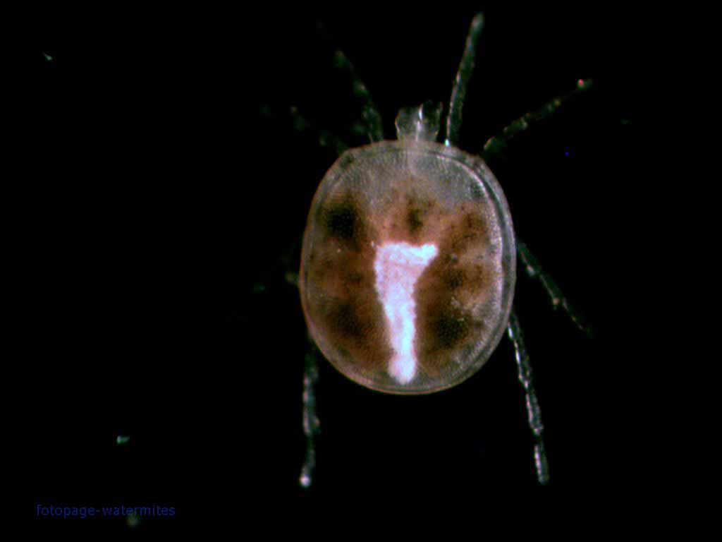
Water mites of the clade Hydrachnidiae are one of the few groups of arachnids that have not only adopted an aquatic lifestyle but have thrived and diversified there. Over fifty families are currently recognised within this clade, some of which can be found in almost every body of fresh water worldwide. Others, however, are notable for their restricted ranges. One of these latter examples is the Omartacaridae.

Ventral view of female Omartacarus elongatus, from Cook (1963).


Omartacaridae is a small family currently recognised as including only two genera, Omartacarus and Maharashtracarus. They have a somewhat elongated body with a soft integument, contrasting with the more globular form of many other water mites. They are also distinguished by the arrangement of the coxae (the basal segments of the legs on the underside of the body) which are clustered together with the medial edges of the anterior pairs much longer than those of the posterior pairs (Walter et al. 2009) so the third pair of coxae are triangular in shape. As far as is known, omartacarids are restricted to interstitial habitats or the hyporheic zone of sediment beneath and alongside stream beds. I am unaware of any direct observations of omartacarid behaviour but they are presumably predators like other water mites. Most of the (rather limited) attention that has been given to omartacarids has focused on discussions of their distribution. Species of Omartacarus are found in South and southern North America, as well as in Australia. Maharashtracarus species are known from India and Costa Rica. It has been presumed that this reflects an ancestral Gondwanan distribution, spreading into North America from South America as the continents joined.

The larval stage of omartacarids is, to date, unknown. Larvae of other water mites live as parasites of water-associated insects such as midges and omartacarid larvae are presumably also parasitic. But in what capacity? Do mature omartacarids emerge from their subterranean habitats at some particular time of year in search of a host for their eggs? Do they somehow manage to find a host while remaining safely sequestered underground? The secret remains to be uncovered.

REFERENCE

Walter, D. E., E. E. Lindquist, I. M. Smith, D. R. Cook & G. W. Krantz. 2009. Order Trombidiformes. In: Krantz, G. W., & D. E. Walter (eds) A Manual of Acarology 3rd ed. pp. 233-420. Texas Tech University Press.

The Ornithocheyletiini: Making a Living off Birds

In an earlier post, I commented on the carnivorous mites of the family Cheyletidae. These rapacious micropredators are commonly associated with the nests and burrows of terrestrial vertebrates, attacking debris-feeders drawn in by the host's leavings. With such a close association already in place, it should come as little surprise that some lineages within the Cheyletidae have learnt to bypass scavenger predation and go directly to the source, becoming parasites of the vertebrate hosts themselves.

Slide-mounted female of Bakericheyla chanayi (left; scale bar = 50 µm) and nest webs on the skin of a heavily parasitised chaffinch Fringilla coelebs (right), from Filimonova (2013).


One such lineage is the Ornithocheyletiini, members of which are parasites of birds. Like other parasitic cheyletids, ornithocheyletiins have a relatively small, simple gnathosoma (the 'head' of the mite), no eyes, and lack the large, pectinate, claw-like setae found on the palps of free-living predatory cheyletids (instead, the setae at this position are small and smooth though they do still have hooked ends). Ornithocheyletiins are further distinguished from other cheyletids by having particularly large claws at the end of each leg that are overhung by a well developed knob on the end of the tarsus (Bochkov & Fain 2001).

Ornithocheyletiins live on the skin of their bird hosts. In the genera Ornithocheyletia and Bakericheyla, the mites spin a protective web beneath which they live and feed. Members of the tribe have been recorded from a number of bird orders, mostly smaller land birds (Passeriformes, Columbiformes, Piciformes, Coraciiformes, Psittaciformes and Apodiformes). At least one species of Ornithocheyletia was described from the Natal spurfowl Pternistis natalensis, a galliform. Members of the genus Apodicheles are restricted to species of Apodiformes (swifts) but other genera are found on a wider range of hosts. One cosmopolitan species, Bakericheyla chanayi, has been found on hosts of both the orders Passeriformes and Coraciiformes. The exact method of exploiting their host may vary: species of Bakericheyla feed on blood whereas Ornithocheyletia species feed on lymph fluid.

Historically, parasitic cheyletids were treated as a separate family Cheyletiellidae but are now classified with their free-living relatives. The exact relationships between free-living and parasitic cheyletids remain open to question. A morphological phylogenetic analysis of cheyletids by Bochkov & Fain (2001) did recover the parasitic forms as a clade but this result was questioned by the authors themselves. Instead, they suggested that the various parasitic tribes of Cheyletidae represented independent lineages whose shared features represented convergent adaptations to the parasitic lifestyle. The Ornithocheyletiini might, for instance, be compared to tribes such as the Cheletosomatini that inhabit the quills of bird feathers but feed on other quill-inhabiting mites rather than the birds themselves. Did ornithocheyletiins evolve from using birds as hunting grounds to using birds as food, or did they carry their parasitic habits with them from some other host?

REFERENCE

Bochkov, A. V., & A. Fain. 2001. Phylogeny and system of the Cheyletidae (Acari: Prostigmata) with special reference to their host-parasite associations. Bulletin de l’Institut Royal des Sciences Naturelles de Belgique, Entomologie 71: 5–36.

Mites of Marine Sands

Mites may be the most ecologically diverse group of animals on the planet. It is something of a challenge to think of a habitat supporting complex life in which mites may not be found. Nevertheless, it can fairly be said that the marine environment has provided them with a challenge. Though a wide variety of mites can be found in habitats along the shoreline, few lineages have learnt to make a life for themselves beyond the littoral fringe. The most diverse group of truly marine mites is the Halacaridae, of which the genus Simognathus is a representative.

Simognathus sp., from Banks (1915).


Halacarids are notably armoured mites, their bodies protected by an array of reticulate plates. They are found in a wide range of marine habitats and pursue the gamut of lifestyles: representatives of halacarids include algal grazers, micropredators, and parasites. Despite their aquatic lifestyle, they are not swimmers. Instead, they cling to their substrate and crawl slowly on legs bearing large claws. The diversity of halacarid morphologies is reflected in their classification with over a dozen subfamilies currently recognised.

Simognathus is a genus of halacarids found around the world though the greater diversity of species are known from the Southern Hemisphere. They are found at depths ranging from near the low tide mark to around 500 m, and from the full range of tropical, warm-temperate and cold-temperate waters. Bartsch (2005) speculated that the only reason they are not known from even colder waters may be a question of sampling effort rather than true absence. Most Simognathus species are known to live among coarse sand, or in other interstitial microhabitats such as among coral rubble, among colonies of sessile animals such as barnacles or tubeworms, or within algal holdfasts. I haven't come across any specific comments on their diet but their robust chelicerae and grasping fore legs leads me to suspect that Simognathus species are probably micropredators.

Simognathus and the closely related genus Acaromantis form the subfamily Simognathinae. Simognathines differ from other halacarids in their spindle-shaped body with short rostrum, reflecting their interstitial habitat. The first leg ends in a pincer arrangement formed from the terminal claw and a spine on the underside of the tibia. Acaromantis species have a two-segmented palp, no lateral claws at the end of the first leg, and a spinose seta on the genu (the segment between the femur and tibia) of the first leg. Simognathus species have a three-segmented palp, a pair of slender lateral claws on the first leg as well as the terminal claw, and no spinose seta on the first genu. The defining features of Simognathus are all likely to be primitive relative to those of Acaromantis and it has been suggested for some time that Acaromantis may be a derived subgroup of Simognathus. This suggestion is bolstered by a recent molecular analysis of halacarids by Pepato et al. (2018) which found the two Simognathus representatives included to be paraphyletic to the included species of Acaromantis.

REFERENCES

Bartsch, I. 2005. Lohmannella and Simognathus (Halacaridae: Acari) from Western Australia: description of two new species and reflections on the distribution of these genera. Records of the Western Australian Museum 22: 293–307.

Pepato, A. R., T. H. D. A. Vidigal & P. B. Klimov. 2018. Molecular phylogeny of marine mites (Acariformes: Halacaridae), the oldest radiation of extant secondarily marine animals. Molecular Phylogenetics and Evolution 129: 182–188.

The Tiny Lurking Fear

The world of micro-organisms can be a cut-throat one. Minute grazers are under constant threat from minute predators. It can be an existence red in tooth and claw or, in the case of today's subjects, haemolymph-covered in chelicera and grasping seta.

The domestic cheyletid Cheyletus eruditus, from here.


The Cheyletini are one of fifteen tribes recognised by Bochkov & Fain (2001) in the mite family Cheyletidae. Cheyletids are small mites, generally less than half a millimetre in length, that are close relatives of the follicle mites seen on this site previously. Many cheyletids (including most Cheyletini) are, nonetheless, voracious predators of other mites. Other members of the family live as parasites on birds or mammals. In the past, such parasitic forms were recognised as a distinct family Cheyletiellidae but it is now recognised that they are descended from predatory ancestors, possibly on more than one occasion.

Predatory cheyletids are not to be sniffed at: the Hemicheyletia wellsina nymph on the left has managed to bring down another much larger predatory mite Metaseiulus occidentalis. Copyright Haleigh Ray.


The Cheyletini can be considered representative of this ancestral form; indeed, as members of the tribe are distinguished from others in the family solely by their retention of features likely to be primitive, it is likely to be non-monophyletic (Bochkov & Fain 2001). Cheyletini have more or less oval or oblong bodies with moderate-length legs, shorter than the length of the body, all tipped by a claw. The gnathosoma (the front section of the body bearing the chelicerae and palps) is well developed and generally makes up a full third of the body length. The palps are the real business end of a cheyletin, though. In many groups of prostigmatic mites, the last segment of the palp (the tarsus) is offset from the main line of the appendage and opposed to a large claw at the end of the tibia, the two of them together functioning to grab whatever the mite wishes to grab. Predatory cheyletids have the tibial claw and offset tarsus but the tarsus also bears a number of intimidating enlarged, claw-like setae that add to the mite's grabbing power. In the Cheyletini, there are four such setae at the end of the tarsus, a pair of comb-like setae dorsally and a pair of sickle-shaped setae ventrally. The mite will generally sit in place, motionless, with its palps held open. Should a potential prey animal come close enough to the predator, the palps will swing together and the prey will be caught.

Cheyletini are diverse in habitat. Many genera are free-ranging hunters on trees but others show preferences for more constrained locales. In particular, a group of genera centred around the type genus Cheyletus includes species living in the nests and burrows of mammals and birds. Most of these species benefit their hosts by hunting down potential parasites and the like or cleaning up organic residue. One genus, Cheletophyes, is found in the nests of carpenter bees Xylocopa and can actually be transported between nests by the host bee in special pockets on the thorax called acarinaria. However, it is not that big a step to take from feeding on shed organic particles in the host's nest to feeding more directly on the host itself and this is presumably how some cheyletids made the switch to parasitism. One member of the Cheyletini, Pavlovskicheyla platydemae, is an ectoparasite of tenebrionid beetles, attaching to them in spots concealed beneath the host elytra (Walter et al. 2009).

Female Hemicheyletia wellsina patrolling near her batch of eggs (in the upper left, under a protective silk covering she has woven for them), copyright Haleigh Ray.


Other Cheyletus species are known from human-associated habitats such as in houses or grain stores where, again, they are usually considered a net benefit due to their controlling effect on pests such as dust mites or flour mites. Indeed, the common species Cheyletus eruditus has been commercially marketed for control of stored product pests under the name Cheyletin. Females of this species in domestic habitats lay their eggs in crevices or other such concealed spaces and remain to guard their brood, driving away other animals that may pose a threat. However, hatching offspring need to disperse quickly, as if they hang around the nesting site too long they may be eaten by the mother herself (Walter & Krantz 2009).

REFERENCES

Bochkov, A. V., & A. Fain. 2001. Phylogeny and system of the Cheyletidae (Acari: Prostigmata) with special reference to their host-parasite associations. Bulletin de l’Institut Royal des Sciences Naturelles de Belgique, Entomologie 71: 5–36.

Walter, D. E., & G. W. Krantz. 2009. Oviposition and life stages. In: Krantz, G. W., & D. E. Walter (eds) A Manual of Acarology 3rd ed. pp. 57–63. Texas Tech University Press.

Walter, D. E., E. E. Lindquist, I. M. Smith, D. R. Cook & G. W. Krantz. 2009. Order Trombidiformes. In: Krantz, G. W., & D. E. Walter (eds) A Manual of Acarology 3rd ed. pp. 233–420. Texas Tech University Press.

False Spider Mites

Among the enormous diversity of the world's mites, some families are particularly notorious for the damage that they inflict on commercial plant crops. Among such Acari non grata are the spider mites of the family Tetranychidae or the gall mites of the Eriophyidae. But a third, equally notorious group is the false spider mites or flat mites of the Tenuipalpidae.

Red and black flat mite Brevipalpus phoenicis, false-colour SEM by Christopher Pooley.


False spider mites include about 800 known species of more or less flattened, slow-moving mites. They are closely related to the true spider mites and both families have the chelicerae modified into a pair of long, whip-like retractable stylets that are used to pierce and suck fluids from plant tissues. In the case of the false spider mites, though, their commercial infamy comes not only from the direct damage caused by the feeding mites themselves but also from the effects of transmitted viruses. Viruses transmitted by false spider mites include the causative agents of diseases such as citrus leprosis and coffee ring spot and may cause significant reductions in the yield and lifespan of infected plants.

Morphologically, false spider mites differ from true spider mites in the absence of what is called the 'thumb-claw' process, an arrangement of the tarsus of the pedipalp alongside a claw on the tibia (hence the family name which means 'slender palp'). The palps are often reduced, with some species having only the barest remnant. Some species also show reduction in the fourth pair of legs, and females of a number of species are six-legged as adults. This merely stands as another example of how mite morphology functions purely to play silly buggers with anything one might learn in basic animal biology.

Hebe stem gall mites Dolichotetranychus ancistrus inside an open gall, copyright Plant and Food Research.


Parthenogenesis is also common in false spider mites. Species found in cooler climes will often overwinter as females, with a new generation of males not appearing until the next spring. In some species, eggs produced parthenogenetically will hatch into males; in others, they will produce females. A few species almost entirely lack functional males. A small group of these species in the genus Brevipalpus is unique among animals in being both parthenogenetic and genetically haploid.

Almost all forms of seed plant seem to be vulnerable to some form of flat mite or another; some mite species are very catholic in their tastes and will latch onto almost anything green and photosynthesising. Others are more discerning. How false spider mites make their way from one host plant to another is little known but they may be passively carried through the air on the wind. Alternatively, they may be inadvertently carried from place to place by feeding herbivores, or by the very human horticulturalists that suffer so much from their presence.

REFERENCE

Walter, D. E., E. E. Lindquist, I. M. Smith, D. R. Cook & G. W. Krantz. 2009. Order Trombidiformes. In: Krantz, G. W., & D. E. Walter (eds) A Manual of Acarology 3rd ed. pp. 233-420. Texas Tech University Press.

Mites in Red Velvet

Adult Platytrombidium fasciatum, copyright Walter Pfliegler.


Mites in red velvet,
decorated with stripes.
Completing their diet,
hunting down eggs from flies*.

*With apologies to Justin Hayward.

Among the mites most likely to be seen by the casual observer are the various species of active predators known as red velvet mites. They grow to relatively large sizes for a mite (the species in the photo above can get up to 2.5 mm long), they are brightly coloured and they can often be seen moving about in search of food. As well as the colour, the name 'red velvet mite' refers to their dense covering of setae giving them almost a teddy-bearish appearance. There aren't many mites that could be described as cuddly, but these are arguably among them (at least as adults, as explained below).

Red velvet mites form a number of families in the mite clade Parasitengonina. Earlier posts on this site (here and here) have already described the somewhat complicated life cycles of parasitengonines, but to recap briefly: parasitengonines start their lives as parasitic larvae, followed by a dormant 'pupa-like' stage, followed by an active predatory nymph, then another dormant 'pupa', and finally the active predatory adult. Whereas differences between the active nymphs and adults are slight (kind of raising the question as to why the intervening dormant phase), differences between adults and larvae are significant. From their appearance alone, there is no way of telling whether a given larval form corresponds to a given adult, and connecting the two requires challenging indirect methods such as brood-rearing. Nevertheless, both forms are commonly encountered: not only are adults significant micro-predators, the larvae are often found attachned to insects and other arthropods. Some larval species, commonly known as chiggers, attack vertebrates such as humans and so are even more well-studied. Because of the resulting need to classify both adults and larvae without an easy way to connect the two, a kind of double taxonomy has developed with many parasitengonines. Adults and larvae are treated as if they were separate 'genera' and 'species', with separate names for each. Sometimes a larval 'species' may be successfully connected to an adult 'species' and the two can be synonymised, but many taxa remain that are known only from one or the other.

The genus Platytrombidium, belonging to the velvet mite family Microtrombidiidae, was established in 1936 on the basis of adults, but its larval form was not described until 2005. A number of species have been assigned to this genus from various parts of the world but, as a result of obtaining better descriptions of both adult and larva, GabryÅ› et al. (2005) restricted it to three species known from the Palaearctic region (Europe and northern Asia). Adult Platytrombidium are characterised by an even covering of stout, uniform setae covered with delicate setules; when alive, they are even more readily recognised from their transverse white stripes across the body. As adults and active nymphs, Platytrombidium fasciatum (the best-known species in the genus and the only one with known larvae) feed on fly eggs. Their larvae are also parasites on drosophilids and similar small flies, most often found attached to the dorsal surface of the abdomen (GabryÅ› et al. recorded one larva found attached to its host's eye).

Most of the confusion about the taxonomy of Platytrombidium has revolved around its relationship to the very similar genus Atractothrombium. For a long time, the only recognised difference between the two was whether the setae on the body were pointed (Platytrombidium) or blunt (Atractothrombium). Needless to say, this was not a very clear character, and might appear to vary even over the surface of a single individual. Nevertheless, GabryÅ› et al. (2005) found that they were able to distinguish the type species of the two genera by features of the adult palps and larval claws (Atractothrombium sylvaticum is also evenly dark red, lacking the white stripes of Platytrombidium fasciatum). They also differ in habits: both are predators and parasites of flies but whereas P. fasciatum is found in drier habitats such as gardens and parks, A. sylvaticum prefers damp habitats that flood regularly, such as reed beds and salt marshes.

REFERENCES

GabryÅ›, G., A. Wohltmann & J. MÄ…kol. 2005. A redescription of Platytrombidium fasciatum (C. L. Koch, 1836) and Atractothrombium sylvaticum (C. L. Koch, 1835) (Acari: Parasitengona: Microtrombidiidae) with notes on synonymy, biology and life cycle. Annales Zoologici 55 (3): 477–496.

Hunter Balls: The Hydrachna Water-Mites

Hydrachna sp., copyright J. C. Schou.


Hydrachnae are among the most rapacious of living animals, bold, fierce and cruel, the natural and inveterate enemies of all their congenera; they are no less hostile to each other, against which is waged a permanent war of extermination. Neither do they hesitate in attacking such animals as are suitable to their appetites, though double the size of their assailant.

This lurid description was applied to the water-mites by John Graham Dalyell in his 1851 book, The Powers of the Creator displayed in the Creation, or observations on life amidst the various forms of the humbler tribes of animated nature. The water-mites are a diverse group found mostly in fresh waters around the world; Dalyell probably intended the name Hydrachna to cover all water-mites, but modern authors recognise a large number of genera and families in addition to Hydrachna (which is distinctive enough that it is placed in its own family. Whether Hydrachna proper deserves the full force of Dalyell's description may be debatable, but there is no denying that they are predators.

As explained in an earlier post, water-mites belong to a group of mites, variously referred as Parasitengonae, Parasitengonina or some variation thereof (depending where you look), that is characterised by a life cycle including parasitic larvae and predatory adults. In the case of Hydrachna, the adults, which are nearly spherical in shape and bright red in colouration, feed on the eggs of aquatic bugs such as water boatmen or backswimmers that they find attached to submerged plants (so for all his charcterisation of Hydrachna as 'bold, fierce and cruel', Dalyell probably committed no less horrific an act of cruelty to chickens when he sat down to a fried breakfast). Despite their aquatic habits, Hydrachna are only clumsy swimmers themselves. After all, it does not require any great athleticism to hunt down an egg.

Larvae of Hydrachna, copyright Pfliegler Walter.


As well as finding their food on submerged plants, female Hydrachna lay their own eggs in them. They have needle-like chelicerae that they use to cut into the plant's stem, and then lay their eggs in air spaces within the plant cells (up to 1500 at a time: Walter et al. 2009). When the eggs hatch, the emerging larvae (which are kind of rugby ball-shaped) swim in search of a suitable host. Usually, this is a water-bug of much the same sort whose eggs were being devoured by the larvae's parents, though some Hydrachna species have also been recorded parasitising aquatic beetles. While some water-mites are quite picky about where exactly they choose to attach to a host, Hydrachna are not so: they may attach pretty much anywhere. They also do not exclude each other: a single host insect may end up with a large number of Hydrachna larvae attached to it (enough to have a serious impact on the host's health). The palps on either side of the chelicerae are used to initially hold on to the host before the larva cuts into the host cuticle with its chelicerae; once its hold with the chelicerae is firm, the palps are folded out of the way (Redmond & Lanciani 1982). Once attached, feeding on the host's haemolymph may not commence immediately: if the larva has found itself on a host that has not yet reached maturity, it will often wait until after the host has moulted. This is because the feeding larva becomes massively engorged and may swell up to hundreds of times its original size. In this swollen state, it obviously becomes immobile (one cannot walk if one's legs no longer touch the ground); should the host shed its cuticle with the engorged larva attached, the larva would be unable to reattach itself to the host.

After about two weeks of feeding, the larva is ready to mature, but this does not necessarily mean leaving the host. In parasitengonines, the first nymphal instar (the protonymph) after the larval stage is dormant as the mite metamorphoses into something closer to its adult form, the first of the two 'pupal' stages that the mite will go through in its life (the second comes between the active deutonymphal instar and maturity, but involves less of a radical change in morphology). Hydrachna passes this 'pupal' stage while still attached to the host, only detaching when it becomes an active deutonymph. As well as saving the larva the inconvenience (and danger) of dropping off the host while in an engorged state, this helps ensure that the deutonymph emerges in a suitable habitat. Hydrachna prefers still waters, such as ponds and lakes. Some species of Hydrachna prefer to breed in temporary seasonal pools, and may remain attached to the host for several months while their home pools are dry. Somehow they can tell the difference between the temporary pools and the more permanent waters in which the hosts spend the rest of their time.

REFERENCES

Redmond, B. L., & C. A. Lanciani. 1982. Attachment and engorgement of a water mite, Hydrachna virella (Acari: Parasitengona), parasitic on Buenoa scimitra (Hemiptera: Notonectidae). Transactions of the American Microscopical Society 101 (4): 388-394.

Smith, I. M., D. R. Cook & B. P. Smith. 2010. Water mites (Hydrachnidiae) and other arachnids. In: Thorp, J. T., & A. P. Covich (eds) Ecology and Classification of North American Freshwater Invertebrates, pp. 485-586. Academic Press.

Walter, D. E., E. E. Lindquist, I. M. Smith, D. R. Cook & G. W. Krantz. 2009. Order Trombidiformes. In: Krantz, G. W., & D. E. Walter (eds) A Manual of Acarology, 3rd ed., pp. 233-420. Texas Tech University Press.

Continued Adventures with Rake-legged Mites

Dorsal view of Neocaeculus kinnearae; photo modified for Taylor (2014).


It's been a noteworthy couple of weeks here at chez CoO. My contract at the university has reached its end, and I've become a Free Agent ('free' as in 'I don't get paid for any of this stuff'). That bit in the sidebar where I describe myself as "an entomologist working on the identification of terrestrial invertebrates" is, for the nonce, more of an aspiration. Or, to put another way, a lie. And my lunch breaks have gotten a lot more generous. Time will tell how long this state of affairs will continue, but in the meantime, there's still research to be done and papers to produce. Which segue's nicely into the subject of today's post: my newest paper, "Two further Neocaeculus species (Acari: Prostigmata: Caeculidae) from Barrow Island, Western Australia".

Back in December, I commented on the publication by myself and my colleagues of the rake-legged mite Neocaeculus imperfectus. In the comments for that post, I indicated that N. imperfectus was not the only new rake-legged mite species that I had on hand*. As it turns out, there was a total of five different species of caeculid in our collection (including N. imperfectus). Two of these were species that had already been described by Coineau & Enns (1969), but two more were new. The first of these I have dubbed Neocaeculus kinnearae, after my colleague Adrianne Kinnear, to whom I owe all of my understanding of mites. Adrianne first identified many of the Barrow Island mites, and trained me in mite identification prior to her recent retirement. Neocaeculus kinnearae is very similar to a species originally described from the Kimberley region of northern Western Australia, N. knoepffleri (some of you not familiar with Australian geography may still know the Kimberley as one of the few regions that diamonds come from). The two primarily differ in size (N. kinnearae is distinctly smaller) and while the large leg-spines in N. knoepffleri end in sharp points, those of N. kinnearae are blunter. Interestingly, N. knoepffleri is also present on Barrow Island, which did lead me to wonder if the specimens I ended up assigning to N. kinnearae might be just smaller individuals of N. knoepffleri. But I have seen several specimens of both from Barrow by now, and I'm yet to see any overlap between the two, so I do think that they are both good species.

*You may wonder why, if I knew that there was more than one species present, I didn't just put them all in the one paper. The reason was that, because I had never prepared a mite taxonomic paper before, I wanted to just do the one species at first as a test run, and then do the others once I felt a bit more confident that I knew what I was doing.

Neocaeculus knoepffleri also has a particular claim on my affections in that I've seen it alive. This is a bigger deal than it sounds: caeculids are cryptic and slow-moving, so observing them in the field is notoriously difficult. I already explained in my earlier post how I've never seen Neocaeculus imperfectus out and about, despite it turning up in samples in numbers that suggest absolute plagues of the things. But on my last trip to Barrow back in March, we were out collecting at night near the shore when I spotted a small grey point on a grey rock move slightly. Closer inspection revealed a caeculid sitting on the rock with spiny front legs outstretched, in the classic caeculid ambush pose. If I moved my forceps close to it, the mite would turn towards them as if warding them off. Having found one, I looked a bit further, and found several more, all perfectly camouflaged against the rock.

Dorsal view of Neocaeculus nudonates; original version of photo used in Taylor (2014).


The second new species was the smallest caeculid I had seen from Barrow so far, but was none the less remarkable. For a start, it didn't have the slender spines on its front legs of other rake-legged mites; instead, the spines were modified into rounded paddles. There are a few other caeculids known to have this feature (one of them, Neocaeculus bornemisszai, is another Kimberley species now also known from Barrow). Where their habits are known, it seems to be an adaptation for digging in sand, something that I can assure you Barrow Island has no shortage of. The other interesting feature of the new species is that the dorsal plates that usually cover the rear half of the body in other caeculids are unusually small. It was the appearance given by these small plates that inspired the name I gave this species: nudonates, from the Latin nudus, naked, and nates, buttocks. This is, literally, the bare-arsed mite.

So Barrow Island is now officially home to five different species of caeculid mite (and shortly after submitting this paper, I came across specimens of a sixth). While only one of these species has actually been observed alive, we can still infer some things about the likely habits of the others. Two, Neocaeculus bornemisszai and N. nudonates, are probably diggers of some sort. They may prefer different substrates: while N. nudonates has the afore-mentioned reduced plates, N. bornemisszai is more heavily armoured than usual. There may be a difference in preferred substrate between N. knoepffleri and N. kinnearae as well, to explain the blunter leg spines of the latter. In the paper, I suggested that N. imperfectus was probably a climber on vegetation, to explain it mostly being found in suction samples while the other species all came from pitfall traps. Since the paper was submitted, I have seen a few N. kinnearae specimens in suction samples, but I still think that it is most likely not a climber because these have been few and far between.

The other main point to be made is that there are probably a lot more caeculids out there than we realise. Though only eight species of caeculid have been recorded from Australia so far, Barrow Island is home to at least six (one of which may or may not be a further undescribed species). Caeculids are generally regarded as associated with warmer, drier habitats, and Australia is almost entirely warmer, drier habitat. I would not be surprised if, once we looked further, we would find a lot more undescribed caeculids out there.

The Eupodoidea: Earth Mites and Silk-weaving Mites

Two cocceupodids, possibly Linopodes motatorius, photographed by Christophe Quintin.


For today's post, I'd like to introduce you to the Eupodoidea. These are a group of mites in the Prostigmata (you can find a whirlwind tour of the Prostigmata here) that can be found in soil or vegetation. Various species of eupodoids are predators or plant-eaters; as far as is known, the group doesn't include members symbiotic with other animals. They are mostly soft-bodied (except for members of the family Penthalodidae) and generally small, though members of the Australian genus Eriorhynchus can grow to nearly two millimetres in length (yes, that's big for a mite). Eupodoids are particularly diverse in cooler climates, though once again there are notable exceptions: the Hawaiian species Hawaiieupodes thermophilus inhabits volcanic steam vents with temperatures of up to 41°C (Walter et al. 2009). The first pair of legs are often used as sensory organs, being held in front of the body while the mite walks on the remaining three pairs.

The eupodoids are currently divided between nine families, some of which have only been established recently. The Cocceupodidae was establised by K. Jesionowska in 2010 for the genera Cocceupodes, Linopodes and Filieupodes, and the South African species Dendrodus acarus was described as its own family by P. A. S. Olivier in 2008 (unfortunately, he named this family the 'Dendrochaetidae', which is an invalid name because it was not based on an included genus). The genera Pilorhagidia (known from Hawaii and Europe) and Eriorhynchus are also placed in separate small families, as is the South African species Pentapalpus unguempodius. The habits of these various small families are little-known, but Pilorhagidia and Pentapalpus are probably predatory, and the large hairy Eriorhynchus may be herbivorous. Similarly uncertain in habits are the sclerotised mites of the Penthalodidae, whose two genera Penthalodes and Stereotydeus may be quite colourful and intricately ornamented.

Winter grain mite Penthaleus major, photographed by Scott Justis.


The Penthaleidae is a family of plant-feeding mites that is most notorious for including some significant agricultural pests. These include the winter grain mite Penthaleus major and the red-legged earth mite Halotydeus destructor (it's all in the name, really). Both these species feed on a range of crops. They both have dark central bodies with bright red legs, but the winter grain mite also has a reddish patch on its back marking the dorsal position of the anus (where the drop of liquid is being extruded in the photo above). Red-legged earth mites lack such a patch, and their anus is terminal in position.

The remaining two families are the most diverse. The Eupodidae often have the femora of the hind legs larger than the other legs, allowing them to rapidly jump backwards when threatened (such enlarged femora are also present in the Cocceupodidae). A number of Eupodes species feed on micro-algae. One eupodid, Benoinyssus najae was originally thought to be an animal symbiont as it was collected from the nasal fossae of a cobra. This species has since been found in soil and leaf litter, and it is thought that its original collection site was accidental.

Rhagidiid mite, photographed by Amir Weinstein.


The final family is the Rhagidiidae, a cosmopolitan group of carnivorous mites. Rhagidiids feed on other small arthropods, and are found in a wide range of habitats, including some that are restricted to caves. Members of the Rhagidiidae spin silk from glands near the chelicerae that they use for protection. Nymphs of Rhagidia species spin a web around themselves before they moult. In at least one species, Rhagidia longisensilla, the web is also used to capture prey: springtails that run into the web become entangled in it, and the mite is thus able to capture springtails twice its own size (Ehrnsberger 1979). I am not aware of any other animal outside spiders that use silk in this way, and I'd be interested if anyone else is.

REFERENCES

Ehrnsberger, R. 1979. Spinnvermögen bei Rhagidiidae (Acari, Prostigmata). Osnabrücker naturw. Mitt. 6: 45-72.

Jesionowska, K. 2010. Cocceupodidae, a new family of eupodoid mites, with description of a new genus and two new species from Poland. Part I. (Acari: Prostigmata: Eupodoidea). Genus 21 (4): 637-658.

Olivier, P. A. S. 2008. Dendrochaetidae, a new family of mites (Acari: Prostigmata), with descriptions of a new genus and species from South Africa. African Zoology 43 (1): 16-24.

Walter, D. E., E. E. Lindquist, I. M. Smith, D. R. Cook & G. W. Krantz. 2009. Order Trombidiformes. In: Krantz, G. W., & D. E. Walter (eds) A Manual of Acarology, 3rd ed., pp. 233-420. Texas Tech University Press.

Meet Australia's Newest Rake-legged Mite

Dorsal view of Neocaeculus imperfectus, as shown in Taylor et al. (2013).


Taylor, C. K., N. R. Gunawardene & A. Kinnear. 2013. A new species of Neocaeculus (Acari: Prostigmata: Caeculidae) from Barrow Island, Western Australia, with a checklist of world Caeculidae. Acarologia 53 (4): 439-452.

And, just in time for Christmas, here comes another publication for our lab! The paper is freely available for download, and represents my first foray into the world of mite taxonomy! So let me introduce to you the Barrow Island rake-legged mite, Neocaeculus imperfectus.

One of the most productive methods that we use for our collections on Barrow Island is suction sampling. This is exactly what it sounds like: we run a blower-vac over the vegetation, slurping up any bugs that might be sitting there. This usually provides us with a ton of material relatively quickly. The method's main caveat is that it provides mostly smaller stuff (larger insects are more likely to fly away before the blower-vac reaches them, or be strong enough to hang onto the vegetation as the vacuum passes over them). You also tend to confuse on-lookers who cannot work out why you are vacuuming the scrub.

Vital equipment for any entomologist.


So the average suction sample from Barrow Island will contain a lot of micro-wasps, a lot of leafhoppers... and one heck of a lot of one particular species of mite. The entire island seems to be crawling with these guys (or girls, rather: we've not yet identified a male, and it seems likely that this species is parthenogenetic). Some samples look to contain specimens numbering in the hundreds.

Like this.


It might seem surprising for the single most abundant species in a region to also be a completely new species to science, but this is indeed what we found. This particular mite belongs to a family called the Caeculidae, the rake-legged mites. The name refers to the presence of long spines on the front legs of these mites, which they use in capturing prey. Otto (1993) observed live individuals of another species, Microcaeculus pica, and found that they would stand in once place, immobile, with their front legs raised above the ground. When a small animal such as a springtail walked underneath the raised legs, the mite would drop them down, and the spines would act like a cage, trapping the victim. This habit of remaining perfectly still goes some way to explaining another interesting detail about the Barrow Island caeculids: despite their apparent abundance, we've never seen them alive in the field (I have occasionally seen one walking about in the lab when sorting samples). Presumably their motionlessness makes them almost invisible.

Unidentified caeculid, photographed in South Africa by Jon Richfield.


Caeculids are common in many parts of the world, particularly in arid regions, but they've never gotten much love; relatively little has been published on them. A large part of this is that they're difficult to work with under a microscope, being dark and heavily sclerotised. Their aforementioned crypsis also means that they are often overlooked and may be difficult to collect. The last person to work extensively on caeculids, the now-retired French researcher Yves Coineau, reported that he collected specimens by filling a tray with leaf litter and then blowing tobacco smoke over it to make the mites move (Coineau 1974). This method would probably not be recommended today. Because of the difficulty in finding information on caeculids, we also included in our paper a key to the genera and a complete checklist of the species of the world.

Finally, in case you were wondering, 'imperfectus' is Latin for 'undeveloped'. Neocaeculus imperfectus is something of an apparently neotenous form compared to other caeculids, with adults retaining a number of juvenile features such as a low number of dorsal setae.

It's just the cutest little baby face.


REFERENCES

Coineau, Y. 1974. Éléments pour une monographie morphologique, écologique et biologique des Caeculidae (Acariens). Mémoires du Muséum National d’Histoire Naturelle, Série A, Zoologie 81: 1-299, pls 1-24.

Otto, J. C. 1993. A new species of Microcaeculus from Australia (Acarina: Caeculidae), with notes on its biology and behavior. International Journal of Acarology 19 (1): 3-13.

The Mites of Springs

The Euroepan groundwater-inhabiting Stygohydracarus subterraneus, from here.


Arachnids might not normally be thought of as aquatic animals, but there are a number of arachnid lineages that have taken up a wetter way of life. Among these, it should come as no surprise that a number of them can be found among the mites, because mites are everywhere*. The Athienemanniidae, the subject of today's post, are a family in one of the most diverse lineages of aquatic mites, the Hydrachnidia.

*Including on you. Right now.

Hydrachnidians are themselves one of the major subgroups of the mite lineage known as the Parasitengonina (briefly referred to in this post). The larval stage of the Parasitengonina, as the name of the group suggests, are parasitic on other animals, while the second nymphal and adult instars are free-living predators. Between each of these stages (i.e. at the first and third nymphal instars) is a non-feeding instar that could be thought of as a 'pupal' stage. As with an insect's pupal stage, parasitengoninans go through a significant physical transformation between stages (and I'm not just talking about the addition of an extra pair of legs between larva and nymph—all mites do that). Mature hydrachnidians are often heavily sclerotised, but larval hydrachnidians (like female ticks) are soft and membranous around the sides so they can swell up full of their host's internal juices.

A North American spring mite, Chelomideopsis besselingi, photographed by I. M. Smith.


The Athienemanniidae are one of the smaller mite families, with only fourteen currently recognised genera (Walter et al. 2009). Distinctive features of the family include a dorsoventrally flattened capitulum (the 'head-like' region of the animal incorporating the chelicerae and the pedipalps) and a rotated pedipalp, with the tarsus (the terminal segment) not visible from the side. The lifestyle of most athienemanniids remain little studied, but where known the larvae are parasites of midge larvae (for reasons about to become clear, this is probably not the case for all species). A number of species of Athienemanniidae are found in streams and springs, but the family is also diverse in the hyporheic zone, the region of sediment beneath and alongside streams where the water contained in the stream mixes with the groundwater. With their flattened, armoured bodies, athienemanniids would be well suited for crawling among the pockets of water between sand and gravel grains. There are also a number of species that have become adapted to live in the groundwater itself and may be found deep below the surface.

Despite the relatively small number of recognised species, the Athienemanniidae have been divided between four subfamilies. The great majority of species belong to the Athienemanniinae, which is also the most widespread subfamily. Species of Athienemanniinae are known from surface to ground waters in Eurasia, Africa and North America, and a single species Anamundamella zelandica is known from groundwater in New Zealand. The other subfamilies all contain small numbers of species in more restricted areas. The genera Stygameracarus and Africasia are each placed in their own subfamily; Stygameracarus is found in hyporheic gravels in North America, and Africasia in streams in southeastern Eurasia and tropical Africa (Walter et al. 2009). The subfamily Notomundamellinae contains four genera found in hyporheic gravels in Australia (Smit 2007).

The world diversity of Athienemanniidae is undoubtedly underestimated, perhaps significantly so. Sampling hyporheic and groundwater habitats can be a difficult process, and species found in these habitats generally exist at very low population densities. Even sampling previously productive localities may not be guaranteed to yield a result (Smit [2007] named one of the Australian athienemanniid species Janszoonia difficilis because of the difficulty in collecting the type specimen). And even if you do find them, you still have to wonder what it is that they're finding to eat down there.

REFERENCES

Smit, H. 2007. New records of hyporheic water mites from Australia, with a description of two new genera and ten new species (Acari: Hydrachnidia). Records of the Australian Museum 59: 97-116.

Walter, D. E., E. E. Lindquist, I. M. Smith, D. R. Cook & G. W. Krantz. 2009. Order Trombidiformes. In: Krantz, G. W., & D. E. Walter (eds) A Manual of Acarology, 3rd ed., pp. 233-420. Texas Tech University Press.

The Prostigmata: Endless Forms

Water mite, possibly Piona coccinea, photographed by Roger Key.


Some groups are just so diverse that it is difficult just to know where to start in introducing them. My topic for today, the mites of the Prostigmata, are definitely one of those groups. Even though few would doubt the coherence of the Prostigmata, their morphological diversity is such that it is difficult to identify features that characterise them all. The majority are small and/or poorly sclerotised mites, but some species are extremely large (by mite standards, at least) and others are heavily armoured. The name 'Prostigmata' refers to the presence in many species of tracheae with the spiracle openings between the cheliceral bases, but many lack tracheae. In one group, the Heterostigmatina, the males and juveniles usually lack tracheae but adult females have tracheae with the spiracles placed at the front of the sides of the body, outside the chelicerae. Prostigmatans include predators, plant-feeders and parasites; their chelicerae, accordingly, may be pincer-like like those of other mite groups, or they may be variously modified. Many groups have the chelicerae fused into a puncturing stylophore; others have the mobile finger adapted into a protruding blade or stylet. The plant-feeding spider mites of the Tetranychoidea have the mobile fingers modified into long thin whips that can be retracted right back into the body, or extended to form the two halves of a sap-sucking tube. Even such features as the number of legs can't always be relied upon: while most Prostigmata have eight legs as is usual for arachnids (though, as with other mites, the fourth pair of legs only develops in the post-larval instars), the hind pairs are reduced or lost in a number of parasitic groups. The gall-forming plant mites of the Eriophyoidea have only four legs at the very front of the body, with an anal sucker at the end of the body to hold them in place.

The labidostomatid Sellnickia, from Macromite.


The phylogeny of the Prostigmata remains poorly known. Six major groups ('cohorts' or 'supercohorts') were recognised in the Prostigmata by Walter et al. (2009), but Dabert et al. (2010) found in their (admittedly somewhat preliminary) molecular analyses that relationships between the groups were not stable with regard to analysis method. These groups are the Labidostomatidae, Eupodides, Anystina, Parasitengonina, Raphignathina and Heterostigmatina. The Labidostomatidae are a small group of heavily armoured predatory mites with chelate chelicerae, found living on or in soil or leaf litter. The other groups, in contrast, are all more diverse.

Bdellid mite feeding on a psocodean, photographed by John J. Kent.


The Eupodides are mostly soft-bodied forms with striated integument. Most have a pair of specialised sensory setae called bothridia on the prodorsum, but these are missing in the Eriphyoidea and the marine Halacaroidea. The snout mites of the Bdelloidea are predatory mites with the chelicerae extended into an elongate proboscis; other members of the Eupodides include plant-feeders, fungivores and parasites.

Rake-legged mite Microcaeculus, photographed by Walter P. Pfiegler.


The Anystina are mostly predatory mites; some species of the families Caeculidae and Anystidae are relatively large, over a millimetre in length. Most Anystina, as well as members of the Parasitengonina and Raphignathina, have the pedipalp developed into what is called a 'thumb-claw process': the tarsus of the pedipalp is offset on the tibia, which has a terminal claw-like seta (sometimes more than one). The tibial 'claw' and the tarsus work together for grasping prey. The Caeculidae, rake-legged mites, are currently particular favourites of mine as my colleagues and I are currently in the process of preparing a description of a new species of one. These heavily sclerotised mites have a double ventral row of large spine-like setae on the forelegs; they sit in place with the forelegs raised until a springtail or some other small animal walks underneath them, at which point they drop the legs like a cage.

Trombidiid velvet mite taking down a micro-wasp, photographed by Jason Green.

The Parasitengonina are most notable for their complex life cycles, with parasitic larvae and free-living predatory adults. The group includes both terrestrial and aquatic species; the aquatic Hydrachnidiae are particularly diverse and often heavily armoured. Differences between larvae and adults are so great that taxonomists have often had no choice but to establish separate classifications for both, with relatively few larval 'species' as yet connected to their corresponding adults. Some of the terrestrial species are particularly large: velvet mites of the Trombidiidae may be over a centimetre in length.

Peacock mite Tuckerella, photographed by Christopher Pooley.

The Raphignathina are another ecologically diverse group: the Tetranychoidea are plant parasites, while other species are animal parasites or free-living predators. Raphignathina may be armoured or soft-bodied; the prodorsum lacks bothridial setae. Vertebrate-associated members of the Raphignathina include the Demodex mites that many people have peacefully living in their hair follicles. Other members of the Raphignathina include the Syringophilidae, bird parasites that live inside the quills of feathers, and the Cloacaridae that can be found in the mucous membranes of a turtle's cloaca.

Broad mite Polyphagotarsonemus latus, from here. Note the oddly stick-like modified hind legs, which are used by the male to carry larval females until they moult to maturity, as also done by the Scutacaridae.


The unusual tracheal system of the Heterostigmatina has already been referred to; this group also includes both free-living and parasitic species, with many species found in association with insects. Most species have a dorsal covering of sclerotised plates, and the palps are often greatly reduced. Heterostigmate mites described in previous posts are the Pygmephoroidea and Acarophenax.

REFERENCES

Dabert, M., W. Witalinski, A. Kazmierski, Z. Olszanowski & J. Dabert. 2010. Molecular phylogeny of acariform mites (Acari, Arachnida): strong conflict between phylogenetic signal and long-branch attraction artifacts. Molecular Phylogenetics and Evolution 56: 222-241.

Walter, D. E., E. E. Lindquist, I. M. Smith, D. R. Cook & G. W. Krantz. 2009. Order Trombidiformes. In: Krantz, G. W., & D. E. Walter (eds) A Manual of Acarology, 3rd ed., pp. 233-420. Texas Tech University Press.

The Pygmephoroidea: Lives of Phoresy and Fungi

Slide-mounted specimen of pygmephorid, Siteroptes sp., photographed by Qing Hai Fan.


Today's subjects, the Pygmephoroidea, are an assemblage of mites that are mostly free-living feeders on fungi, though exceptions occur. The taxonomic coverage of Pygmephoroidea does vary a bit between systems: Walter et al. (2009) divide the taxa covered in this post between two superfamilies Pygmephoroidea and Scutacaroidea, but Khaustov & Ermilov (2011) combine them all in the one superfamily. Taxonomy of the pygmephoroids is further complicated by presence in many species of distinct adult female forms, some of which were different enough to have been placed by past authors into different families. The normal adult female is a fairly sedentary individual, but other females are what are referred to as 'phoretomorphs', specialised to be able to disperse by hitching a ride on other animals (most commonly larger arthropods), a process known as 'phoresy'. Phoretomorphs tend to be more heavily sclerotised than the normal females, and may also differ in features such as fusion of the tibia and tarsus of the first pair of legs, and enlargement of the claws on the second and third pairs. For the most part, phoretic mites are only passengers of their hosts, and do not cause them any noticeable harm. However, some pygmephoroids in the family Microdispidae have become parasites.


Video of the life cycle of the pygmephorid Pediculaster mesembrinae.

Like ants and wasps, pygmephoroids have what is called a haplodiploid system of sex determination where unfertilised eggs hatch into haploid males and fertilised eggs into diploid females (Camerik et al. 2006). Only larvae and adult females feed; adult males live only to mate. In between the active larval and adult stages of the pygmephoroid life cycle is a quiescent nymphal stage (comparable to the pupal stage in insect development). Adult males of the family Scutacaridae will pick up a quiescent female nymph and carry her until she moults to adulthood and is ready for mating. After mating, normal females of Pygmephoridae and Microdispidae become physogastric: the hind part of their body swells up to relatively enormous size as their eggs develop within them; females of the pygmephorid Siteroptes ceralium may produce up to 500 offspring each (Scutacaridae swell slightly, but not to the extent of the other two families; phoretic females do not generally become physogastric until after they've left their dispersal host). In the microdispid genera Glyphidomastax and Perperipes, that feed on the larvae or eggs of the army ants they live with, the elongate physogastric body may mimic a young ant larva (Walter et al. 2009). In at least some species of Pygmephoridae, the progeny of a female will develop through the larval stage while still within the mother, and emerge at or close to maturity (a process that often involves the mother literally bursting open; even if it doesn't, pygmephoroid females do not survive long past egg-laying). As previously discussed in the related mite Acarophenax, males and females may even copulate while still inside their mother. In the pygmephorid Xenaster longiabdominalis, copulation occurs outside the mother, but the fully developed males are already grabbing their unmated sisters and carrying them away at the time of birth (Walter et al. 2009).

SEM of the scutacarid Imparipes, by Ernst Ebermann. In species of this family, one of the dorsal shields has become extended forward to cover the front of the body.


Some pygmephoroids such as the microdispid Microdispus lambi and the pygmephorid Pediculaster flechtmanni can cause damage to commercial mushroom crops. Other species are vectors of plant diseases, such as Siteroptes cerealium spreading carnation bud rot. The phoretomorphs of many pygmephorids possess structures on their underside to carry spores of their preferred food fungus with them as they travel; in a number of cases, the fungus in question happens to be a plant pathogen.

REFERENCES

Camerik, A. M., E. de Lillo & C. Lalkhan. 2006. The neotype of Pediculaster mesembrinae (Canestrini, 1881) (Acari: Siteroptidae) and the description of all life stages. International Journal of Acarology 32 (1): 45-67.

Khaustov, A. A., & S. G. Ermilov. 2011. A new species of the genus Siteroptes (Acari, Heterostigmata, Pygmephoridae) from European Russia. Zoologicheskii Zhurnal 90 (6): 756-760 (transl. Entomological Review 91 (4): 528-532.

Walter, D. E., E. E. Lindquist, I. M. Smith, D. R. Cook & G. W. Krantz. 2009. Order Trombidiformes. In: Krantz, G. W., & D. E. Walter (eds) A Manual of Acarology, 3rd ed., pp. 233-420. Texas Tech University Press.

Most Unbelievable Organisms Evah!

Last week I asked for nominations for the title of Most Incredible Organism Ever. Thank you very much to those of you who responded with your selections. Some of them were organisms I'd already selected myself, some of you reminded me of amazing organisms that were even better than the ones that I'd considered*. Certainly, getting the list down to ten top nominations was not easy, and I'm sure anyone else would have chosen differently from myself. Allen Hazen pointed out that, strictly speaking, "incredible" means "inspires disbelief", and certainly some of the things I have lined up do exactly that.

*As an aside, something that never fails to amuse is looking up what Google search terms have brought people to Catalogue of Organisms. Trust me, "amazing organism" is bound to bring in the punters.

Honorable mentions should be given to those organisms that people nominated that I didn't end up using, because they're certainly all incredible. Allen Hazen suggested the platypus, while Alan nominated the aye-aye. Dave Coulter was all for the Osage orange, while Amie Roman asked me to "pick an onychophoran, any onychophoran".

But I'm afraid I ended up passing over these wonders. In no particular order, here are my nominations for "Most Incredible Organism" (click on the pictures to be taken to their source):


Homo sapiens Linnaeus, 1758: Both myself and Mike Keesey agreed on this one. As much as I hate to stoke this species' notoriously smug satisfaction, it has to be admitted that humans are pretty amazing. Douglas Adams once explained that "The History of every major Galactic Civilization tends to pass through three distinct and recognizable phases, those of Survival, Inquiry and Sophistication, otherwise known as the How, Why and Where phases. For instance, the first phase is characterized by the question How can we eat? the second by the question Why do we eat? and the third by the question Where shall we have lunch?" As far as we know, Homo sapiens is the only species on this planet to have reached Adam's second stage, let alone the third.


Polyascus polygenea (Lützen and Takahashi, 1997): Polyascus polygenea is a member of the Rhizocephala, notorious crustacean parasites of crabs. The larval rhizocephalan looks very similar to the larva of a barnacle (to which it is closely related), but when it finds a decapod host it burrows in and transforms into an almost fungus-like mass spreading through the hosts body. The only externally visible part of the parasite is its large egg-sac (the orange tube in the picture above, which does not show a Polyascus but another rhizocephalan species, Peltogaster paguri). The rhizocephalan egg-sac grows at the base of the crab's tail, where it would normally hold its own eggs. In order to make sure this spot is free, the rhizocephalan chemically castrates its host, preventing it from ever reproducing. It also affects its host's behaviour so that the crab lovingly tends the parasite's egg-sac as if it were its own. So powerful is the parasite's mental ju-ju that even male hosts that would not naturally produce eggs will tend the parasite just as a female would.

Vasha nominated the best-known rhizocephalan, Sacculina carcini, but I've decided to go with Polyascus polygenea because this species adds a further twist to the tale. A single Sacculina larva will give rise to a single egg-sac. But Polyascus reproduces within the host asexually by budding, so that one larva will give rise to multiple egg-sacs (Glenner et al., 2003).

Polyascus is also acting as the stand-in for all mind-controlling parasites. As we learn more about the natural history of parasitic organisms, it turns out that behavioral control of parasites over their hosts is not uncommon. Parasitic wasps make caterpillars guard the wasp's cocoons. Horsehair worms make crickets drown themselves so the aquatic adult worm can emerge. Tanya reminded me about Cordyceps unilateralis, a fungal parasite of ants that, when it's ready to produce spores, makes its host climb to the highest available point so that the spores will spread as far as possible. The ways of parasites are disturbing. And speaking of disturbing...


Acarophenax tribolii Newstead & Duvall, 1918: It is not uncommon for pregnant women to express delight at feeling their baby kick inside them. But what if it was doing more than just kicking? Mites of the genus Acarophenax are parasites of beetles that can claim to have perhaps the just-plain-ickiest life history of any animal. The sex ratio of this genus is highly skewed - depending on the species, a brood may contain up to thirty females, but usually only a single male. These offspring reach sexual maturity before they are even born, and the male proceeds to fertilise all of his sisters while still within their mother. In fact, the male doesn't even survive to become free-living - by the time the already-fertilised females emerge from their parent, the male has reached the end of his short (but extremely busy) lifespan. The advantage to the mite in this twisted incestuous life cycle? An exceedingly short generation time, of course - Acarophenax mahunkai, for instance, has a generation time of only three to five days (Steinkraus & Cross, 1993).


Mites of the closely related family Pyemotidae have a similar life cycle - the offspring reach full sexual maturity while in their mother, and begin copulating the instant that they emerge from their proud parent. Females of Pyemotes herfsi (shown in the picture above), known as "itch mites" and facultative biters of humans, can produce more than 250 fully mature offspring.


Welwitschia mirabilis Hook.f.: I also have to thank Tanya for reminding me of the wonder that is Welwitschia. Welwitschia mirabilis is unique to the Namib Desert in Angola and Namibia, and is a member of the gymnosperm order Gnetales along with the genera Ephedra and Gnetum. The Gnetales have received a lot of attention due to their much-debated phylogeny (morphological characters suggest they are the living sister group to angiosperms, while molecular analyses place them closer to conifers), but that's not what's so amazing about Welwitschia. It's not even the bright pink, insect-pollinated cones. What makes this plant so incredible is the way it grows. Welwitschia mirabilis only ever produces two adult leaves, followed by the death of the plant's apical meristem (growing tip). The two strap-like leaves, however, continue to grow indefinitely, and can reach lengths of over eight metres (most individuals look like they have more than two leaves, but this is only because of the leaves splitting as the ends get frayed). Welwitschia is very slow-growing, and individual specimens can live to be hundreds, if not thousands of years old.


Argentinosaurus huinculensis Bonaparte & Coria, 1993: There's no other way to say it - sauropods were just stupidly huge. And Argentinosaurus was one of the most ridiculous of all, being the largest well-characterised sauropod (potentially outdone only by such almost-apocryphal taxa as Amphicoelias fragillimus and Bruhathkayosaurus matleyi). With an estimated total length of nearly thirty metres, and potential weight of up to 80 tonnes... well, there's nothing much that can be said in response except "Whoa".

Sauropods are so huge that when a popular blog was set up dedicated to them, the site authors couldn't fit in the entire animal and were forced to dedicate themselves to a single section. I refer, of course, to the famed Sauropod Vertebra Picture Of the Week - SV-POW!. Rumour has it, however, that a second site is in the works devoted to sauropod crania, to be called "Sauropod Heads - Anatomy, Zoology And Morphology".


Rhizanthella slateri (Rupp) M. A. Clem. & P. J. Cribb, 1984: Rhizanthella is a small genus of three orchid species unique to Australia. What makes Rhizanthella so amazing is that its entire life cycle is spent underground. The plant is saprophytic, dependent on an associated fungus for nutrition, and its stems are entirely subterrean. Even the flowers do not have to break the surface - they are pollinated by minute gnats that can reach them through tiny cracks in the covering litter. The first known Rhizanthella specimens were discovered in 1928 when they were brought up by a farmer's plough, and only intermittent finds were made for a long time afterwards. Even today, their obscure habits mean that Rhizanthella species are poorly known. Sad to say, they are also all regarded as endangered. They are only known from restricted, scattered ranges, limited by the presence of their associated fungus and the tree of which it is in turn connected to mycorrhizally (in Rhizanthella gardneri, the tree is Melaleuca uncinata, but the associations of Rhizanthella slateri are still unknown).

Vasha reminded me of Rhizanthella by telling me of the American saprophytic plant Thismia americana, which also spends most of its life underground with only the minute flowers emerging above the surface. Thismia americana has not been recorded since 1916, and is feared to be extinct, though it is hard to know for certain. As described at the link, an intensive search in the early 1990s failed to find any specimens, but a concurrent dummy run using scattered white beads about the same size as T. americana flowers was also a failure.


Puccinia monoica Arthur, 1912: The object of the photo above is not a flower. It grew from a flowering plant, but it's not a flower. Puccinia monoica is a fungus parasitic on Brassicaceae (mustard) species. Like rhizocephalans on their crabs, Puccinia monoica changes the reproductive biology of its host, preventing it from growing its own flowers. Instead, it makes the host plant grow a tight whorl of leaves, which are covered by the bright yellow sporangia of the fungus. Not only does the fungus-induced 'false flower' look like a real flower, it even produces nectar and scent like a real flower, attracting insect pollinators just like a real flower would (Raguso & Roy, 1998). And just like pollen from a real flower, these pollinators carry spores from fungus to fungus, cross-fertilising the fungi as they do so.


Deinococcus radiodurans (ex Raj et al. 1960) Brooks and Murray 1981: A dose of radiation of 10 joules per kilogram will kill a human being. Sixty joules per kilogram will kill Escherichia coli. Deinococcus radiodurans may look like a fairly unremarkable bacterium at first glance, but it can withstand a radiactive dose of 5000 joules per kilogram and not even blink (that is, if it had eyes they wouldn't blink). It can withstand radiation so strong that its genome is simply blasted to pieces, stoically knitting the fragments back together again afterwards. Deinococcus can withstand extreme heat, extreme cold, and strong acidity. In a pun so bad that it demands to be repeated, this organism has been dubbed Conan the Bacterium. Pavlov et al. (2006) went so far as to suggest that Deinococcus' incredible resilience to radiation indicated an extraterrestrial origin, carried from Mars on an asteroid, but it seems more likely to be a by-product of resilience to other stressors such as desiccation (Cox & Battista, 2005). Still, one can't help wondering if, even if it didn't come from Mars in the first place, it has managed to make it over there on one of Earth's probes.

So resistant is Deinococcus to everything possibly imaginable, in fact, that we still have no idea where it lives naturally. It was first isolated from cans of irradiated beef, and has not yet been found to be abundant in any particular environment. Phylogenetically, Deinococcus forms a clade with the thermophilic bacterium Thermus (one species of which, Thermus aquaticus, is of enormous significance to molecular biology as the source of the Taq enzyme used in PCR). This clade is most commonly referred to (rather unimaginatively) as the Deinococcus-Thermus group, but I personally prefer the name given to them by Cavalier-Smith (2002) - Hadobacteria, the bacteria of Hades.


Proteus anguinus anguinus Laurenti, 1768: The white olm, the only truly cave-dwelling tetrapod (the closely related black olm, Proteus anguinus parkelj, is a surface-dweller). [Update: Much to my chagrinn, Nick Sly has reminded me that there are other cave-dwelling salamanders out there.] I've included the olm not only for its own sake, but as a representative of the entire world of troglobitic and stygobitic fauna (troglobitic animals are those that live in actual caves while stygobitic taxa live buried in the ground, usually in aquifers). In this strange, silent world, animals are almost entirely dependent on food particles washing down from the surface, so life underground is slow, and patient. Troglobites can go for incredible amounts of time without eating - Darren Naish informs us of an olm that was supposedly kept at the Faculty of Biotechnology in Ljubljana without food for twelve years! If that is what a large, complex vertebrate is capable of, imagine what is possible for the smaller invertebrates with their lower metabolic requirements.

And last, but certainly not least:


Wasmannia auropunctata (Roger, 1863): Commonly known as the little fire ant or electric ant (the latter name has been promoted in recent years to dissuade confusion with the larger, not closely related fire ants of the genus Solenopsis), Wasmannia auropunctata is regarded as one of the world's worst invasive organisms. It has been linked with decreases in biodiversity in locations to which it has been introduced, and has a painful sting to boot. It also has one of the world's most remarkable reproductive systems (Fournier et al., 2005). Like other ants, Wasmannia has both haploid males and diploid females, with the females divided between reproductive queens and non-reproductive workers. Genetically, though, Wasmannia is a little different from other ants. While males appear to mate with queens the normal way, only workers are produced by male fertilisation. Any new queens that are produced are genetically identical to their mothers. Still, the male lineage doesn't disappear - somehow, the male genes are able to eliminate the female genes from some of the eggs, and the resulting male Wasmannia are genetically identical to their fathers.

Wasmannia is one of very few organisms that exhibit androgenesis - clonally reproducing males. The only other known natural habitual cases are a cypress species, Cupressus dupreziana, and freshwater bivalves in the genus Corbicula, though odd cases of androgenesis have been recorded in laboratory and cultivated organisms (Hedtke et al., 2008). Effectively, the male and female Wasmannia are reproductively isolated from each other - they are separate species.

REFERENCES

Cox, M. M., & J. R. Battista. 2005. Deinococcus radiodurans — the consummate survivor. Nature Reviews: Microbiology 3 (11): 882–892.

Fournier, D., A. Estoup, J. Orivel, J. Foucaud, H. Jourdan, J. Le Breton & L. Keller. 2005. Clonal reproduction by males and females in the little fire ant. Nature 435: 1230-1234.

Glenner, H., J. Lützen & T. Takahashi. 2003. Molecular and morphological evidence for a monophyletic clade of asexually reproducing Rhizocephala: Polyascus, new genus (Cirripedia). Journal of Crustacean Biology 23: 548-557.

Hedtke, S. M., K. Stanger-Hall, R. J. Baker & D. M. Hillis. 2008. All-male asexuality: origin and maintenance of androgenesis in the Asian clam Corbicula. Evolution 62 (5): 1119-1136.

Pavlov, A. K., V. L. Kalinin, A. N. Konstantinov, V. N. Shelegedin & A. A. Pavlov. 2006. Was Earth ever infected by martian biota? Clues from radioresistant bacteria. Astrobiology 6 (6): 911-918.

Raguso, R. A., & B. A. Roy. 1998. 'Floral' scent production by Puccinia rust fungi that mimic flowers. Molecular Ecology 7 (9): 1127-1136.

Steinkraus, D. C, & E. A. Cross. 1993. Description and life history of Acarophenax mahunkai, n. sp. (Acari, Tarsonemina: Acarophenacidae), an egg parasite of the lesser mealworm (Coleoptera: Tenebrionidae). Annals of the Entomological Society of America 86 (3): 239-249.