Field of Science

Showing posts with label Sar. Show all posts

Scaleyness is Next to Diatom-ness

The last few decades have seen significant advances in our understanding of microbial diversity. Consistent improvements in available technologies and methods for study, both molecular and ultrastructural, have allowed researchers to look further and deeper than they ever could before. Not only have they identified taxa that were previously unknown, they have been able to develop a much better understanding of how microbial taxa relate to each other. Among the fields that has seen particularly remarkable advances has been the study of the picoplankton, that component of the marine plankton comprising organisms less than two or three microns in size. Much of the picoplankton, of course, is made up of bacteria but another significant component is species of microalgae belonging to the group known as heterokonts or stramenopiles.

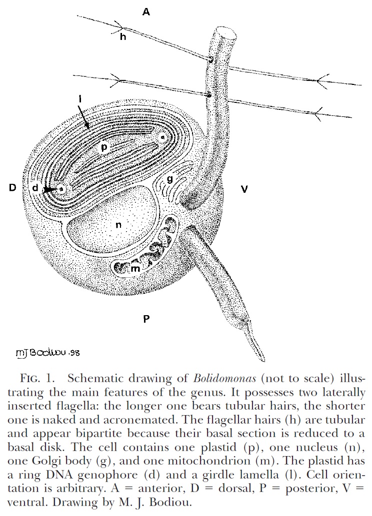
Schematic diagram of motile bolidophyte cell, from Guillou et al. (1999).


Heterokonts are a major clade of eukaryotes that are commonly characterised by cells bearing anterior pairs of morphologically distinct cilia. One of the cilia is longer and bears rows of hairs referred to as mastigonemes; the other, shorter cilium is usually smooth. Many heterokont species are photosynthetic and belong to a subclade of the heterokonts known as the ochrophytes. For most people, the best known ochrophytes will be the often-decidedly-not-microbial brown algae such as kelps. However, ochrophytes also include a broad diversity of microbial forms. Most ochrophyte cells share a characteristic golden-brown coloration owing to the presence of yellowish pigments such as fucoxanthin as well as the more standard chlorophyll.

Recent molecular studies have supported a division of the ochrophytes between two major clades. On one side are the brown algae and their closer microbial relatives. In the other clade are those ochrophytes more closely related to the diatoms. Appropriately enough, this latter clade was dubbed the Diatomista by Derelle et al. (2016). Other than the diatoms themselves, most representatives of the Diatomista belong to the picoplankton. For the most part, diatoms have lost the cilia otherwise associated with heterokonts. The only exceptions are the reproductive sperm cells which have a single anterior cilium bearing mastigonemes (Adl et al. 2019). The remaining Diatomista commonly have cells bearing one or two anterior cilia (if only one cilium is present, it will typically have mastigonemes). Nevertheless, the basal apparatus of the cilia is reduced, lacking microtubular roots or a rhizoplast, suggestive of an intermediate stage towards total loss (Guillou et al. 1999). Many also bear a covering of silica scales; enlargement of individual scales may have lead to the evolution of diatom-style frustules.
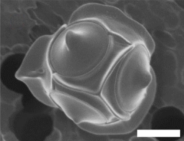
Non-motile cell of Triparma laevis f. inornata, from Kuwata et al. (1987).


The closest known relatives of diatoms are currently classified as the class Bolidophyceae. Motile cells of the Bolidophyceae were first described in 1999 (Guillou et al. 1999). They possessed two cilia, with the haired cilium directed anteriorly and the smooth cilium directed posteriorly, and lacked silica scales. Nevertheless, they were identified as the sister group to diatoms by molecular data. This was corroborated by the absence of a transitional helix structure at the base of each cilium, a feature shared with diatom sperm cells. Guillou et al. (1999) commented on the relatively high mobility of the bolidophytes, in contrast to the general expectation that picoplankton should exhibit a reduction in individual cell mobility owing to the difficulty in meeting energy demands.

The concept of bolidophytes shifted somewhat in the 2010s with the isolation in culture of the Parmales, a group of minute eukaryotes that had first been recognised in the 1980s but had long eluded detailed characterisation. These were non-motile cells enclosed within ornate silica scales. Once molecular data become available, researchers realised that 'Parmales' were not just closely related to 'bolidophytes', they were close enough that the two forms could reasonably be included in a single genus (Kuwata et al. 2018). The exact details of their connection, however, remain uncertain. It seems likely that the flagellate and non-flagellate forms represent alternate forms of single species. But whether we are looking at alternate generations of the life cycle, or whether the flagellate cells are generated in response to particular conditions, remains to be determined.

Skeleton of silicoflagellate Dictyocha speculum, copyright Proyecto Agua.


The remaining members of the Diatomista form a clade currently treated as including three classes, the Dictyochophyceae, Pelagophyceae and Pinguiophyceae. Together they are a diverse array of minute organisms, whether ciliated or amoeboid, naked or carrying organic scales, photosynthetic or heterotrophic or some combination of both. Among the representatives of the Dictyochophyceae are the so-called silicoflagellates, ciliated cells reinforced with a skeleton of (duh) silica. Though only a few species of silicoflagellate are recognised in the modern environment, they have an extensive fossil record extending back to the Middle Cretaceous (Kristiansen 1990). In some places, their preserved skeletons may dominate rock formations. Silicoflagellates appear to have reached their peak in the Miocene, followed by a decline to their modern condition. The exact interpretation of the silicoflagellate fossil record is a long-standing challenge (whether differences in morphology are taxonomic or environmental, for instance) but they hold the potential to tell us much about the history of our seas.

REFERENCES

Adl, S. M., D. Bass, C. E. Lane, J. Lukeš, C. L. Schoch, A. Smirnov, S. Agatha, C. Berney, M. W. Brown, F. Burki, P. Cárdenas, I. Čepička, L. Chistyakova, J. del Campo, M. Dunthorn, B. Edvardsen, Y. Eglit, L. Guillou, V. Hampl, A. A. Heiss, M. Hoppenrath, T. Y. James, A. Karnkowska, S. Karpov, E. Kim, M. Kolisko, A. Kudryavtsev, D. J. G. Lahr, E. Lara, L. Le Gall, D. H. Lynn, D. G. Mann, R. Massana, E. A. D. Mitchell, C. Morrow, J. S. Park, J. W. Pawlowski, M. J. Powell, D. J. Richter, S. Rueckert, L. Shadwick, S. Shimano, F. W. Spiegel, G. Torruella, N. Youssef, V. Zlatogursky & Q. Zhang. 2019. Revisions to the classification, nomenclature, and diversity of eukaryotes. Journal of Eukaryotic Microbiology 66: 4–119.

Derelle, R., P. López-García, H. Timpano & D. Moreira. 2016. A phylogenomic framework to study the diversity and evolution of stramenopiles (=heterokonts). Molecular Biology and Evolution 33 (11): 2890–2898.

Guillou, L., M.-J. Chrétiennot-Dinet, L. K. Medlin, H. Claustre, S. Loiseaux-de Goër & D. Vaulot. 1999. Bolidomonas: a new genus with two species belonging to a new algal class, the Bolidophyceae (Heterokonta). Journal of Phycology 35: 368–381.

Kristiansen, J. 1990. Phylum Chrysophyta. In: Margulis, L., J. O. Corliss, M. Melkonian & D. J. Chapman (eds) Handbook of Protoctista. The structure, cultivation, habitats and life histories of the eukaryotic microorganisms and their descendants exclusive of animals, plants and fungi. A guide to the algae, ciliates, foraminifera, sporozoa, water molds, slime molds and the other protoctists pp. 438–453. Jones & Bartlett Publishers: Boston. Kuwata, A., K. Yamada, M. Ichinomiya, S. Yoshikawa, M. Tragin, D. Vaulot & A. Lopes de Santos. 2018. Bolidophyceae, a sister picoplanktonic group of diatoms—a review. Frontiers in Marine Science 5: 370.
Lifestyles of the Rosalinidae

Among the modern foraminiferans, one of the most prominent radiations is among members of the Rotaliida, characterised by globose chambers and calcareous, hyaline test walls. Among the numerous families making up the Rotaliida are members of the Rosalinidae.

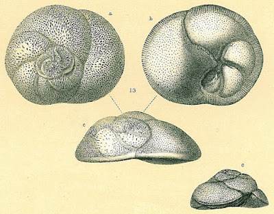
Benthic form of Rosalina globularis, from Brady (1884).


Rosalinids may be regarded as fairly typical-looking marine rotaliids with the test growing freely as a low trochospire (so a flattened cone or dish shape). The aperture of the test is a low slit on the interior margin along the umbilicus (Hansen & Revets 1992). Rosalinids have a complex life cycle involving both benthic and planktonic stages (Sliter 1965). The asexually reproducing diploid stage is benthic. Depending on conditions, diploid individuals may divide to produce other diploid individuals, resulting in several asexual generations. Eventually, however, the diploid generation will undergo meiosis to produce the haploid sexual generation (in the common species Rosalina globularis, this is induced by exposure to warmer water). In the sexual generation, a large globular chamber forms at maturity that covers the umbilical side of the test. This float chamber becomes filled with gas, allowing the foram to disperse planktonically before releasing gametes to produce the next diploid generation. Planktonic individuals are distinct enough in appearance from their benthic counterparts that they were long mistaken for distinct taxa before their identity was revealed by lab cultures.

Life cycle of Rosalina globularis, from Sliter (1965).


The majority of forams are particulate feeders. A network of filamentous pseudopodia radiating outwards from the cell body captures micro-organisms and other organic particles. However, one genus of rosalinids, Hyrrokkin, lives as parasites on sessile invertebrates (Cedhagen 1994). Species of this genus have variously been found on sponges, corals and bivalves. On sponges, they settle on the inhalent surface of the sponge and dissolve the underlying tissues. On bivalves, they form pits on the shell surface from which they bore holes through to the body cavity. Pseudopodia extended through this hole allow the foram to feed on host tissue. Infested hosts may bear multiple scars from the foram moving about on the outer surface. The forams may also feed on other animals such as polychaete worms or bryozoans attached to the surface of their primary host. In such cases, Hyrrokkin remains in its original pit but develops an irregularly shaped chamber with its aperture directed towards the alternate prey. Hyrrokkin species evidently do well from their rapacious lifestyle: whereas other rosalinids are only a fraction of a millimetre in diameter, Hyrrokkin sarcophaga is an absolute giant reaching around six millimetres across and with protoplasm containing thousands of nuclei. Proving once again that one may make a great deal of profit from the labour of others.

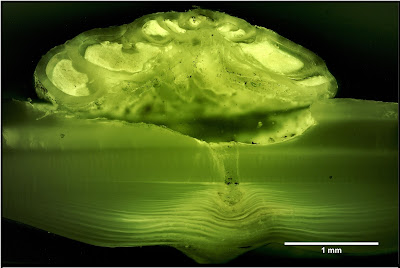
Cross-section of Hyrrokkin sarcophaga boring into shell of file clam Acesta excavata, from Schleinkofer et al. (2021).


REFERENCES

Cedhagen, T. 1994. Taxonomy and biology of Hyrrokkin sarcophaga gen. et sp. n., a parasitic foraminiferan (Rosalinidae). Sarsia 79: 65–82.

Hansen, H. J., & S. A. Revets. 1992. A revision and reclassification of the Discorbidae, Rosalinidae, and Rotaliidae. Journal of Foraminiferal Research 22 (2): 166–180.

Sliter, W. V. 1965. Laboratory experiments on the life cycle and ecologic controls of Rosalina globularis d'Orbigny. Journal of Protozoology 12 (2): 210–215.

The Bolivinitids

The Cretaceous was a period of significant innovation in the evolution of Foraminifera with a number of distinct new lineages making their appearance during this period. Among those, appearing in the latter part of the Cretaceous, were the first members of the modern family Bolivinitidae.

Bolivinita costifera, from the Smithsonian National Museum of Natural History.


The Bolivinitidae are free-living benthic forams with a calcareous, hyaline (glassy) test. The overall shape of the test is elongate with chambers arranged in biserial coils (that is, there are two chambers per loop). The terminal aperture is usually loop-shaped with a surrounding lip. Inside the chamber, a tooth plate (an inner protrusion of the test) runs from the aperture to the opening of the previous chamber and may protrude through the aperture (Revets 1996).

Representatives of the Bolivinitidae are found in a wide range of depths, from the shallow waters of the ocean to the bathyal zone. They may be among the most abundant forams in areas of low oxygen concentrations and are commonly associated with sustained organic matter input (Erdem & Schönfeld 2017). In other words, these are muck-lovers. Individuals growing in low oxygen conditions tend to show less pronounced surface sculpture on the test than those where the oxygen levels are higher. Conversely, individuals at deeper levels tend to be larger overall than those in shallower waters (Brun et al. 1984). As such, bolivinitids have received their fair share of attention as potential indicators of changes in environmental condition over time.

REFERENCES

Brun, L., M. A. Chierici & M. Meijer. 1984. Evolution and morphological variations of the principal species of Bolivinitidae in the Tertiary of the Gulf of Guinea. Géologie Méditerranéenne 11 (1): 13–57.

Erdem, Z., & J. Schönfeld. 2017. Pleistocene to Holocene benthic foraminiferal assemblages from the Peruvian continental margin. Palaeontologica Electronica 20.2.35A: 1–32.

Revets, S. A. 1996. The generic revision of the Bolivinitidae Cushman, 1927. Cushman Foundation for Foraminiferal Research Special Publication 34: 1–55.

Dictyotales

Most of the various 'seaweeds' found around the world can be assigned to one of three major groups, each named for their most characteristic pigments: green algae, red algae and brown algae. Of these, green algae are the closest relatives of land plants, and red algae are the most taxonomically diverse. But for many people, the most familiar of the three will be brown algae. Owing to their often relatively large size and predilection for growing in visible locations, brown algae are likely to be the first examples to come to mind when one thinks of seaweed. For this post, I'm examining a particular subgroup of the brown algae, the family Dictyotaceae.

Forkweed Dictyota dichotoma, copyright Ria Tan.


Representatives of the Dictyotaceae can be found around the world but are more diverse in warmer tropical and subtropical waters. They seem to be particularly diverse in the Australasian region. Dictyotaceae are moderately sized seaweeds with flattened thalli that may grow as branching ribbons or radiating fans. One fan-shaped species of Dictyotaceae, Padina pavonica, has earned itself the vernacular name of 'peacock's tail'(this species is also notable for being one of the few calcified brown algae). These thalli grow apically from meristematic cells. Dictyotaceae have an isomorphic life cycle with the alternating sexually and asexually reproducing generations being similar in overall appearance. Sporangia in asexual individuals grow as superficial nodules scattered over the surface of the thallus; the resulting spores usually differ from those of other brown algae in lacking flagella. The less abundant sexual individuals are mostly divided between separate males and females (Bittner et al. 2008).

Peacock's tail Padina pavonica, copyright Diego Delso.


Dictyotaceae are distinct enough from other brown algae to have consistently been treated as their own order (indeed, their sporangia are unique enough that some very early authors did not even regard them as brown algae). Two species found around Australasia, Dictyotopsis propagulifera and Scoresbyella profunda, have previously been considered distinct enough to warrant their own separate families within this order Dictyotales. Dictyotopsis propagulifera has a monostromatic thallus (that is, the thallus is only one layer of cells thick). Scoresbyella profunda has an apical growing cell that divides lengthwise to the thallus instead of transversely as in other Dictyotales. However, molecular data have indicated that these two genera are nested within Dictyotaceae and so only the single family is currently recognised. Dictyotaceae has also been divided in the past between tribes Dictyoteae and Zonarieae based on the nature of the apical growing cells (Dictyoteae have a single meristematic cell whereas Zonarieae have a cluster or row of cells) and some authors have even treated them as distinct families. Again, however, molecular data have not corroborated this division (Bittner et al. 2008).

Lobophora variegata, copyright John Turnbull.


For most species of Dictyotaceae, their greatest significance to humans probably comes from the role they play in providing habitats to fish and other marine animals. As with other algae, Dictyotaceae produce a range of secondary metabolites that serve functions such as protecting them from grazers, and some of these may prove to have economic applications. Some species of Dictyotaceae, on the other hand, have become significant invasive species. A dramatic recent example has been provided by the northern Pacific species Rugulopteryx okamurae which was probably first imported to the Mediterranean as a contaminant on farmed oysters (García-Gómez et al. 2020). This species was recorded on the southern coast of France in 2002 and was later recorded on the coast of Ceuta in 2015. Within a year of the latter record, its presence in Ceuta had reached absolute plague proportions. Most of the illuminated rocky sea bottom was covered by R okamurae, up to about 90% coverage at optimal depths about ten to twenty metres. Over 5000 tons of washed-up seaweed was removed from the beaches of Ceuta in 2016. Needless to say, native seaweeds, and other sessile marine organisms such as corals, would have been severely impacted by this spread.

Rugulopteryx okamurae in Morocco, from El Aamri et al. (2018).


What caused this dramatic invasion? It would have certainly been a factor that defensive metabolites produced by Rugulopteryx okamurae had a negative impact on competitors. But perhaps even more significant a factor was climate change. Rising sea temperatures in the Straits of Gibraltar would have made things uncomfortable for native marine life used to cooler conditions. Meanwhile, the subtropical immigrant would have found things increasingly to its liking. With its competition hobbled and nothing to hold it back, R. okamurae was set to take over.

REFERENCES

Bittner, L., C. E. Payri, A. Couloux, C. Cruaud, B. de Reviers & F. Rousseau. 2008. Molecular phylogeny of the Dictyotales and their position within the Phaeophyceae, based on nuclear, plastid and mitochondrial DNA sequence data. Molecular Phylogenetics and Evolution 49: 211–226.

García-Gómez, J. C., J. Sempere-Valverde, A. R. González, M. Martínez-Chacón, L. Olaya-Ponzone, E. Sánchez-Moyano, E. Ostalé-Valriberas & C. Megina. 2020. From exotic to invasive in record time: the extreme impact of Rugulopteryx okamurae (Dictyotales, Ochrophyta) in the strait of Gibraltar. Science of the Total Environment 704: 135408.

Naviculi, Navicula

Diatoms are one of the most prominent groups of micro-algae in aquatic environments, perhaps more abundant than any other major group of aquatic organisms except bacteria. As such, they are a key component in many of the environmental processes that we ultimately depend on: food for aquatic animals, producers of oxygen, et cetera et cetera. To those who study them, they are also known for the intricate architecture of their silica walls. As well as being aesthetically pleasing, this architecture forms a key component of diatom classification. One of the most diverse groups of diatoms recognised has been the mega-genus Navicula.

Light microscope view of Navicula tripunctata, copyright Kristian Peters.

Historically, over one thousand species have been assigned to Navicula. Though more recent authors have restricted the name to a smaller, more tightly defined concept than before, it still contains some 200 or so species (Bruder & Medlin 2008). Species assigned to this genus are an elongate diamond or pill shape. Though the term 'navicula' can be translated from Latin as a small boat, and this is often assumed to be the name's origin, this is incorrect. Its original author, the French naturalist Jean-Baptiste Geneviève Marcellin Bory de Saint-Vincent, derived the name from the French term for a weaver's spindle (navette de tisserand; Cox 1999). A long fissure, the raphe, runs down the midline of each valve of the diatom wall; the diatom moves by extruding secretions through the raphe. In Navicula, the raphe is largely straight though it may be hooked at the ends of the valve. Perpendicular to or radiating from the raphe are striae formed of rows of openings (areolae); in Navicula, these areolae are more or less elongate with their long axes perpendicular to the line of the stria. In some species historically included in Navicula, the striae may be biseriate with two rows of areolae. Some authors have proposed recognising species with biseriate striae as a distinct genus Hippodonta. Cox (1999) disputed whether this distinction was enough to warrant a separate genus but Bruder & Medlin (2008) conducted a molecular phylogenetic analysis of naviculoid diatoms in which the one Hippodonta species included was placed as the sister taxon to Navicula sensu stricto. In distinguishing the genus Sellaphora from Navicula, Mann (1989) also identified a number of cytoplasmic features characteristic of Navicula sensu stricto, such as the possession of two distinct plastids per cell with rod-like pyrenoids.

SEM view of Navicula dobrinatemniskovae, from Van de Vijver et al. (2011). Scale bar = 1 µm.


Ecologically, the majority of species of Navicula sensu stricto (about 150 species) are found in freshwater environments (Bruder & Medlin 2008). In temperate and tropical regions, they are a diverse element of benthic diatom communities, but they are less predominant in coldwater habitats (Van de Vijver et al. 2011). They are most characteristic of meso- to eutrophic lakes and permanent waterways and Van de Vijver et al. (2011) therefore suggested that they might be less suited for the damp soils and temporary pools that dominate freshwater habitats in the frozen South. Nevertheless, these authors still managed to identify five previously unknown species from just this inhospitable region, giving some indication of what still remains to be discovered of this already diverse genus.

REFERENCES

Bruder, K., & L. K. Medlin. 2008. Morphological and molecular investigations of naviculoid diatoms. III. Hippodonta and Navicula s. s. Diatom Research 23 (2): 331–347.

Cox, E. J. 1999. Studies on the diatom genus Navicula Bory. VIII. Variation in valve morphology in relation to the generic diagnosis based on Navicula tripunctata (O. F. Müller) Bory. Diatom Research 14 (2): 207–237.

Mann, D. G. 1989. The diatom genus Sellaphora: separation from Navicula. British Phycological Journal 24 (1): 1–20.

Van de Vijver, B., R. Zidarova, M. Sterken, E. Verleyen, M. de Haan, W. Vyverman, F. Hinz & K. Sabbe. 2011. Revision of the genus Navicula s.s. (Bacillariophyceae) in inland waters of the sub-Antarctic and Antarctic with the description of five new species. Phycologia 50 (3): 281–297.

Silicon Rockets

In a previous post, I spoke of the radiolarians, marine protists renowned for their intricate skeletons, and the major radiolarian group known as the Spumellaria. Standing in contrast to the spumellarians is another major group, the Nassellaria. Like spumellarians, nassellarians have a skeleton of silica but whereas the basic shape of spumellarian skeleton is a sphere, that of nassellarians is a cone, bell or some similar shape, arranged along a longitudinal axis. The origination point of the skeleton is at or near the top of the cone and is known as the cephalis (from the Greek for 'head'). There may be an apical spine rising above the cephalis. Below it, the skeleton is commonly divided into recognisable sections referred to as the thorax, abdomen and post-abdominal segments (if present). The nucleus of the cell is more or less associated with the cephalis, contained within it at least during the juvenile stage of development though it may shift below the cephalis as the cell matures (Suzuki et al. 2009).

Skeleton of a Eucyrtidium sp., copyright Picturepest.


As is commonly the case with unicellular organisms, radiolarian taxonomy has been influenced by disagreements about which features should be regarded as more significant. Some would arrange taxa based on the overal formation of the skeleton. Others would focus on the development of the initial embryonic spicule around which the cephalis develops. A recent phylogenetic analysis of living nassellarians by Sandin et al. (2019), based on both morphological and molecular data, found that overall skeleton morphology was a much better indication of relationships than the internal structure. One well supported subgroup of the Nassellaria is the superfamily Eucyrtidioidea.

Eucyrtidioids have a fossil record going back to the Triassic (Afanasieva et al. 2005). The cephalis is spherical and clearly distinguished from the following segments by a constricted basal aperture. The test is usually multi-segmented; members of the subfamily Theocotylinae may have just two segments but other members of Eucyrtidiidae have up to ten segments. Fossil families assigned to Eucyrtidioidea by Afanasieva et al. (2005) may have up to twenty (but as Afanasieva et al.'s concept of Eucyrtidioidea was not found to be monophyletic by Sandin et al., the affinities of these fossil families perhaps warrant re-investigation). Segments are commonly divided by distinct inner rings. The skeleton lacks feet, the term used for protruding spines around the basal aperture of the skeleton found in many other nassellarians.

The phylogeny of nassellarians indicated by Sandin et al. (2019) places the Eucyrtidiidae as the sister taxon to other living nassellarians. Other living families included in the Eucyrtidioidea by Afanasieva et al. (2005) were placed in more nested positions. The implication is that the multi-segmented condition may be ancestral for crown Nassellaria. Segments are added progressively during the life of the radiolarian, leading the organism to look quite different at different ages. Indeed, this metamorphosis is pronounced enough that one of the earliest influential researchers on radiolarians, Ernst Haeckel (he of Kunstformen der Natur fame), made the mistake of classifying different ages as different species, genera and even families. Our understanding may be better than in Haeckel's time but there may still be a lot to learn about these intricate organisms.

REFERENCES

Afanasieva, M. S., E. O. Amon, Y. V. Agarkov & D. S. Boltovskoy. 2005. Radiolarians in the geological record. Paleontological Journal 39 (Suppl. 3): S135–S392.

Sandin, M. M., L. Pillet, T. Biard, C. Poirier, E. Bigeard, S. Romac, N. Suzuki & F. Not. 2019. Time calibrated morpho-molecular classification of Nassellaria (Radiolaria). Protist 170: 187–208.

Suzuki, N., K. Ogane, Y. Aita, M. Kato, S. Sakai, T. Kurihara, A. Matsuoka, S. Ohtsuka, A. Go, K. Nakaguchi, S. Yamaguchi, T. Takahashi & A. Tuji. 2009. Distribution patterns of the radiolarian nuclei and symbionts using DAPI-fluorescence. Bulletin of the National Museum of Nature and Science, Series B 35 (4): 169–182.

The Glandulinid Position

In an earlier post, I described how the majority of modern multi-chambered foraminiferans can be divided between two lineages, the Tubothalamea and Globothalamea. The two groups generally differ in the shape of the first chamber following the proloculus (the central embryonic chamber of the test): in one, this chamber is tubular whereas in the other it is globular or crescent-shaped (guess which is which). But there is a third notable group of multi-chambered forams: the Nodosariata. In both tubothalameans and globothalameans, the chambers more or less coil around the proloculus to form a spiral. In the Nodosariata, the test is more or less linear with apical chamber apertures. The chambers may be successively stacked one after the other to form a uniserial test, or they may be arranged in a zig-zag or twirling arrangement to form biserial, triserial, etc. arrangments. In living Nodosariata, the wall of the test is made of a single layer of hyaline calcite though some earlier representatives (up to the end of the Jurassic) had differing wall make-ups (Rigaud et al. 2016). Among the numerous notable representatives of the Nodosariata in the modern fauna are representatives of the family Glandulinidae.

Series of Glandulina ovula, from Brady (1884).


Species have been assigned to the Glandulinidae going back to the Jurassic with the modern genus Glandulina recognisable in the Palaeocene (Loeblich & Tappan 1964). The test may be uniserial, biserial or polymorphine (more than two series); a common arrangement is for the test to start out biserial or polymorphine then become uniserial as the individual chambers become larger. In Glandulina, the microspheric generation starts biserial but the megalospheric form is uniserial throughout (Taylor et al. 1985). As the test grows, the internal walls between chambers may be resorbed. The terminal aperture of the test may be radial or slit-like. The most characteristic feature of the family is a tube running into the chamber from the inside of the aperture, referred to as the entosolenian tube. Some glandulinids have been described as lacking an entosolenian tube but such absences are likely artefacts of preservation: the delicate tube is easily dislodged during the fossilisation process (Taylor et al. 1985).

The overall relationships of the Nodosariata remain a question open to investigation. The classification of forams by Loeblich & Tappan (1964) included both multi-chambered and single-chambered (unilocular) forms within the Glandulinidae, with the unilocular forms placed in a subfamily Oolininae. Oolinines resemble glandulinids proper in a number of features including wall structure and the presence of an entosolenian tube. More recent authors, however, have rejected this relationship. Rigaud et al. (2016) entirely excluded unilocular forms from the Nodosariata as a whole, regarding it as improbable that single-chambered forms could have evolved from multi-chambered ancestors (as would seemingly be required by their relative appearances in the fossil record). Do the similarities between glandulinids and oolinines reflect a common ancestry, or are they the result of simple convergence? Unfortunately, with so few significant characters available to inform our understanding of foram higher relationships, the answer you prefer may come down to no more than your own personal feelings about which indicators are more reliable.

REFERENCES

Loeblich, A. R., Jr, & H. Tappan. 1964. Treatise on Invertebrate Paleontology pt C. Protista 2. Sarcodina: chiefly "thecamoebians" and Foraminiferida vol. 2. The Geological Society of America, and The University of Kansas Press.

Rigaud, S., D. Vachard, F. Schlagintweit & R. Martini. 2016. New lineage of Triassic aragonitic Foraminifera and reassessment of the class Nodosariata. Journal of Systematic Palaeontology 14 (11): 919–938.

Taylor, S. H., R. T. Patterson & H.-W. Choi. 1985. Occurrence and reliability of internal morphologic features in some Glandulinidae (Foraminiferida). Journal of Foraminiferal Research 15 (1): 18–23.

Pyrgoidae

I have referred in the past to there being something of a divide in approaches to the classification of the Foraminifera. This divide arises from disagreements such as the relative significance of various character complexes. One taxon that stands as an example of such disagreements is the subject of this post, the family Pyrgoidae as recognised by Mikhalevich (2005).

Pyrgo williamsoni, copyright Michael.


Pyrgoids are members of the group of forams generally recognised as the Miliolida, the porcelaneous forams. In this group, the wall of the test is composed of calcite but the calcite crystals are not regularly lined up with each other so the wall is not transparent. As a result, the wall of the test resembles porcelain in appearance. Most miliolidans have the chambers of the test coiling in a single plane. The Pyrgoidae were distinguished from other miliolidans by Mikhalevich (2005) by the overall structure of the test which is primarily biloculine (with the whorls of the test composed of two chambers). The family was divided into subfamilies by the nature of the test aperture: single with an inner tooth in Pyrgoinae, single with a flap in Biloculinellinae, and multiple (at least when mature) in Cribropyrgoinae and Idalininae. Idalininae also differed from other subfamilies in that the very last chamber was further enlarged to envelop the entire test. Members of the Pyrgoidae are known from the fossil record going back to the Jurassic period.

In the system of Loeblich & Tappan (1964), however, the pyrgoids were not recognised as a single group. Instead, they were dispersed among separate subfamilies of the family Miliolidae. Part of the reason was simply that Loeblich & Tappan did not divide the miliolidan families as finely as Mikhalevich later would but a bigger difference was one of priority. Loeblich & Tappan regarded the nature as an aperture as a more important feature taxonomically than the arrangement of chambers. Both classifications seem to have been constructed more from a diagnostic viewpoint than necessarily intended to reflect phylogenetic relationships.

Cribropyrgo aspergillum, from the National Museum of Natural History.


As with most other forams, pyrgoids exist in what are called megalosphaeric and microsphaeric forms. These forms represent alternate generations in the foram life cycle: microsphaeric forams are the sexually reproducing generation whereas megalosphaeric forams reproduce asexually. The names refer not to the overall size of the individuals but to the size of the proloculus, the very first embryonic chamber that sits at the center of the test. In megalosphaeric pyrgoids, the developing test is biloculine from the very start. In microsphaeric individuals, the earliest stages of the test are quinqueloculine (with five chambers per whorl) then become triloculine then finally biloculine (with a further progression for the idalinines, of course). The significance of the differences between the two forms has historically been the subject of discussion with some authors arguing that the microsphaeric forms represented a retention and overwriting of ancestral forms, or an expression of the trajectory the lineage might evolve along in the future (Loeblich & Tappan 1964). The most likely explanation, though, seems to me to be the simplest. The size of the proloculus correlates with the amount of cytoplasm in the young foram. Megalosphaeric pyrgoids start with fewer chambers per volution from the start for the simple reason that they don't have the space to pack in more.

REFERENCES

Loeblich, A. R., Jr, & H. Tappan. 1964. Treatise on Invertebrate Paleontology pt C. Protista 2. Sarcodina: chiefly "thecamoebians" and Foraminiferida vol. 1. The Geological Society of America, and The University of Kansas Press.

Mikhalevich, V. 2005. The new system of the superfamily Quinqueloculinoidea Cushman, 1917 (Foraminifera). Acta Palaeontologica Romaniae 5: 303–310.

Rugosofusulinids?

We return once again to the fusulinoids, large, complex Foraminifera of the late Palaeozoic. For this post, I'm taking a look at the Rugosofusulinidae, a group known from the last part of the Carboniferous and the earliest part of the Permian. Or to put it more technically, from the Gzhelian and Asselian epochs; their numbers collapsed at the end of the Asselian (Leven 2003).

Axial section of Rugosofusulina prisca, from Loeblich & Tappan (1964).


In an earlier post, I referred to a historical divide that has existed between American and Russian classifications of fusulinoids, with the Russian system recognising a more divided arrangement of taxa. The rugosofusulinids are one example of this: whereas Rauzer-Chernousova et al. (1996) recognise them as a distinct family in the order Schwagerinida, Loeblich & Tappan (1964) treated the entire group of 'schwagerinidans' as a subfamily Schwagerininae in the Fusulinidae (I believe more recent western authors might be inclined to at least treat Schwagerinidae as a separate family but would probably still not separate the rugosofusulinids). Whatever level you wish to place them at, the most distinctive feature of rugosofusulinids as a group is a distinct rugosity of the outer wall of the chambers. This may be due to undulations in the entire chamber wall or rugosity of the outer surface only. When first described, it was thought that this unevenness reflected ridges on the outer surface, but it was later observed that the rugosity looked much the same whatever angle the foram was cut at (remember, fusulinoids are most commonly studied in thin sections rather than as entire separated fossils) so probably represented more discrete ornaments. Skinner & Wilde (1966) suggested that "the outer surface [of Rugosofusulina] is scored by numerous sharp furrows which are directed both axially and sagittally, resulting in a surface which resembles a miniature cobblestone pavement".

The question of whether you wish to recognise rugosofusulinids as a distinct family is definitely not helped by a question hanging over recognition of the name Rugosofusulina. The problem is not really with Rugosofusulina itself but with another genus, Pseudofusulina, recognised in the Rauzer-Chernousova et al. (2007) system as type of another family of Schwagerinida, Pseudofusulinidae, and its type species P. huecoensis. Classically, this genus and family has been supposed to have a smooth rather than rugose outer tectum. However, the type specimen of P. huecoensis was re-examined by Skinner & Wilde (1966) who found that it did indeed have 'Rugosofusulina'-type external rugosities. They consequently synonymised the two genera with Pseudofusulina standing as the older name. The response of Russian authors to this challenge to their system, it seems, was generally to ignore it. Pseudofusulina and Rugosofusulina may still potentially be distinguishable as genera by degree of rugosity (Zhang et al. 2013) but this seems a weak basis for a full family distinction. Even if 'Rugosofusulina' is okay, 'Rugosofusulinidae' may not be.

REFERENcES

Leven, E. J. 2003. The Permian stratigraphy and fusulinids of the Tethys. Rivista Italiana di Paleontologia e Stratigrafia 109 (2): 267–280.

Loeblich, A. R., Jr, & H. Tappan. 1964. Treatise on Invertebrate Paleontology pt C. Protista 2. Sarcodina: chiefly "thecamoebians" and Foraminiferida vol. 1. The Geological Society of America, and The University of Kansas Press.

Rauzer-Chernousova, D. M., F. R. Bensh, M. V. Vdovenko, N. B. Gibshman, E. Y. Leven, O. A. Lipina, E. A. Reitlinger, M. N. Solovieva & I. O. Chedija. 1996. Spravočnik po Sistematike Foraminifer Paleozoâ (Èndotiroidy, Fuzulinoidy). Rossijskaâ Akademiâ Nauk, Geologičeskij Institut, Moskva "Nauka".

Skinner, J. W., & G. L. Wilde. 1966. Type species of Pseudofusulina Dunbar & Skinner. University of Kansas Paleontological Contributions 13: 1–7.

Zhang, Y.-C., Y. Wang, Y.-J. Zhang & D.-X. Yuan. 2013. Artinskian (Early Permian) fusuline fauna from the Rongma area in northern Tibet: palaeoclimatic and palaeobiogeographic implications. Alcheringa 37 (4): 529–546.

Chilostomellidae: Deep Forams

Holotype of Chilostomella serrata, from the Smithsonian National Museum of Natural History.


The specimen in the figure above is a fairly typical representative of the Chilostomellidae, a cosmopolitan family of forams known from the Jurassic to the present day. Members of this family have a translucent calcareous test with chambers arranged in a trochospiral (broad conical) or planispiral (flat spiral) pattern. The chambers of each spiral are expanded to cover over the prior spirals so only the outermost spiral is generally visible. The aperture of the test in the final chamber is a narrow slit along the margin with the underlying chamber (Loeblich & Tappan 1964).

Despite their long history and wide distribution, I get the general impression that chilostomellids are not usually abundant. They are generally restricted to deeper waters, more than 100 m below the surface (Cushman et al. 1954). Members of the genus Chilostomella, at least, have commonly been regarded as associated with low-oxygen environments. However, it has also been suggested that their favoured conditions are not so much a question of low oxygen as high organic flux (Jorissen 2002). Perhaps the best location to find chilostomellids would be around sites where dead animals and seaweeds have fallen to the deeper waters below.

REFERENCES

Cushman, J. A., R. Todd & R. J. Post. 1954. Recent Foraminifera of the Marshall Islands. Bikini and nearby atolls, part 2, oceanography (biologic). Geological Survey Professional Paper 260-H: 319–384, pls 82–93.

Jorissen, F. J. 2002. Benthic foraminiferal microhabitats below the sediment-water interface. In: Sen Gupta, B. K. (ed.) Modern Foraminifera pp. 161–179. Kluwer Academic Publishers: Dordrecht.

Loeblich, A. R., Jr & H. Tappan. 1964. Treatise on Invertebrate Paleontology pt C. Protista 2. Sarcodina: chiefly "thecamoebians" and Foraminiferida vol. 2. The Geological Society of America, and The University of Kansas Press.

The Model Tetrahymenidans

Ciliates have long been one of the most (if not the most) confidently recognised groups of unicellular eukaryotes owing to their distinctive array of features, in particular locomotion by means of more or less dense tracts of small cilia that often run the length of the organism. And of all ciliates, perhaps none have been more extensively studied than species of the genus Tetrahymena such as T. thermophila. Being easily cultured in the laboratory, Tetrahymena species have become model organisms for the study of a great many genetic and cellular systems such as cell division and gene function. At least two Nobel prizes have been awarded for work based on Tetrahymena that established the functions of telomeres and ribozymes. But Tetrahymena is just one genus of larger group of ciliates, the Tetrahymenida.

Tetrahymena thermophila, from Robinson 2006.


In general, tetrahymenidans are more or less 'typical'-looking ciliates with an ovoid body form and a well-developed 'mouth' at one end. The name Tetrahymena, meaning 'four membranes', refers to the presence of four membrane-like structures inside the oral cavity, a larger, ciliated undulating membrane on the left and three membranelles (formed from polykinetids, complex arrays of cilia and associated basal bodies and fibrils). Most tetrahymenidans possess some variation of this arrangement with the exception of Curimostoma, a genus of parasites of freshwater flatworms and molluscs that lack oral structures (Lynn & Small 2002). Life cycles may contain a number of morphologically differentiated stages. A more mobile theront stage will seek out food sources then transform into a feeding trophont. Mature trophonts may divide asexually or reproduce through conjugation. Cellular multiplication often involves successive divisions so a single parent cell may give rise to four daughter cells. In a number of species, resistant resting cysts may form under adverse conditions.

Glaucoma scintillans, another well-studied tetrahymenidan, copyright Proyecto Agua.


Tetrahymenidans are also ecologically diverse, occupying a range of freshwater habitats. They may be free-living, feeding on bacteria, or they may be parasitic or histophagous, feeding on the tissues of invertebrates. Some species may switch between one or the other depending on circumstances. A few Tetrahymena species have even been cultured in the laboratory axenically: that is, absorbing nutrients directly from a culture broth without requiring a bacterial food supply. Recently, the first confirmed case of a tetrahymenidan containing endosymbiotic algae was described by Pitsch et al. (2016). The species Tetrahymena utriculariae inhabits the bladders of the carnivorous bladderwort Utricularia reflexa. Endosymbiotic green algae provide it with oxygen, allowing the ciliate to survive within the anoxic environment of the bladders.

REFERENCES

Lynn, D. H., & E. B. Small. 2002. Phylum Ciliophora Doflein, 1901. In: Lee, J. J., G. F. Leedale & P. Bradbury (eds) An Illustrated Guide to the Protozoa: Organisms traditionally referred to as Protozoa, or newly discovered groups 2nd ed. vol. 1 pp. 371–656. Society of Protozoologists: Lawrence (Kansas).

Pitsch, G., L. Adamec, S. Dirren, F. Nitsche, K. Šimek, D. Sirová & T. Posch. 2016. The green Tetrahymena utriculariae n. sp. (Ciliophora, Oligohymenophorea) with its endosymbiotic algae (Micractinium sp.), living in traps of a carnivorous aquatic plant. Journal of Eukaryotic Microbiology 64: 322–335.

Of Crosses and Clubs

One of the major groups of eukaryotes that has been somewhat under-represented on this site has been the Cercozoa. This is a diverse clade of unicellular organisms, distantly related to the foraminiferans and radiolarians, that has only been recognised within the last few decades with the introduction of molecular phylogenetic analyses. It has become increasingly clear that cercozoans form a major part of the world's microscopic biota but this diversity is poorly known as most cercozoans have little direct effect on human industry. One subgroup of the cercozoans that does make itself known in this regard, however, is the Phytomyxea.

Club roots of a rape plant infected by Plasmodiophora brassicae, photographed by Leafhopper65.


The Phytomyxea include parasites of plants, algae and other aquatic micro-organisms. The best known phytomyxean species, Plasmodiophora brassicae, causes a condition known as 'club root' in cabbages; another, Spongospora subterranea, is responsible for powdery scab on potatoes. They form multinucleate 'plasmodia' when growing within the cells of their host. Nuclei divide within the plasmodium in a characteristic cruciform pattern: the nucleolus does not break down during division but instead stretches elongately before pinching in two. While stretched, the nucleolus is oriented perpendicularly to the separating chromatin, forming a cross (Dylewski 1990). Owing to a superficial resemblance between phytomyxean plasmodia and those formed by the plasmodial slime moulds, phytomyxeans were historically also treated as slime moulds and hence as fungi (alternative historical names for the group, such as Plasmodiophoromycota or Plasmodiophoromycetes, reflect this supposed affinity). However, whereas the amoeboid plasmodia of slime moulds are capable of active movement and ingestion of food particles via phagocytosis, the phytomyxean plasmodium is more or less incapable of moving of its own volition, instead moving within the host cell by means of the host's own cytoplasmic streaming, and do not engulf host tissue in vacuoles. Slime moulds are no longer regarded as a single evolutionary lineage, and no 'slime moulds' are directly related to fungi.

Nuclei undergoing cruciform division in plasmodium of Tetramyxa parasitica, copyright James P. Braselton.


Over 40 species of Phytomyxea have been recognised to date but, not surprisingly, studies on the group have focused heavily on those species of economic importance to humans (Neuhauser et al. 2011). Terrestrial phytomyxeans produce thick-walled resting cysts, often aggregated in clumps known as cystosori, that may persist in soil for several years. These cysts hatch into biflagellate primary zoospores that seek out a suitable host. Upon finding one, the spore ceases swimming and adheres to the host cell before piercing the cell wall and injecting its cytoplasm which grows into the aforementioned plasmodium. Nuclei divide by mitosis and are eventually parcelled into sporangia that release secondary zoospores that escape from the host cell. These secondary spores generally do not disperse far; instead, they tend to cycle back and re-infect the original host to form new plasmodia. When these secondary plasmodia reach maturity, their nuclei divide meiotically and are divvied into new resting cysts. Presumably, the haploid nuclei produced in this manner fuse at some point with another to return to diploidy but it is unknown when exactly this happens. The cysts, when formed, each contain two nuclei but later only one, so it is possible that this reduction results from fusion. However, it might seem more likely that one of the nuclei breaks down without issue and the cyst remains haploid through to excystment with fusion occurring at the primary zoospore phase, thus allowing greater scope for cross-fertilisation. Marine phytomyxeans have long been thought not to produce resting cysts but recent observations of variations in zoospore morphology and sporangial wall thickness in the brown algal parasite Maullinia ectocarpii suggest the possibility of similarly complex life cycles (Neuhauser et al. 2011). The length of the phytomyxean life cycle can vary from about a month for Plasmodiophora brassicae to as little as one or two days for the brown algal parasite Phagomyxa algarum.

Diagram of the life cycle of Plasmodiophora brassicae, from Auer & Ludwig-Müller (2015).


For most phytomyxean species, infection by plasmodia causes physiological changes in the host, commonly taking the form of galls or other excesses of growth. Club root disease of Brassica results from Plasmodiophora brassicae plasmodia producing growth hormones that cause nutrients to be concentrated in the roots at the expense of leaf growth, thus increasing their availability to the parasite. Other alterations may be related to parasite dispersal. Ligniera junci, a parasite of rushes, causes a proliferation in the growth of root hairs in which the resting cysts form, providing an extra protective sheath. Plasmodiophora bicaudata is a parasite of marine Zostera eelgrass that produces galls at internodes together with reduced root growth. As a result, the eelgrass is easily uprooted by water movement, potentially being carried to new areas where the next generation of phytomyxeans can find new eelgrasses to infect.

REFERENCES

Dylewski, D. P. 1990. Phylum Plasmodiophoromycota. In: Margulis, L., J. O. Corliss, M. Melkonian & D. J. Chapman (eds) Handbook of Protoctista. The structure, cultivation, habitats and life histories of the eukaryotic microorganisms and their descendants exclusive of animals, plants and fungi. A guide to the algae, ciliates, foraminifera, sporozoa, water molds, slime molds and the other protoctists pp. 399–416. Jones & Bartlett Publishers: Boston.

Neuhauser, S., M. Kirchmair & F. H. Gleason. 2011. The ecological potentials of Phytomyxea ("plasmodiophorids") in aquatic food webs. Hydrobiologia 659: 23–35.

We've Got a Thing that's called Foram Love

Pileolina patelliformis, from Brady (1884).


It's been a while since we last had a foram post, so why don't we have one today? Ladies and gentlemen, I present to you: the Glabratellidae.

Glabratellids are a group of forams found in littoral habitats, first appearing in the fossil record in the Eocene (Loeblich & Tappan 1964). They secrete a calcareous test with a hyaline (glass-like) microstructure. By foram standards, glabratellids can be quite small: the smallest are well under 100 µm in diameter. They have a trochospiral body shape—that is, the body chambers are arranged in such a way that they spiral like a trochus or top shell—with a flat base. At the centre of the underside is an aperture or umbilicus. The spire may be fairly low, giving them what I always think of as a 'jelly mould' shape, or it may be high so their overall appearance is conical. In the genus Schackoinella, the test bears a spine on the outside of each of the body chambers.

The glory that is Schackoinella sarmatica, from the Geological Survey of Austria.


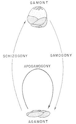
The most distinctive feature of glabratellids, perhaps, is their life cycle. We know the life cycles of relatively few foram species but as a rule they show a clear alternation of generations, with both well-developed haploid and diploid individuals. Haploid individuals (gamonts) produce gametes by nuclear mitosis that fuse to form zygotes that grow into mature diploid individuals (schizonts or agamonts); these latter produce haploid embryos via meiosis. The two generations may differ somewhat in appearance, and many foram species have had their gamonts and schizonts mistaken in the past for separate species. The most consistent difference between generations in all chambered forams is that the gamonts have a larger first chamber as a result of growing from larger embryos than the schizonts. In glabratellids, gamonts are also smaller and relatively higher-spired than schizonts, and the former are sinitrally coiled (to the left) while the latter are dextrally coiled (to the right).

The life cycle of Glabratellidae was described in detail by Loeblich and Tappan (1964) (the figure to the the left from therein shows the lifecycle of Pileolina patelliformis). Schizonts herald the production of offspring by wrapping themselves in a protective cover of dead diatoms and other rubbish. Young gamonts are formed by nuclei dividing in the test and each becoming surrounded by their own individual cell membranes. After they form, the embryonic offspring crawl around in the parent test feeding on any leftover cytoplasm and also on the test itself. By the time they grow to about two or three chambers in size, the gamonts dissolve the umbilical wall of the parent test and escape through the aperture.

As the gamonts themselves reach maturity, their thoughts no doubt turn to their own posterity. Whereas in some other forams the haploid generation simply releases their gametes into the water column to find their own way to fusion, sexual reproduction in glabratellids is a somewhat more intimate affair. Mature gamonts form into pairs, joined to each other via their umbilical surfaces from which they will resorb the test. Locked in their embrace, the pair become cemented to the substrate. Gametes, again, are formed by the production of plasma membranes around individual nuclei; these gametes move by means of three flagella instead of by pseudopodia. The two parents exchange gametes of which only about a tenth fuse to form zygotes; the remainder provide a food source for their developed siblings. Again, the young schizonts grow to about two or three chambers in size before being released by the dissolution of the cement holding the parent tests together.

This cosy mode of reproduction means that glabratellids may have the potential for greater population differentiation than other broadcast-spawning Foraminifera. Tsuchiya et al. (2003), in a study genetic diversity in representatives of the genus Planoglabratella collected around Japan, found evidence for cryptic speciation in P. opercularis. Some individuals of this 'species' were closer genetically to individuals of another species P. nakamurai than to other P. opercularis, and closer inspection revealed certain details of their morphology that were more nakamurai-like than opercularis-like. It may be that we have underestimated the diversity of glabratellids, and many more species of this group remain to be discovered.

REFERENCES

Loeblich, A. R., Jr & H. Tappan. 1964. Treatise on Invertebrate Paleontology pt C. Protista 2. Sarcodina: chiefly "thecamoebians" and Foraminiferida. The Geological Society of America, and The University of Kansas Press.

Tsuchiya, M., H. Kitazato & J. Pawlowski. 2003. Analysis of internal transcribed spacer of ribosomal DNA reveals cryptic speciation in Planoglabratella opercularis. Journal of Foraminiferal Research 33 (4): 285–293.

Water Moulds

Salmonid infected with Saprolegnia, from the Scottish Government.


In the 1970s and 1980s, stocks of salmon and trout around the North Atlantic Ocean took a sizeable hit. Mature fish entering fresh water had their skin break out in lesions that eventually became covered in a slimy, cottony growth. With the lesions eventually eating into the underlying tissue, many fish died from these infections before they could spawn.

The disease became known as ulcerative dermal necrosis, and its underlying cause remains unknown. The cottony growth so often associated with the disease, however, was made up of a mould-like organism called Saprolegnia. Saprolegnia belongs to a family Saprolegniaceae in a group of organisms known as the Oomycetes, commonly referred to as 'water moulds'. Most Saprolegniaceae function as saprobes, living off decaying organic matter. A few, however, can occasionally function as pathogens. In the case of the aforementioned necrosis outbreak, the Saprolegnia would have been a secondary infection that exacerbated the progress of the disease. Another genus, Aphanomycese, includes species that can cause root rot in vegetables such as peas or beets (Johnson et al. 2002).

Mature and developing oogonia of Saprolegnia, copyright George Barron.


In habit and lifestyle, water moulds resemble fungi, and were long classified as such. When they were first described in the 1700s, however, they were identified as algae due to similarities in their cell and spore morphology to freshwater algae such as Vaucheria. In recent decades, it has become clear that it was these original observers that were closer to the mark. Oomycetes are not directly related to the true fungi, but belong to a lineage known as the heterokonts or stramenopiles. Most heterokonts are microbial, but they also include algal forms such as the brown algae and (yes) Vaucheria. The heterokont affinities of water moulds become apparent during asexual reproduction when they produce motile zoospores bearing a pair of flagella (though many 'water moulds' are terrestrial rather than aquatic, these zoospores do require water to spread). As is typical of heterokonts, these two flagella differ in appearance: the anterior flagellum bears a series of lateral side-branches whereas the posterior flagellum in smooth. Other significant differences between oomycetes and true fungi are that oomycetes are diploid through the greater part of their life cycle (fungi are haploid), and their cell walls are composed not of chitin but of other compounds such as glucans and/or cellulose.

Drawing of zoospores of Saprolegnia, showing divergent flagella, from here.


Characteristic features of the Saprolegniaceae in particular include their possession of relatively broad hyphae, up to 150 µm in some cases (Dick 2001), that are not divided into cells by septae. Other distinguishing features relate to the production of reproductive cells. Most oomycetes are capable of both asexual and sexual reproduction, though one genus of Saprolegniaceae, Aplanopsis, is only known to reproduce sexually. In asexual reproduction, the motile zoospores are produced within a distinct zoosporangium (some other oomycetes do not separate the zoosporangium from the adjoining hypha until after zoospore formation). When first released, the zoospores move relatively little and soon transform into an immotile cyst. This cyst will eventually revert back into a zoospore, and it is at this stage that the greater part of dispersal happens. This secondary zoospore will then transform again into a cyst, from which will grow the mature hyphae.

Hyphae of an Achlya-like oomycete, with clusters of encysted zoospores at the ends of emptied zoosporangia, from here.


Sexual reproduction involves the production of distinct oogonia and antheridia, with the latter fertilising the former to produce oospores (some species can produce oospores parthenogenetically). These differ from zoospores in being aflagellate and immobile, with thick walls that make them more resistant to adverse conditions. Oospores of Saprolegniaceae contain oil globules that probably function as an energy store (like the endosperm of a plant seed). Depending on the species, the distribution of oil globules may vary between numerous small globules evenly distributed around the periphery of the centrally located cytoplasm (referred to as 'centric'), or one large globule pushing the cytoplasm off to one side ('eccentric'). An oospore may geminate into hyphae alone, or it may produce hyphae topped by zoosporangia.

Oogonium of Saprolegnia, with associated antheridium, copyright George Barron.


The genera of Saprolegniaceae have been primarily distinguished by features of the zoosporangia, such as the manner of release of the zoospores. In some genera, the initial zoospores may have already progressed to encystment or the secondary zoospore stage by the time they fully emerge. In genera such as Achlya, the spores are released from a single terminal opening and form a clump at the end of the emptied sporangium. In others such as Saprolegnia, they disperse individually as soon as they escape. And in genera such as Dictyuchus, the zoosporangium wall opens in multiple places and the spores are all sent out by their own distinct orifice. However, more recent phylogenetic studies have cast doubt on the integrity of some of these genera: the Achlya type of zoospore dispersal, for instance, is probably basal for the Saprolegniaceae as a whole and this genus is polyphyletic.

REFERENCES

Dick, M. W. 2001. Straminipilous Fungi: Systematics of the Peronosporomycetes including accounts of the marine straminipilous protists, the plasmodiophorids and other similar organisms. Kluwer Academic Publishers.

Johnson, T. W., Jr, R. L. Seymour & D. E. Padgett. 2002. Biology and systematics of the Saprolegniaceae. http://dl.uncw.edu/digilib/biology/fungi/taxonomy%20and%20systematics/padgett%20book/.

Let's You and Me Enter Syzygy

Finally, you and your beloved are together. For the two of you, there are no others; all the world is yours alone. You gaze into each other's eyes, and then you pull your beloved into an embrace. Your lips touch in a passionate kiss. Your arms and legs intertwine in a firm hold. As you press so close to one another, it almost feels like you can no longer tell where the dividing line is between you. The excitement builds, and then... the two of you explode, each dissolving into a cascading avalanche of twitching gobbets of flesh.

Life cycle of Lecudina, from Clopton (2002).


This, roughly, is syzygy, a key event in the life cycle of many of the invertebrate-gut-parasitising protists known as eugregarines. Originally, the term 'syzygy' referred to the conjunction of two heavenly bodies, and provides a very poetic label for the process by which two of these unicellular organisms conjoin, rotating around one another as they produce and outer membrane to contain themselves within a single gametocyst. Once within the gametocyst, each divides into numerous gametes (which are produced through straight mitotic division, as eugregarines are haploid at maturity rather than diploid like ourselves). The resulting gametes will then be released from the gametocyst to fuse with one another in the production of diploid zygotes. Each zygote encloses itself in a resistant oocyst, in which state it may be passed out of the host's digestive system and be swallowed by a new host. While within the oocyst, the zygote divides to produce a number of new haploid individuals. Once the oocyst is in a suitable host, the new eugregarines are released, ready to feed and hopefully to eventually find a syzygy of their own.

Mature individuals of Blabericola in association, copyright R. E. Clopton.


Eugregarines have been referred to at this site before. As described in that post, they are part of the group of protists known as gregarines. Eugregarines differ from the other two major subgroups of gregarines, the archigregarines and neogregarines, in that they do not include an extensive asexually reproducing phase in their life cycle in addition to the sexual phase. All known eugregarines are parasites of invertebrates: their hosts include arthropods, molluscs, annelids and tunicates. Most eugregarines parasitise only a single host species over the course of their life cycle. The only known exception is members of the family Porosporidae, which are believed to spend part of their life cycle in a crustacean, and part in a mollusc. However, the porosporid life cycle has only been observed in its entirety once in 1940, when H. F. Prytherch fed infective spores from an oyster to crabs. It has been suggested that Prytherch may have conflated two separate parasites, with the eugregarine infection observed in the crabs after feeding them the spores actually representing a pre-existing infection that they had been carrying before the start of the experiment (Clopton 2002).

Individual of Schneideria quadrinotatus, from Clopton (2002); scale bar = 100 µm. Offhand, I can't be the only one who can't help seeing the nuclei in these sort of drawings as eyes. And for some reason, they always seem to look a bit wistful.


The eugregarines are usually divided between three suborders. Two of these, the Septatorina and Aseptatorina, include the great majority of species and are distinguished (as their names suggest) by the presence or absence of septae dividing the cell into sections. The third suborder includes the single small genus Siedlickia, parasites of marine annelids, which differs from other eugregarines in that it does not go through syzygy; instead, reproductive cells are budded directly off the mature feeding cells. The relationships between the three suborders are largely unknown; the Aseptatorina in particular seems to be defined largely by plesiomorphies. Clopton (2009) argued for a marine ancestry of eugregarines as a whole, and that the radiation of the septate eugregarines had been driven by adaptations of the gametocyst allowing their transmission in freshwater and terrestrial habitats. However, both the aseptate and septate eugregarines include parasites of marine, freshwater and terrestrial hosts. The fact that Clopton did not refer in 2009 to the marine members of the Septatorina (in the Porosporidae and various families of the Gregarinoidea) is somewhat bemusing as he himself had reviewed them some years earlier in his 2002 chapter on the eugregarines for The Illustrated Guide to the Protozoa. It is possible that he simply assumed the marine species to sit outside the terrestrial-freshwater clade, but it would have been nice for hime to say so.

Multiple syzygy in Hyalospora roscoviana, from Clopton (2002). The one in front doesn't look like it was quite expecting this.


Ignorance of the marine eugregarines does seem to be a theme, though: they're definitely less well-studied than the parasites of terrestrial species. Not that the latter can claim to have been exhaustively studied either: as noted by Clopton (2002), while over 1600 species of eugregarine have been described, only a fraction (much less than one percent) of potential hosts have been investigated for their presence. As almost every investigation of a new host results in the description of new parasite species, it is possible that the total number of eugregarine species out there ranks in the millions. Eugregarines are morphologically and behaviourally diverse. Attachment to the cells of the host's intestinal lining is via a structure called the epimerite, which may be a simple nubbin or may be a complex branching, fingered, collared or dart-like structure. When not attached to the host cell, most eugregarines move by gliding, but the worm-like Selenidiidae move by nondirectional swinging or thrashing. Many taxa are all distinguished by the characteristics of their syzygy. They may connect end to end, or they may lie top-to-tail. Members of the septate superfamily Gregarinoidea form associations some time before entering actual syzygy, so they are often found connected (whereas other taxa that do not become conjoined until the point of syzygy are more often found as isolated cells). Syzygy is most often between two individuals, but some Gregarinoidea regularly form associations of three or more. At least one species, Hirmocystis polymorpha, has been found in head-to-tail chains of up to twelve individuals. Whether such polygamous associations lead to all the individuals involved combining to form one gametocyst, or whether some form of competition occurs to whittle them down to a single victorious pair, is something I haven't yet discovered.

REFERENCES

Clopton, R. E. 2002. Order Eugregarinorida Léger, 1900. In: Lee, J. J., G. Leedale, D. Patterson & P. C. Bradbury (eds) Illustrated Guide to the Protozoa, 2nd ed., vol. 1 pp. 205–288. Society of Protozoologists: Lawrence (Kansas).

Clopton, R. E. 2009. Phylogenetic relationships, evolution, and systematic revision of the septate gregarines (Apicomplexa: Eugregarinorida: Septatorina). Comp. Parasitol. 76 (2): 167–190.