Field of Science

Showing posts with label problematica. Show all posts

Seriously, What Is This Thing?

So there weren't too many people speculating about the identity of that mysterious figure (hi, Adam!) As it happens, there was a reason I'd put it out there: the reason being, I really don't have any idea what it is either.

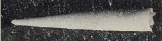
Spinita spp., from Kordè in Koren' (2003). 1: S. sanashticgolica, 2: S. cryptosa, 3: S. spinoglobosa.


The figure comes from a Russian book, Атлас ископаемой фауны и флоры палеозоя Республики Бурятия ('Atlas of the Palaeozoic fossil fauna and flora of the Republic of Buryatia'), edited by T. N. Koren' and published in 2003 in Ulan-Udè. Buryatia is a Russian republic in south-eastern Siberia, wrapping around the eastern and southern coasts of Lake Baikal. The fossils shown above come from the Lower Cambrian (the Botomian stage in the Russian system) of the Eastern Sayan Mountains. Going by the appearance of the figures, I presume they're being examined as thin sections, a commonly used method for studying Palaeozoic microfossils. Though as microfossils go, these are definitely on the large side: the specimen figured as 1a is a centimetre long and three millimetres wide. The other specimens are smaller, about half a centimetre in length.

When I saw these figures, I was just mystified. Their describer, K. B. Kordè, regarded them as a new class of 'Nemathelminthes', claiming that 'the first impression that is created from the described material is that they are representatives of the Kinorhyncha or Gastrotricha'. I'm not sure that I would agree with that. I found myself wondering if they were even animals, though I was hard pressed to think what else they might be. Not being familiar with the interpretation of thin sections, the thought did cross my mind to ask how certain can we be that these are even fossils, but I think that may be a bit uncharitable. Kordè also suggested that a break in the apparent cuticle of the S. sanashticgolica specimen about halfway along the flattened side (interpreted as the venter) might be the mouth. If so, that would be very unlike any kinorhynch or gastrotrich I've heard of. Could be a flatworm, I suppose, though Kordè then goes on to read the cluster of spines at one end (as magnified in figure 2b) as marking the anus which would seem to put paid to that! Said spines, or papillae, or whatever, are also supposed to have medial channels that Kordè interprets as nephridia.

All in all, I can't express anything other than confusion about this one. Certainly I haven't been able to find any further commentary on these enigmas; a Google search for Spinita sanashticgolica brings up just one result, an offhand mention in this book which seems to be just referring to it as found in the same formation as another fossil. Confusingly enough, that mention seems to date from 1986, a good seventeen years before Koren' (2003) was even printed: whether that indicates that the latter publication was not actually the first time the description of Spinita saw print, or whether this genus saw time floating around in unpublished communications, I have no idea.

Ice-cream Cones of the Early Palaeozoic

It's time for something I haven't done in a very long time... (credit to Niel from Microecos):


I briefly described tentaculitoids on this site way back in September 2007. These narrowly conical shells of uncertain affinities were prominent members of the marine fauna during the Silurian and the Devonian, only to then disappear without a trace. No direct evidence is available for the soft-body appearance of the animals that produced them nor are we overly certain on their lifestyle. But at least one of the major subgroups of the tentaculitoids, the Dacryoconarida, are held to be of palaeontological significance due to their ubiquity and cosmopolitan distribution at the species level making them of use in biostratigraphy.

Reconstruction of Nowakia elegans, from Berkyová et al. (2007).


Dacryoconarids have generally been presumed to be planktonic in some way, owing to the aforementioned tendency of individual species to be found more or less worldwide, together with their small size (generally about the centimetre range). Dacryoconarids are distinguished from other tentaculitoids by the apical portion of their shell ending in a small globular bulb, presumed to represent the embryonic or larval shell of the original animal (Farsan 2005). A more or less distinct constriction or 'neck' separates this embryonic bulb from the remainder of the shell. In those forms with more heavily ornamented shells such as the genus Nowakia, a distinct juvenile section of the shell is visible immediately following the embryonic bulb in which the adult ornament is absent or weakly developed; said adult ornament, when it appears, takes the form of rounded transverse ridges and troughs, often associated with longitudinal and/or transverse striae. In other forms, such as the genus Styliolina, the outside of the shell is flat and ridgeless, with at most the only ornamentation present being striae. The inside of the shell may be rippled to follow the exterior ornamentation or it may be perfectly smooth (Fisher 1962).

Dacryoconarids are first recorded from the Late Ordovician but they remained at relatively low diversity until the Devonian which saw a notable radiation (Wittmer & Miller 2011). Nevertheless, they declined rapidly towards the end of the Devonian. It has been suggested that their extinction by the end of that period may be related to the appearance of more actively swimming predatory fish before which the tentaculitoids may have been relatively defenceless. Other early Palaeozoic planktic groups such as the graptoloids experienced a similar collapse at about this time, though the disappearance of the dacryoconarids may have lagged behind that of the graptoloids.

Styliolina clavulus, from Fisher (1962).


Over the years, a wide range of suggestions have been made about the affinities of the tentaculitoids, ranging from jellyfish to annelids. Perhaps the most persistent association has been made with molluscs but there really is little to support such a premise than the possession of a calcareous shell, a feature that is hardly unique to molluscs even among living animals. The structure of the tentaculitoid shell is most similar to that of some brachiopods (Fisher 1962) and some sort of brachiozoan affinity is perhaps the currently most favoured concept. As noted above, we know nothing about the tentaculitoid anatomy other than what we can infer from the nature of the shells themselves. In some larger tentaculitoids (though not among the dacryoconarids so far as we know) the apical parts of the shell may become walled off by solid septa so the living animal presumably didn't occupy the entire shell. Fisher (1962) described the tentaculitoids as "presumably tentacle-bearing" but I have no idea on what basis he made that statement (as I've noted before, the name 'tentaculitoid' itself comes not from a belief that they possess tentacles but from the mistaken interpretation of the first specimens named as being themselves the tentacles of larger animal). Tentacles would be a not unreasonable method of capturing the smaller micro-plankton on which the dacryoconarids presumably fed but it is not impossible that some other structure served this purpose.

REFERENCES

Farsan, N. M. 2005. Description of the early ontogenetic part of the tentaculitids, with implications for classification. Lethaia 38: 255–270.

Fisher, D. W. 1962. Small conoidal shells of uncertain affinities. In: Moore, R. C. (ed.) Treatise on Invertebrate Paleontology pt W. Miscellanea: Conodonts, Conoidal Shells of Uncertain Affinities, Worms, Trace Fossils and Problematica pp. W98–W143. Geological Society of America, and University of Kansas Press.

Wittmer, J. M., & A. I. Miller. 2011. Dissecting the global diversity trajectory of an enigmatic group: the paleogeographic history of tentaculitoids. Palaeogeography, Palaeoclimatology, Palaeoecology 312: 54–65.

The Mongolian Death Worm

This would have been a comment on a recent post by Darren Naish at Tetrapod Zoology on the behaviour of amphisbaenians, but the commenting system they have at Scientific American these days means that any comment on a post more than a couple of days old will never be seen by anyone. As such, I'm posting it up here:

PhilJTerry's comment in response to Darren's post: "As I love introducing cryptozoology into the conversation wherever possible - are Amphisbaenians a likely influence for the Mongolian Death Worm? Can they live in desert environments?"

Image from National Geographic.


For those not already aware, the "Mongolian death worm" or "olgoi-khorkhoi" is a supposedly incredibly dangerous animal found in the deserts of Mongolia. It's first mention in Western literature came in Roy Chapman Andrew's (1926) On the Trail of Ancient Man. Andrews heard about the animal in a meeting with Mongolian officials:

Then the Premier asked that, if it were possible, I should capture for the Mongolian government a specimen of the allergorhai-horhai. I doubt whether any of my scientific readers can identify this animal. I could, because I had heard of it often. None of those present ever had seen the creature, but they all firmly believed in its existence and described it minutely. It is shaped like a sausage about two feet long, has no head nor legs and is so poisonous that merely to touch it means instant death. It lives in the most desolate parts of the Gobi Desert, whither we were going. To the Mongols it seems to be what the dragon is to the Chinese. The Premier said that, although he had never seen it himself, he knew a man who had and had lived to tell the tale. Then a Cabinet Minister stated that "the cousin of his late wife's sister" had also seen it. I promised to produce the allergorhai-horhai if we chanced to cross its path, and explained how it could be seized by means of long steel collecting forceps; moreover, I could wear dark glasses, so that the disastrous effects of even looking at so poisonous a creature would be neutralized. The meeting adjourned with the best of feeling; for we had a common interest in capturing the allergorhai-horhai.


Since then, there have been a number of expeditions have been conducted in search of the Premier's "allergorhai-horhai"; all have come up fruitless. Various opinions have been expressed as to what the stories may have been based on, with the most popular suggestions being some sort of reptile (Darren says in his response to the above comment on the original post that he "could buy that the stories are based on exaggerated tales of erycine boas or something"). For my part, I suspect that the question of the 'original identity' of the Mongolian death worm may be a futile one. When I first heard Andrews' account, I was not reminded of an amphisbaenian or a boa; I was immediately put in mind of a drop bear.


I feel almost certain that Andrews was being told a local tall tale, a popular joke at the expense of visiting travellers. The nature of Andrews' response to the officials suggests that he was in on the joke and more than happy to play his part in communicating it. Admittedly, other accounts of the Mongolian death worm have been recorded at more recent dates. And in the same way, I've never seen a drop bear myself, but I can assure you that my cousin did once and got the fright of his life. Be careful. They're out there.

Linnaeus' Infernal Fury

The starting point of modern zoological nomenclature (Clerck notwithstanding) has been established as the tenth edition of Linnaeus' Systema Naturae, published in 1758. Linnaeus divided the animal kingdom between six classes, with vertebrates making up four (Mammalia, Aves, Amphibia and Pisces) and invertebrates assigned to just two. One of these, Insecta, essentially corresponded to modern arthropods, and all other invertebrates were included in the class Vermes, 'worms'. Linnaeus' concept and arrangement of Vermes bears little resemblance to anything that exists in modern zoological classifications; with the study of invertebrate anatomy still in its absolute infancy, he was largely classifying animals based on their overall external appearance alone. One of Linnaeus' orders of Vermes, the 'Intestina', defined as 'simple, shell-less and limb-less', included animals now classified as annelids, nematodes, molluscs and even a chordate (the hagfish Myxine glutinosa). It also included a species whose identity would be debated for the next several decades: the 'infernal fury', Furia infernalis.

A reconstruction of Furia infernalis, from Piter Kehoma Boll.


Furia infernalis was described by Linnaeus as "Corpus filiforme, continuum, aequale, utrinque ciliatum: aculeis reflexis corpori appressis" ('body thread-like, continuous, uniform, ciliated on both sides with reflexed spinules appressed to the body'). It was found in marshes of southern Sweden and Finland. Linnaeus went on to record that F. infernalis was, "Pessima omnium, ex aethere decidua in corpora animalium, ea momento citius penetrat, intra horae quadrantem dolore atrocissimo occidit": the 'worst of all, falling from the sky onto the bodies of animals, into which it rapidly penetrates within a moment, striking [the victim] down with the most atrocious pain within quarter of an hour'. Linnaeus had good reason to highlight this animal's unpleasantness: he had been attacked by one himself when collecting botanical specimens in 1728, and barely escaped the resulting ailment with his life. A more detailed description of "der Höllenwurm" was compiled by Jördens (1802): it was a very slender worm, about the length of a nail, of a pale yellow or fleshy colour (other authors described it as greyish), with one end black. It climbed up standing vegetation, from whence it was carried by the breeze onto the exposed skin of humans and animals into which it rapidly burrowed. For victims, the first sign of its presence was usually a sudden pain in the afflicted spot, like the stab of a needle, and a small black spot marking the worm's entry point. A violent itching followed that developed into severe and extensive inflammation, often accompanied by fever; in the majority of cases, the affliction was so violent that the victim was dead within a matter of days if immediate action was not taken. If applied quickly enough, the worm could sometimes be drawn out with a poultice of fresh cheese curds. Otherwise, treatment required the careful dissection of the worms from between the muscle tissue into which they had entered, a process that (considering the surgical facilities available at the time) must have nearly as hazardous as the original infection.

As can be imagined, the attacks of this animal were greatly feared. In 1823–1824, an epidemic of Furia attacks spread through herds of livestock in Swedish and Finnish Lapland; thousands of head of reindeer perished, as well as countless cattle and sheep. Scavengers such as wolves feeding on the carcasses themselves sickened and died. One account from the time involves a young woman who was shearing wool from a recently deceased sheep (on a waste not, want not principle, I suppose) when she felt the tell-tale sting on a knuckle. Her life was saved by her master who was working nearby, when he quickly chopped off the affected finger with an axe. So great was the devastation that Norway, which had hitherto been free of the worm, passed an edict banning the import of animal furs from affected areas (Brooke 1827).

There were some, however, who greeted the description of Furia infernalis with skepticism. The idea of a tiny worm that somehow flew through the air and caused almost instantaneous mortality seemed fantastic. Even more problematic was the dearth of specimens. Many had seen the wounds caused by the worm and observed its effects; very few had seen the worm itself. Linnaeus himself had only seen a single, very poorly preserved specimen submitted to him by a church pastor. Most of the details about the worm's supposed appearance came from a single source, an article written by Solander, a student of Linnaeus'. The Academy of Sciences at Stockholm, naturally keen to discover all they could about such a scourge afflicting their country, offered generous rewards to anyone who could procure them a genuine specimen; no such specimen was forthcoming. Eventually, a consensus was reached: the worm Furia infernalis was an entirely fabulous animal, with no place in the annals of physical zoology. By 1827, notwithstanding the epidemic of only a few years previously, Brooke was able to comment that one could quite easily accept that something had affected the supposed victims of Furia without presuming that that something had to be the Furia itself. Even Linnaeus eventually came to accept that his inclusion of Furia in the Systema Naturae had been an error.

That Furia infernalis never existed outside the realms of fantasy remains the accepted wisdom to this day. But in that case, what did afflict Linnaeus and other unfortunates wandering the marshes of Sweden in the early 1700s? One thing that struck me was how much I was reminded of the more recent phenomenon here in Australia of 'white-tailed spider bites'. In recent decades, many people (including many medical professionals) have attributed serious ulcerative skin lesions, sometimes so serious that treatments such as skin grafts are required, to the bite of white-tailed spiders Lampona spp., common ground-running spiders often encountered near human dwellings. The actual evidence linking white-tailed spiders to such injuries is minimal; indeed, a clinical survey of 130 confirmed white-tail bites by Isbister & Gray (2003) found not a single incidence of one leading to ulceration. In both the 'Furia attacks' and the 'white-tailed spider bites', it seems likely that the primary culprit is bacterial infections resulting from opportunistic pathogens such as Streptococcus and Staphylococcus species. The initial wound may indeed have been caused by something like an animal bite or sting, or for that matter a splinter or pin-prick. Germ theory would not become widely accepted until the mid- to late 1800s; when Linnaeus compiled the Systema Naturae, flying worms probably seemed as good an explanation as any. The first 'attack' recorded by Furia victims may have simply been the first moment they noticed the infection's symptoms. And the 'worms' dissected out of advanced victims? Personally, I'm inclined to suspect that they may have been small pieces of tissue from the unfortunate sufferers themselves.

The exact causes of the 1823 epidemic are probably lost to history. Brooke (1827) stated that faculty at the Stockholm academy "had been led to consider the disorder by which [the reindeer] were attacked as a particular variety of hydrophobia". He also mentioned another possibility: reindeer were known to be vulnerable to inflammation of the brain, and dissections of the brains of deer killed by this condition sometimes revealed the presence of "a small vesicular worm". We can now recognise these vesicles as the cysts of hydatid tapeworms, which can hatch to cause tapeworm infections in any predator that eats the flesh of their host. So perhaps the 1823 epidemic was caused by a worm after all—just not the worm that was blamed.

REFERENCES

Brooke, A. de C. 1827. A Winter in Lapland and Sweden, with various observations relating to Finmark and its inhabitants; made during a residence at Hammerfest, near the North Cape. John Murray: London.

Isbister, G. K., & M. R. Gray. 2003. White-tail spider bite: a prospective study of 130 definite bites by Lampona species. Medical Journal of Australia 179: 199–202.

Jördens, J. H. 1802. Entomologie und Helminthologie des Menschlichen Körpers, oder Beschreibung und Abbildung der Bewohner und Feinde desselben unter den Insekten und Würmern vol. 2. Gottfried Adolph Grau: Hof.

Linnaeus, C. 1758. Systema Naturae per regna tria naturae, secundum classes, ordines, genera, species, cum characteribus, differentiis, synonymis, locis 10th ed., revised, vol 1. Laurentius Salvius: Copehagen.

Typhloesus: The 'Alien Goldfish' of Bear Gulch

The following post first appeared on May 19th on my Patreon page, available only to subscribers. If you would like to show your support for Catalogue of Organisms, and potentially gain access to future Patreon-exclusive content, please become a subscriber for as little as $1 a month!

***


Above: Typhloesus wellsi, external appearance, and the same with major anatomical details shown. Based on figure of specimen U.M. 6027 in Conway Morris (1990).


Recently, the interwebs became all agog at the suggestion that the hitherto-mysterious Carboniferous fossil Tullimonstrum gregarium could possibly represent a vertebrate, distantly related to modern lampreys. But there are other fossil animals whose relationships remain inexplicable and one of these is another child of the Carboniferous, the so-called 'alien goldfish' Typhloesus wellsi.

When I announced my plan to write this post, I referred to Typhloesus as coming from Mazon Creek, the fossil deposit from whence comes Tullimonstrum. This, as it turns out, was a mistake on my part: Typhloesus actually comes from a different deposit, Bear Gulch in Montana. Bear Gulch is perhaps most famous for its fossils of early fish, such as symmoriiform sharks (the ones with the weird shoebrush headgear) and heavily armoured palaeoniscoids. Indeed, compared to other Carboniferous deposits, Bear Gulch is unusual for its preponderance of swimming rather than benthic animals. Typhloesus is represented in the deposit by a number of individuals in varying states of preservation.

In some ways, Typhloesus is more famous for what it is not than for what it is. It was one of the first body fossils found in association with conodonts, minute teeth-like fossils that had been subject to much speculation as to what sort of animal they might have come from. Initially, there was much excitement that the conodont animal may have finally been found, but it did not take very long for questions to be raised about the nature of this association. By the time Typhloesus was reviewed in detail by Conway Morris (1990), it was clear that the conodont fossils had been preserved within its gut, not its mouth, and Typhloesus was a conodont-eater rather than a conodont-bearer (it has since been found that conodont animals were eel-like chordates).

Externally, Typhloesus was a fairly simple, cigar-shaped animal, with its body laterally compressed and higher than wide. It grew to a decent size, with the largest specimens being a little under ten centimetres in length. There is no sign of eyes or any other prominent sensory structure, and so far as is known the external skin or cuticle was smooth and unornamented. The most distinctive external feature is a large 'tail-fin' at the rear. This fin was supported by an arrangement of criss-crossing rods or fibres, and would have been fairly stiff in life. Another pair of folds or fins ran along most of the underside of the body with a noticeable gap towards the rear. Typhloesus probably swam in a not dissimilar manner to an active modern fish, using sweeps of the tail-fin to provide thrust; the ventral fins may have provided stability and steerage. The visible line of the foregut comes to a halt slightly before reaching the front of the body, and it seems that the mouth would have been slightly ventral and contained within a 'hood'. Though its overall conformation and known gut-contents (most commonly conodonts, but sometimes worm jaws or fish scales) suggest an active predator, I am at a loss to understand how it located its prey without eyes. Perhaps the hood contained some sort of chemical sensors in life.

When it was first found, it was thought that its overall appearance suggested a relationship of Typhloesus to the chordates. However, Conway Morris (1990) saw its internal anatomy as incompatible with this view. Fossils of this animal show a narrow foregut leading into a voluminous, sack-like midgut. Below the midgut is a pair of dark, disc-shaped organs showing a concentration of iron deposits called the ferrodiscus; though a striking element of all Typhloesus fossils, the function of this structure is completely unknown. What Conway Morris found conspicuous by its absence, however, was an anus: there appeared to be no sign of any gut structures in the rear of the animal. The gut was a blind sack, with the only way out being the same as the way in. The absence of a through-gut would be unprecedented in a chordate, or indeed in many animals except jellyfish or flatworms. Conway Morris was also unable to identify other chordate-specific structures such as muscle-blocks, gill openings or a notochord; though he confessed that the first two might be obscured by the vagaries of decay, he felt that the third at least should have left more of a sign. It was this combination of an overall fish-like appearance with a very un-fish-like anatomy that led Conway Morris to later dub Typhloesus the 'alien goldfish'.

With the exclusion of a chordate connection as a possibility, Conway Morris found himself at a loss as to just where Typhloesus fitted into animal evolutionary history. Finned swimmers are also known among molluscs, nemerteans and chaetognaths, but Typhloesus is no more like any of these than it is like a chordate. Conway Morris felt himself compelled to declare the affinities of Typhloesus completely unknown. Personally, though, I can't help wondering if the 'alien goldfish' might not be so alien after all: maybe it is a chordate. The overall similarities of Typhloesus to a chordate are remarkable; in particular, the hooded mouth is very similar to that of a lancelet. But what about that missing anus, you say? Where is that all-important butthole? To which I respond, is it really missing? Looking at the figures of Typhloesus fossils in Conway Morris (1990) (which is of course a poor competitor to Conway Morris' ability to look directly at the fossils themselves), I see that directly below the midgut is the ferrodiscus. And directly below that is a streak running between the ferrodiscus and the animal's venter. Conway Morris saw this structure (which he called the 'midventral strand') as some sort of connection between the ferrodiscus and the exterior, but could it in fact be the tail-end of the reargut? It is certainly not unknown for the anus in chordates to not be right at the very rear of the animal; in some fish (such as the scorpionfish-like Aploactinidae) it is even moved so far forward as to be almost underneath the head. And the missing notochord? Considering that despite the presence of specimens numbering in the thousands, a notochord was only announced in Tullimonstrum within the past year, maybe on that front Typhloesus could reward a second look.

REFERENCE

Conway Morris, S. 1990. Typhloesus wellsi (Melton and Scott, 1973), a bizarre metazoan from the Carboniferous of Montana, U.S.A. Philosophical Transactions of the Royal Society of London Series B 327: 595–624.

Bitubulites: Yeah, Nach

Several months back, I published a post on a decidedly obscure tubular fossil by the name of Serpularia: once described, completely forgotten. Is it possible to top that for inconsequentiality?

You betcha, because the subject I drew for today's post, Bitubulites irregularis, never even got a sketchy diagram to illustrate it. And once again, we have an early German palaeontologist to thank. In 1820, Ernst Friedrich, Freiherr von Schlotheim, published a book called Die Petrefactenkunde auf ihrem jetzigen Standpunkte durch die Beschreibung seiner Sammlung versteinerter und fossiler Überreste des Thier- und Pflanzenreichs der Vorwelt erläutert, because they don't write book titles like they used to. As far as I can tell, the title basically translates as Got Some Great Fossils Here, Wanna See? As with Münster's Beiträge zur Petrefakten-Kunde that provided the source for the Serpularia post, Schlotheim's publication was basically a description of some of the fossils held in his own collection. His comments on B. irregularis appeared on p. 376, and were as follows:

Bitubulites irregularis.
Aus dem Muschelflötzkalk der Gegend von Weimar.

Einzelne cylinderförmige Stücke, von der Dicke eines mäſsigen Fingers, mit gröſstentheils concaven Durchschnittsflächen, auf welchen sich gewöhnlich ins Dreyeck gestellte kleine Öffnungen zeigen, welche mit durchgehenden Nervenröhren in Verbindung zu stehen scheinen. Äuſserlich ist die Oberfläche fein puncktirt.
Da sich mehrere übereinstimmende Stücke finden, so läſst sich nicht erwarten, daſs wir ein bloſses Naturspiel vor uns hätten. Zuweilen sind auch vier, aber alsdenn noch unregelmäſsiger gestellte Öffnungen, vorhanden. Bis jetzt haben sich, meines Wissens, noch keine ganz vollständigen und recht gut erhaltenen Exemplare aufgefunden.
And I'll be honest, I have very little idea what any of that means, because I don't read German beyond chucking stuff into Google Translate. The genus Bitubulites had established in 1803 by another German scientist, Johann Friedrich Blumenbach, for a different fossil, B. problematicus. Bitubulites problematicus was composed of two small conjoined tubes that Blumenbach illustrated thus:
Schlotheim's B. irregularis was presumably similar to Blumenbach's original; as far as I can work out, each tube was cylindrical and about the thickness of a finger. Small openings down the sides of the main tubes indicated the presence of smaller connecting tubes ("nerve-tubes") between them; the entire outer surface was finely punctate. The second paragraph of Schlotheim's states that he possessed several examples, indicating that the association between the tubes was not a mere accident. I think he says that the tubes were sometimes associated in fours rather than in pairs, but the overall confirmation remained the same. The "Muschelflötzkalk" refers to a Middle Triassic formation; both the two Bitubulites species came from the same formation.

Blumenbach had called his original fossil 'problematicus' because he had little idea what type of animal it represented. Schlotheim was none the wiser, listing Bitubulites among unclassifiable forms with no close analogues in the modern fauna. He did tentatively compare it to a couple of fossil mollusks, such as the straight-shelled cephalopods or the reef-forming hippuritid bivalves. And there Bitubulites lay for over a century, more or less forgotten by all except the most pedantic of cataloguers (ahem...)

In 1962, Walter Häntzschel included Bitubulites in his chapter on problematica for the Treatise on Invertebrate Paleontology. It was tucked away in the end, in a list of "Unrecognized and Unrecognizable "Genera"" that Häntzschel felt largely deserved nothing more than to be cast forever into the outer darkness. Nevertheless, he did suggest a possible identity for Bitubulites: Rhizocorallium, a fossil that can be found in deposits dating from the Cambrian all the way to the present, commonly looking like this (copyright Manuel Flöther):
Rhizocorallium is a trace fossil, a structure created by the activity of some animal and preserved in the geological record. Rhizocorallium takes the form of a U-shaped burrow, with the two arms of the burrow connected by fine cross-lines or fractures referred to as Spreiten (German for "spread"). The Spreiten are the result of the animal digging its burrow further into the substrate, with sediment being taken from the outer side of the tube and packed on the inner side. The burrows often run more or less parallel to what would have been the original surface of the sediment; they may have been primarily dwelling burrows, or they may have been feeding burrows extended as the animal searched for buried organic matter. Rhizocorallium-type burrows were probably made by many different types of animal, such as worms or arthropods, and their presence in a deposit is more indicative of environmental conditions than faunal composition.

At the time that Blumenbach and Schlotheim were writing their books, no-one had yet twigged what these kind of trace fossils were; neither author would have been alone in mistaking a burrow for a body fossil. Many structures that are now recognised as traces were described as fossil algae. It was not until the late 1800s that a Swedish and an American palaeontologist independently noted the similar between a number of these 'algae' and the structures left by marine organisms as they went about their daily life. Even today, this earlier misunderstanding has left its mark in the tradition of referring to particular trace fossils by binomial names, as trace 'genera' and 'species'. Nevertheless, trace fossils often provide us with a window into the past over and above what we can learn from body fossils alone, and they are an invaluable tool in developing a truly rounded understanding of life in ages past.

REFERENCES

Blumenbach, J. F. 1803. Specimen Archaeologicae Telluris terrarumque inprimis Hannoverarum. Henricum Dieterich: Göttingen.

Häntzschel, W. 1962. Trace fossils and problematica. In: Moore, R. C. (ed.) Treatise on Invertebrate Paleontology pt W. Miscellanea: Conodonts, Conoidal Shells of Uncertain Affinities, Worms, Trace Fossils and Problematica pp. W177-W245. Geological Society of America, and University of Kansas Press.

Schlotheim, E. F. von. 1820. Die Petrefactenkunde auf ihrem jetzigen Standpunkte durch die Beschreibung seiner Sammlung versteinerter und fossiler Überreste des Thier- und Pflanzenreichs der Vorwelt erläutert. Becker'schen Buchhandlung: Gotha.

Tully as a Vertebrate

Reconstruction of Tullimonstrum gregarium by Sean McMahon, from McCoy et al. (2016).


McCoy, V. E., E. E. Saupe, J. C. Lamsdell, L. G. Tarhan, S. McMahon, S. Lidgard, P. Mayer, C. D. Whalen, C. Soriano, L. Finney, S. Vogt, E. G. Clark, R. P. Anderson, H. Petermann, E. R. Locatelli & D. E. G. Briggs (in press, 2016) The ‘Tully monster’ is a vertebrate. Nature.

Several years ago, I included the 'Tully monster' Tullimonstrum gregarium in a list of some of the most phylogenetically mysterious organisms on the planet. Multiple suggestions have been made as to its affinities: mollusc, annelid, nemertean (nemerteans and sea cuumbers both having weird histories of problematic fossils assigned to them for little apparent reason), some sort of de-chitinised arthropod relative by way of Opabinia, the Loch Ness monster... A new publication just out by McCoy et al. (2016) adds a further interpretation to the mix.

Tullimonstrum is represented by literally thousands of specimens from the Carboniferous Mazon Creek deposit of Illinois. The organisms preserved in this deposit are contained within nodules, each individual at the centre of a mineral ball that precipitated around it after its death. It had a somewhat elongate, torpedo-shaped body, at the front of which was an elongate proboscis ending in a pincer-like structure. Towards the front of the main body was a dorsal cross-bar with a dark round body at each end; these bodies have most commonly been seen as eyes on the end of stalks but alternative interpretations include statocysts, solid structures that many aquatic animals possess for sensing balance. A fin-like structure was present at the tail end of the animal. Many specimens also show regularly spaced dark cross-lines suggesting some sort of segmental division of the body.

Another structure commonly visible in the Tullimonstrum fossils is a pale, flattened linear structure running down the length of the animal. Most authors have presumed that this represents the gut but McCoy et al. argue that it does not resemble the gut as preserved in other Mazon Creek fossils. In these other fossils, the gut is dark-coloured and is not flattened. Some authors have tried to explain this difference between the 'gut' of Tullimonstrum and that of its associates by suggesting that the Tully monster fed on soft prey such as jellyfish whose remains did not preserve after death, but the dark colour in most Mazon Creek guts does not represent the actual gut contents themselves but minerals that precipitated around the gut contents during the fossilisation process. Presumably, such minerals would be just as likely to condense around jellyfish remains as any other organic tissue. Even more damning, McCoy et al. identified a handful of Tullimonstrum specimens in which the gut was indeed preserved as in other Mazon Creek fossils, and as a separate structure from the pale line that was also present in these same specimens.

An actual fossil of Tullimonstrum in the Museo di Storia Naturale di Milano, copyright Ghedoghedo.


So what was this structure, if not a gut? McCoy et al. note that at least one other fossil from the Mazon Creek preserves a similar structure: the hagfish-like Gilpichthys, in which it represents the notochord. The structure's preservation is consistent with this interpretation: being a fluid-filled tube, the notochord would flatten readily during fossilisation, and it does not accumulate minerals like the gut because it lacks an external connection. And if Tullimonstrum also possesses a notochord, then that makes it also a chordate. And with that in mind, McCoy et al. interpret other structures as supporting chordate, and specifically vertebrate, affinities: the fin-like structures are indeed fins, paired stains bordering the notochord in a few specimens appear to be gill pouches, tooth-like structures within the 'pincer' at the end of the proboscis are keratinous teeth similar to those of lampreys and hagfish, and the apparent 'segments' in some specimens represent vertebrate myomeres (muscle blocks). Including Tullimonstrum in a phylogenetic analysis of basal vertebrates, coded according to these and other interpretations, places it within the stem-lineage of modern lampreys.

So how strong is this re-assignment? The problem with the structural analysis of any problematic fossil is that it is ultimately dependent on finding the right comparative framework, and the more distinct the problematicum is from any living organism the harder it is to be sure you're making the right comparison. That's not a criticism of this particular paper; that's simply the limitation its authors have to work with. In this case, I kind of suspect that the identification of Tullimonstrum as a vertebrate all hinges on whether they've correctly identified that notochord. None of the other 'vertebrate' features identified is sufficiently distinct to clinch the deal on their own. A tail-fin could indicate a vertebrate, or it could indicate a mollusc like a squid. The famous Tullimonstrum proboscis (which, offhand, McCoy et al. interpret as a cartilage-supported structure rigidly bending at set points like an arm rather than curling like a tentacle, based on the regular aspect of its preservation) is unlike anything known from any other vertebrate, but nor does it strongly resemble anything found in any other animal (the aforementioned Opabinia suggestion is right out: as I mentioned in an earlier post on Nectocaris, the Opabinia proboscis contains no direct part of the digestive tract itself). Certainly the placement of Tullimonstrum as a stem-lamprey is the weakest part of the whole deal, as the specific features cited as synapomorphies are either convergently present in other vertebrates (e.g. keratinous teeth) and/or dependent on some admittedly more tentative structural interpretations (e.g. tectal cartilages). There may be a certain element here of Tullimonstrum's intractable weirdness conflicting with the phylogenetic analysis' need to put it somewhere. I also wonder if I should be criticising Sean McMahon's reconstruction (reproduced at the top of this post) for presenting Tullimonstrum as somewhat laterally flattened: the majority of Tullimonstrum specimens are preserved dorsoventrally rather than laterally, which I would suspect indicates that they were probably flatter top-to-bottom than side-to-side.

Those criticisms aside, McCoy et al. have certainly presented one of the more robust reconstructions of Tullimonstrum to date. Most of what I've said comes under the heading of intrigued enquiries rather than actual disagreements, and if they're right about that notochord then they're on pretty firm ground. After all, even if the Tully monster is not specifically a stem-lamprey doesn't exclude it from being any sort of chordate. There are few (if any) problematica as well represented in the fossil record as Tullimonstrum, and we have not heard the last word on it yet.

Serpularia: A Rightly Forgotten Problematicum

I think it may be time to rock out something that hasn't been seen on this site for a while. Horns at the ready...


(Credit, again, to Neil from Microecos). And I'm afraid that may just be the most excitement that we get in this post. While some fossils are problematic because they're so strange that they can't be easily compared to living animals, others are problematic simply because they're rubbish.

In 1840, the palaeontologist Georg Graf zu Münster ('Graf' being a German title that generally gets translated as 'Count') published his Beiträge zur Petrefakten-Kunde, in which he described a number of fossils held in his collection. This book included a section on fossils from the Ordovician Orthoceratite Limestone of the Fichtel Mountains in Bavaria. Which, close to the end, included this little tidbit:
Unter mehreren Bruchstücken einiger mir noch unbekannten Versteinerungen kommen auch einige röhrenformige Korper vor, welche ich anfänglich für den von Murchison aus der 27sten Tafel abgebildeten Myrianites hielt, allein genaue Untersuchung zeigte, dass diese Korper formliche Schalen hatten und daher vielleicht zu den Serpuliten gehört hatten, daher ich sie vorläufig Serpularia genannt habe. Aus der Taf. IX. Fig. 14 und 15 sind zwei Arten von dergleichen Bruchstücken abgebildet; Fig. II. Serpularia crenata; glatt gebogene Röhre, aus dem Rücken crenulirt. Fig. 15. Serpularia bicrenata; glatte etwas zusammengedrückte ganz grade Röhrchen, die an beiden Seiten crenulirt sind.

Translated with the help of Google Translate, I think this means: "Among several fragments of fossils unknown to me occured a tube-like body, which I initially took for Myrianites as figured by Murchison in the 27th plate, until close examination showed that this body had distinct signs of segmentation and was therefore perhaps one of the Serpulidae. Therefore, I have provisionally called it Serpularia. On Plate IX Figs 14 and 15 are shown two types of the like fragments; Fig. 14, Serpularia crenata: smooth curved tube crenulated from the back. Fig. 15, Serpularia bicrenata: smooth, slightly compressed, quite straight tubes that are crenulated on both sides".

Münster's (1840) original figures of the two Serpularia.


As perfunctory as it was, that seems to be all there was to say on the matter. The good Graf's Serpularia has pretty much never been mentioned again*, beyond being cited to cause a name change in a later homonymous gastropod genus, and a brief listing in Howell's (1962) coverage of worm fossils for the Treatise on Invertebrate Paleontology that adds nothing to the original description.

*Though if it were to be mentioned again, it would probably have to be under a different name. The name 'Serpularia' had earlier been used by Fries in 1829 for a genus of slime moulds. At the time, slime moulds were treated as fungi, and hence fell under the purview of botanical rather than zoological names, but with the recognition that they are amoebozoans an increasing number of authors would move them into the field of the Zoological Code.

Münster believed that his fossils belonged to the Serpulidae, a family of annelid worms. Annelids, being mostly soft and squishy things that do not stand up well to decay, have a pretty deplorable fossil record, but serpulids are a bit of an exception. These are sessile worms that secrete a calcareous tube in which they live their lives (modern serpulids appeared on this site in this post). Unfortunately, while these tubes are eminently fossilisable, they are also a bit nondescript, and have little to mark them as uniquely serpulid.

Because of the dominance of annelids among modern worms, there has been a definite tendency in the past to assume that any given worm-like fossil represents an annelid. Howell's (1962) aforementioned list of annelids includes the Ediacaran Spriggina (identity still under debate, but probably not an annelid) and the Cambrian Pikaia (now generally regarded as an early chordate). Similarly, any worm-like tube has been assumed a serpulid. But even among annelids, serpulids are not the only tube-bearing worms. At least two other families, the Sabellidae and the Cirratulidae, include species producing calcareous tubes. There are also other groups of non-annelid worms that, though relatively uncommon or unprepossessing today, may have been more prominent in the past. After all, we are talking here about a period of hundreds of millions of years. We know that vertebrates have gone through a great deal of evolutionary change over that period; why should we assume that worms have not?

So while fossils have been assigned to the serpulids going back as far as the Cambrian (if not beyond), there is little reason to take those assignations at face value. When so-called Palaeozoic serpulids have been examined critically in recent years, they have so far proven to lack features that would definitely confirm their identification (Vinn & Mutvei 2009). Weedon (1994) found that Palaeozoic fossils that had been assigned not only to the Serpulidae, but to the modern genus Spirorbis, had a shell microstructure that suggested a relationship to bryozoans or brachiozoans rather than to annelids. Without a similar close analysis, we could not assume a priori that Münster's Serpularia were not serpulids, but odds would currently be against it.

REFERENCES

Howell, B. F. 1962. Worms. In: Moore, R. C. (ed.) Treatise on Invertebrate Paleontology pt W. Miscellanea: Conodonts, Conoidal Shells of Uncertain Affinities, Worms, Trace Fossils and Problematica pp. W144–W177. Geological Society of America and University of Kansas Press.

Münster, G. 1840. Beiträge zur Petrefacten-Kunde von Herm. v. Meyer und Georg Graf zu Münster vol. 3. In Commission der Buchner'schen Buchhandlung: Bayreuth.

Vinn, O., & H. Mutvei. 2009. Calcareous tubeworms of the Phanerozoic. Estonian Journal of Earth Sciences 58 (4): 286–296.

Weedon, M. J. 1994. Tube microstructure of Recent and Jurassic serpulid polychaets and the question of the Palaeozoic 'spirorbids'. Acta Palaeontologica Polonica 39 (1): 1–15.

Life on Mars: the Cambrian terrestrial environment

The question of when life first moved onto the land has been the subject of speculation for as long as anyone has realised that there was a 'first' to speculate about. Established terrestrial communities were clearly present by the latter part of the Silurian, but was there anything earlier? The reasonable expectation is that there was, at least on some level. Pretty much as soon as there was life inhabiting the oceans in prokaryote form, weather cycles would have been carrying bacteria and their spores onto their land. It is not unreasonable to assume that some of them may have been able to acquire a toehold in some attainable niche, and from there diversify to the surrounding environment. Later, other microbial and simple organisms may have joined them. But such organisms leave little trace in the fossil record. What were they like, how did they live? A paper that has just been published in Palaeontology (Retallack 2011) has described simple terrestrial fossils preserved from the Middle Cambrian, and may provide a rare glimpse of the early Earth.

Reconstruction of Cambrian terrestrial biota from Retallack (2011).


The remains described by Retallack (2011) are extremely simple: flat, thallose impressions called Farghera, subterranean threads known as Prasinema and buried ovoid structures called Erytholus. All of these are described as form taxa: that is, they represent a particular recognisable fossil structure whose relationship to other such fossils is unknown. Different form taxa may even represent different parts of a single organism.

The linear, branching Farghera thalli were an average of just under 2 mm wide, though they could get much wider, and preserved thalli are often several centimetres in length. The living thalli would have been similar to an alga or lichen, either of which they could have been. The thread-like Prasinema are preserved as a central filament less than 1 mm in diameter, surrounded by a dark halo up to about 2.5 mm across. It seems likely that only the central filament represents the original central organism; the halo would have formed by microbes growing around the filaments as they decayed. Prasinema filaments could apparently grow to 30 cm beneath the original soil surface, and probably represent structures similar to fungal hyphae.

Most unusual are the Erytholus, globose structures up to 2 cm in diameter, divided into internal layers with a broad central column. Retallack (2011) suggests a number of possible interpretations for Erytholus: vendobiont or xenophyophore (unlikely because of the terrestrial location), alga (again unlikely, because it is both terrestrial and buried beneath the surface), or fungal or slime mold reproductive structures, comparable to truffles. However, the truffle interpretation is problematic because truffles are produced to disperse spores through being eaten by animals. Obviously, this could not have been the case in the terrestrial Cambrian! A further possibility that I can think of is that Erytholus may have been some sort of resting structure, analogous to a plant bulb or tuber (though note that this interpretation would not necessarily exclude a reproductive function).

As with the Silurian, I think it is important to remember that the environment would have been very different in those days in more ways than one might immediately think. There are parts of the world today where lichens and algae remain the primary ground cover, but we should be careful in assuming that such spots are close analogues of the Cambrian terrestrial environment. Such areas are today arid and/or highly eroded, but in the Cambrian lichens and algae would have also been able to dominate areas in which vascular plants would overshadow them today. I also find myself again wondering what effect the absence of a complex vegetation profile might have had on weather patterns at the time. Would winds have been stronger if there were less low-level wind breaks? Would the effects of rain events have been more catastrophic if water flow was less impeded by ground-cover (if Erytholus was indeed a sort-of-tuber, perhaps it functioned as a source of regrowth if the above-ground component of the organism was destroyed by weather?) If we could see the Cambrian environment for ourselves, there could be no doubt that we would find it utterly alien.

REFERENCE

Retallack, G. J. 2011. Problematic megafossils in Cambrian palaeosols of South Australia. Palaeontology 54 (6): 1223-1242.

From Three to Two

(I've been waiting three and a half years to use Neil's icon.)


The mysterious anabaritids of the Lower Cambrian have been referred to on this site before. In the earlier, somewhat brief post, I referred to their triradial structure and uncertain, though probably coelenterate-grade, relationships. In the time since that post appeared, the anabaritids have been the subject of a review by Kouchinsky et al. (2009) that brought together a lot of the previously scattered information on these animals.


The image of an anabaritid in the previous post showed the best known species, Anabarites trisulcatus. However, this was not the only species in the group. The image just above, from Kouchinsky et al. (2009), shows another species, Anabarites biplicatus, recorded from the Siberian Platform. This species differs from A. trisulcatus in that it started out life triradial (albeit with the internal dividing ridges between the lobes only weak), but as it grew it lost its triradiality and became more bilateral (cross-section below from Kouchinsky et al.):


Some of my readers may remember that Cambrian problematica were something of a cause célèbre during the mid-90s when a lot of journals and magazines ran features on them (probably inspired to a certain degree by Stephen Jay Gould's somewhat dreadful book Wonderful Life). In the more academic corners of this pageant, the triradiality of anabaritids (as well as some other early animals such as Tribrachidium) garnered them a certain degree of attention. It was suggested by some that they might represent a unique animal lineage that was eventually superseded by our own bilateral dynasty. However, the changing symmetry of Anabarites biplicatus serves as a reminder that we should not be too hasty to assign great significance to such features. Indeed, in the modern fauna, nematodes are partially triradial (they have a triradial head structure, with one upper and two lower lips around the mouth). Though the affinities of anabaritids are somewhat debatable, the most popular scenario is that they are related to cnidarians: their tubes bear a certain resemblance to the polyps of some medusozoans. Cnidarians also exhibit a wide variety of symmetries, such as the tetraradial organisation of scyphozoans and the hexaradial organisation of hexacorals. Without preserved soft tissue to inform us what the organisation of the inhabitant animal may have been, it is difficult to say just how much weight the triradiality of the anabaritid tube should be given.

REFERENCE

Kouchinsky, A., S. Bengtson, W. Feng, R. Kutygin & A. Val'kov. 2009. The Lower Cambrian fossil anabaritids: affinities, occurrences and systematics. Journal of Systematic Palaeontology 7 (3): 241-298.

So Nice When People Agree With You


Nectocaris pteryx


Some of you may remember this post from back in May, in which I critiqued Smith & Caron's (2010) interpretation of Nectocaris as an early cephalopod. I was not convinced. Nor am I the only one: a paper recently released by Mazurek & Zatoń (in press) comes to much the same conclusions. They point out that Smith & Caron's proposed model of cephalopod evolution conflicts strongly with what we previously knew from the cephalopod fossil record (and that's no small amount—there are few groups of organisms whose fossil record has been as intensely studied as cephalopods), and that most of the characters supposedly shared between Nectocaris and cephalopods are in fact only shared between Nectocaris and coleoids (modern octopods and squid) that did not appear in the fossil record until during the Mesozoic, considerably later than the Cambrian Nectocaris. Smith & Caron (2010) suggested that the absence of a shell in Nectocaris indicated that the cephalopod shell had been evolved independently to that of other molluscs, but Mazurek & Zatoń point out that the only known cephalopods to completely lack any trace of a shell are octopods, and that octopods are secondarily shell-less is indicated, not only by their phylogenetic position, but also by the presence of a remnant shell in Cretaceous stem-octopods.

What I find particularly interesting about Mazurek & Zatoń's paper, however, is how much it brings up the same points already raised by commenters here. The primary feature cited by Smith & Caron (2010) as connecting Nectocaris with cephalopods, the presence of a funnel, is contradicted by the apparent difference in functional structure between a cephalopod siphon and Nectocaris' 'funnel', as noted by Adam Yates. Aydin Örstan commented on the absence of a radula. Just goes to show that I've got some pretty clever readers here. Mind you, I'm not happy with everything in Mazurek & Zatoń's paper. They make the argument that Nectocaris could not have evolved from a shelled and radula-possessing ancestor because it was 'too early', but the known fossil record of molluscs pre-dates Nectocaris by about twenty million years, more than long enough for shell loss to potentially occur. The absence of a beak in Nectocaris is also of doubtful significance as this is a cephalopod autapomorphy.

I am also not swayed by Mazurek & Zatoń's (provisional) alternative placement for Nectocaris as a dinocarid. Though it is tempting to compare the funnel of Nectocaris to the proboscis of dinocarids such as Opabinia, and dinocarids have the advantage over coleoid cephalopods of being coeval with Nectocaris, no dinocarid has cephalic tentacles like Nectocaris. Also, some of the figures in Smith & Caron (2010) appear as if the pharynx might pass through the funnel; in Opabinia, the proboscis was a separate structure in front of the mouth. I can think of two groups of animals for which cephalic tentacles are definitely known: annelids and molluscs (presuming that the tentacles of Nectocaris are not a unique autapomorphy of its own). The lack of obvious segmentation makes it unlikely that Nectocaris is an annelid. That leaves us with mollusc. While Smith & Caron's identification of a pinhole camera eye in Nectocaris could still connect it to cephalopods, I believe that this is outweighed by the arrangement of the gut. The presence of an apparent through-gut, opening with a terminal anus, was identified by Chen et al. (2005) in Vetustovermis (now recognised as a synonym of Nectocaris)*. Cephalopods, however, have a U-shaped gut, opening in the mantle cavity not too far from the head. This is related to the 90° shift that the cephalopod body plan has gone through during its evolution: the apparent "front-back" axis of a squid actually represents the "top-bottom" axis of other molluscs. A similar U-shaped gut is present in scaphopods, the probable living sister group of cephalopods, so a molluscan Nectocaris would have to sit outside the scaphopod-cephalopod clade. As far as is known, cephalic tentacles are a possible synapomorphy of the clade uniting cephalopods, scaphopods and gastropods**, so a molluscan Nectocaris would probably have to be either a stem representative of this clade, or just possibly a stem gastropod.

*My apologies to the commenter somewhere whose identity I've forgotten who brought my attention to this point.

**Cephalic tentacles are definitely absent in polyplacophorans and tryblidiids***. They are also absent in bivalves, but bivalves don't have a head to have cephalic tentacles on in the first place, so the absence of tentacles is probably best treated as ambiguous for bivalves.

***Or whatever Neopilina and its ilk are going by these days.

REFERENCES

Chen, J-Y., D.-Y. Huang & D. J. Bottjer. 2005. An Early Cambrian problematic fossil: Vetustovermis and its possible affinities. Proceedings of the Royal Society of London B 272: 2003-2007.

Mazurek, D., & M. Zatoń. in press. Is Nectocaris pteryx a cephalopod? Lethaia.

Smith, M. R., & J.-B. Caron. 2010. Primitive soft-bodied cephalopods from the Cambrian. Nature 465: 469-472.

Conical Problematica

Scattered throughout the fossil record are little mysteries, organisms whose remains have been preserved but which are not obviously relatable to any more familiar group. Either their remains are too simple to preserve much evidence of their affinities (as with the 'tubular problematica' I've discussed before), or they are too distinct from other organisms for their affinities to be clear, or both, or some other reason. Unless they are particularly common or otherwise significant, most of these problematica are probably doomed to remain so. Case in point:


The figures above show Asymmetroconus splendidus, described by Korde in 1975 from the Albian (early Cretaceous) of the Crimea. The photos are of thin sections of the fossils; the complete skeleton would have probably been shaped rather like a wine goblet. The largest specimens of Asymmetroconus were just under 8 mm in height. In the same paper, Korde described a number of similar fossils aged from the Albian to the Danian (earliest Palaeocene), assigning them all to the new order Asymmetroconida. Korde attributed the asymmetroconidans to the Hydroconozoa, a group of similar fossils he had himself described previously from the early Cambrian. Hydroconozoa have generally been assigned to the Cnidaria, though their exact position therein remains obscure. Asymmetroconida resembled hydroconozoans in being small and goblet-shaped, with a conical interior to the cup and a basal globular hollow below the point of the cone. However, they differed from Cambrian hydroconozoans in their skeletal microstructure and in the asymmetry of the cup, with one side much thicker than the other. Rozanov & Zhuravlev (1992) later dismissed the idea of Mesozoic hydroconozoans, stating simply that structures described as such had 'little in common with this group'. No alternative identification of the Asymmetroconida has ever been proposed, and they do not appear to have been properly studied since Korde's original description.


Reconstruction of the hydroconozoan Hydroconus mirabilis, from Rozanov & Zhuravlev (1992). Whether actually related or not, the Asymmetroconida would have probably looked superficially similar.


REFERENCES

Korde, K. B. 1975. [Hydroconozoa from Cretaceous and Palaeocene deposits of the Crimea]. In: Shimansky, V. N., & A. N. Soloviev (eds) Razvitie i smena organičeskogo mira na rubeže Mezozoâ i Kajnozoâ. Novye dankye o razvitii fauny pp. 32-38. Nauka: Moscow. [in Russian]

Rozanov, A. Yu., & A. Yu. Zhuravlev. 1992. The lower Cambrian fossil record of the Soviet Union. In: Lipps, J. H., & P. W. Signor (eds) Origin and Early Evolution of the Metazoa pp. 205-282. Plenum Press: New York.

The Trouble with Coelosclerites

A couple of years ago, I posted a brief review of the chancelloriids, mysterious sessile animals from the Cambrian period. As explained in that post (which I'd recommend reading before this one), chancelloriids are remarkable for how much we know about them while still being unable to place them anywhere in the animal family tree. However, there are two main options that are currently supported: one is that chancelloriids are sponge-grade animals, probably in the stem-group of modern Epitheliozoa (the clade including coelenterates and bilaterians, which differ from sponges in having a differentiated external skin around their bodies); the other is that chancelloriids are related to other Cambrian animals such as halkieriids, which have themselves been shown to be closely related to molluscs (Vinther & Nielsen, 2005).


Diagram of coelosclerite microstructure from Porter (2008).


The sponge interpretation of chancelloriids has some strong points marshalling in its favour: chancelloriids lack any sign of bilateral symmetry and no sign has been recognised in them of differentiated organ systems. The main feature associating them with halkieriids is the microstructure of their sclerites. Chancelloriids and halkieriids (and a couple of other Cambrian families) possessed sclerites with a microstructure unknown for any other animal group. Known as coelosclerites, these structures were hollow and would have been secreted as a single unit without any subsequent growth*. The greater part of the sclerite was formed of aragonite fibres, arranged parallel to the axis of the sclerite. External protrusions on the sclerite were formed by aragonite bundles sitting at an angle to the main body. A thin layer, probably originally organic, covered the outer surface of the sclerite (Porter, 2008).

*But see Jakob Vinther's comment on the earlier post.

Porter (2008) felt that the similarity between chancelloriid and halkieriid sclerites was so great that it was unlikely that they had evolved independently. The coelosclerite was far from being the only way to develop such a structure: the Cambrian and subsequent periods have seen the evolution of many other sclerite-possessing animals, all of which exhibited different sclerite microstructures. Nor could any convergence be explained by selective pressures: the sessile chancelloriids and slug- or chiton-like halkieriids would have ecologically very different animals. If the coelosclerite structure arose independently in the two groups, the similarities would have to be accepted as pure coincidence.

However, if we accept that coelosclerites had a single origin, we have to explain the complete absence of apparent bilaterian traits in chancelloriids. Many groups of bilaterians have lost their ancestral bilateral symmetry: tunicates, entoprocts, echinoderms, for instance. None of them, however, have lost all trace of their ancestry to quite the same degree that chancelloriids would have had to. Porter (2008) proposed two options: (1) chancelloriids were indeed highly derived bilaterians forming a clade with halkieriids, or (2) chancelloriids were sponge-grade stem-epitheliozoans; coelosclerites arose in the common ancestor of chancelloriids and bilaterians but were subsequently lost by bilaterians other than halkieriids.

Option 2 might appear tempting if halkieriids were close to the base of bilaterians, but it is well-established that they are not. If halkieriids are interpreted as stem-trochozoans (a fairly conservative interpretation) then coelosclerites would have had to have been lost at least six times, in the ancestors of ctenophores, cnidarians, deuterostomes, ecdysozoans, bryozoans and platyzoans (and that is ignoring more phylogenetically contentious groups such as acoelomorphs and chaetognaths that could potentially increase the number even further). If, as seems more likely, halkieriids are stem-molluscs, we have to factor in another two losses for brachiozoans and annelids (and, again, I'm ignoring phylogenetic renegades such as entoprocts). And in the case of brachiozoans, the greater part of the brachiozoan stem group appear to have themselves possessed sclerites; Porter's hypothesis 2 would require the stem-brachiozoans to have lost coelosclerites, only to re-evolve a distinct new sclerite form shortly afterwards.

So, in my opinion, the only really viable options are coelosclerites evolved convergently in two entirely separate lineages, or coelosclerite-possessing animals formed a single monophyletic clade. My personal inclination would be to favour the latter; the examples of ascidians and others demonstrate that significant re-organisations of the bilaterian body plan are not a priori impossible. Of course, the supporting evidence either way remains shaky, and the whole structure could still come tumbling down tomorrow.

REFERENCES

Porter, S. M. 2008. Skeletal microstructure indicates chancelloriids and halkieriids are closely related. Palaeontology 51 (4): 865-879.

Vinther, J., & C. Nielsen. 2005. The Early Cambrian Halkieria is a mollusc. Zoologica Scripta 34: 81-89.

Nectocaris: Largely Irrelevant to Cephalopods?


Nectocaris pteryx as reconstructed by Marianne Collins in Smith & Caron (2010).


Today's issue of Nature sees the publication of a paper presenting a radical reinterpretation of the Middle Cambrian nektonic animal Nectocaris pteryx (Smith & Caron, 2010). Previously only known from a single specimen, Smith & Caron increase the hypodigm of Nectocaris by a whopping 91 specimens, an absolutely mindblowing advance. Unfortunately (and, I'm sad to say, not uncommonly for a Nature paper), the authors then take this amazing discovery and use it to make some decidedly unwarranted inferences.

Smith & Caron reconstruct Nectocaris as a small squid-like animal with two anterior tentacles, broad lateral fins and a ventral cylindrical funnel close to the head. Based on the similarity of the funnel to the siphon of living cephalopods, the authors infer a relationship between Nectocaris and cephalopods and suggest that the former is representative of the ancestral morphology of the latter. One problem with that - Nectocaris doesn't have a shell and cephalopods have always been assumed to have evolved from shelled ancestors like other mollusc classes. Smith & Caron suggest that this assumption is incorrect and that each of the living mollusc classes acquired shells independently.

This is the representation given by Smith & Caron (2010) of molluscan evolution and the known fossil record of each of the classes:


Smith & Caron (2010): "Arrows indicate the crown groups of 1, molluscs; 2, conchifera; 3, cephalopods. Stars represent the earliest record of mineralization in each lineage (after ref. 23). Clade divergence times (dotted lines) are unconstrained. Early branches follow previous phylogeny (after ref. 20)."


Simple, straightforward and very misleading. The diagram only shows the living classes of mollusc but omits all lineages not directly relatable to one or another of the recent taxa - a category that includes most Cambrian molluscs, including many that are directly relevant to cephalopod ancestry. The phylogenetic positions of Tryblidiida (including modern 'monoplacophorans') and Polyplacophora (chitons) as sister group or serial* sister groups to other molluscs, together with features of putative stem molluscs such as Wiwaxia and their possible nearest living relatives the annelids, suggest that serially-repeated structures were part of the ancestral ground plan for molluscs. The absence of indications of serial structures in many Cambrian 'monoplacophorans' such as helcionelloids suggests that they were (at least) part of the clade including bivalves, gastropods and cephalopods, and the fossil record for helcionelloids extends back to the very earliest Cambrian (Runnegar & Jell, 1976). The supposed absence of an early fossil record for scaphopods overlooks good support for a derivation of scaphopods from the Rostroconchia, another Palaeozoic mollusc group (Peel, 2006) which may take the scaphopod lineage back to the early Cambrian. Smith & Caron dismiss the possibility that Nectocaris may have secondarily lost an ancestral shell by claiming that it is too early in the fossil record and lacks likely predecessors; however, shells have been lost on a large number of occasions in molluscan history; shelled molluscs appeared in the fossil record some twenty million years or so before the earliest known nectocarids; and the relative rarity and simplicity of early molluscan fossils (early molluscs were generally small and fairly delicate) means that it is quite possible that a direct nectocarid ancestor may not have been preserved, nor is there any guarantee that it would be recognised as such if it had.

*No pun intended.

As described in an earlier post, the earliest known stem cephalopods (from the Late Cambrian) possessed shells with large numbers of very tightly packed septa and were unlikely to have been very buoyant. Their generally short conical shape would have been ill-suited for jet-propelled swimming as in modern cephalopods and they were most likely benthic. As other molluscan classes were also ancestrally benthic, it seems unparsimonious that the actively swimming Nectocaris represents the ancestral cephalopod lifestyle.

If Nectocaris is a stem cephalopod (which essentially depends on how strong the siphon is as a supporting apomorphy), then the most likely scenario is that its shell loss and squid-like form is an independent convergence on modern shell-less cephalopods rather than representing the ancestral form for cephalopods as a whole. Nectocaris would not be an ancestor, but a highly specialised side branch of its own.

REFERENCES

Smith, M. R., & J.-B. Caron. 2010. Primitive soft-bodied cephalopods from the Cambrian. Nature 465: 469-472.

Peel, J. S. 2006. Scaphopodization in Palaeozoic molluscs. Palaeontology 49 (6): 1357-1364.

Runnegar, B., & P. A. Jell. 1976. Australian Middle Cambrian molluscs and their bearing on early molluscan evolution. Alcheringa 1 (2): 109-138.

Prototaxites: A Giant that Never Was?


Reconstruction of Prototaxites as columnar perrenial fungus from Hueber (2001), painted by Mary Parrish.


Nearly two years ago, I presented a post on Prototaxites, a mysterious fossil of the late Silurian, the earliest truly large terrestrial organism known from the fossil record. In that post (which I'd recommend reading before this one) I discussed the possibility that Prototaxites might have represented a giant fungus but a recent publication by Graham et al. (2010) presents a new alternative interpretation of Prototaxites. If they are correct, the Silurian may never be the same again.


Thalli of the liverwort Marchantia. Photo from here.


In Graham et al.'s estimation, Prototaxites should not be classed with the fungi but with the liverworts. Liverworts are small, often mosslike plants of moist habitats. Members of one group of liverworts, the thallose liverworts, lack any distinction between leaves and stem but grow as a flattened thallus anchored to the ground by rhizoids (rootlets) on the lower surface. Liverworts are one of the earliest diverging groups of land plants and they or their ancestors would have certainly been part of the Silurian flora. One group of Silurian plant fossils, the nematophytes, possess a microstructure of criscrossing tubular filaments; Graham et al. (2004) demonstrated that this structure was also found in the decaying remains of modern thallose liverworts, as the upper tissue of the thallus rotted away to leave the more resistant rhizoids and connective tissue. The microstructure of Prototaxites is also similar to that of nematophytes, to the extent that some palaeontologists have regarded nematophytes as Prototaxites leaves (this interpretation is not currently supported as nematophytes have never been found actually attached to Prototaxites). But modern liverworts lack strong supporting tissue and would be pushing to reach an inch in height - how could they have produced the eight-metre columns recorded for Prototaxites?


The largest known Prototaxites fossil (at least as of 2001), photographed by Charles Meissner in Saudi Arabia. From Hueber (2001).


A transverse section of Prototaxites shows a ring structure like that found in a tree trunk. Hueber (2001), who interpreted Prototaxites as a perennial fungal fruiting body, felt that this ring structure also resembled tree rings in indicating discontinuous growth by the organism. Graham et al. (2010) interpret the ring structure differently. They suggest that large mats of thallose liverworts covered the Silurian landscape. These mats could become detached from their substrate by agents such as wind and rain, and start to roll up as they decayed. As they rolled, they would form the large columns that, after being compressed by burial and fossilised, would eventually be identified as Prototaxites.


Reconstruction by Kandis Elliot of Silurian liverwort mats being rolled by wind, gravity and/or water movement to form 'Prototaxites'. From Graham et al. (2010).


Under this interpretation of Prototaxites, the fungal hyphal structures identified by Hueber (2001) within Prototaxites sections would be those of fungi growing among the liverwort mats. Boyce et al. (2007) identified significant variations in carbon isotope ratios between Prototaxites individuals as supportive of fungal identification because they suggested heterotrophy (nutrients being obtained from the surrounding environment rather than being produced by the organism itself); however, Graham et al. (2010) establish that thallose liverworts may grow heterotrophically when conditions encourage it. The liverwort interpretation is also more consistent with the size of most Prototaxites filaments (much larger than found in modern fungi) and also explains the occasional discovery of other land plants embedded in Prototaxites columns - these would have been growing among the mats and become swept up when the mats became rolled, like Silurian Cleopatras.

I find this new interpretation intriguing, if a little difficult to accept outright. Prototaxites is represented by a reasonable number of specimens (I don't know the actual number, but thirteen species have been named from numerous localities around the world) - were the conditions that would have lead to mat-rolling common enough to have produced that number of fossils? I wonder if it would be worth investigating how Prototaxites specimens compare in abundance to nematophyte specimens and what that might tell us about the likelihood of 'Prototaxites' formation from liverwort mats. Certainly, the only thing that could be more intriguing than the existence of these giant pillars from so early in the earth's history would be if it turned out that they never existed at all.

REFERENCES

Boyce, C. K., C. L. Hotton, M. L. Fogel, G. D. Cody, R. M. Hazen, A. H. Knoll & F. M. Hueber. 2007. Devonian landscape heterogeneity recorded by a giant fungus. Geology 35: 399–402.

Graham, L. E., M. E. Cook, D. T. Hanson, K. B. Pigg & J. M. Graham. 2010. Structural, physiological, and stable carbon isotopic evidence that the enigmatic Paleozoic fossil Prototaxites formed from rolled liverwort mats. American Journal of Botany 97 (2): 268-275.

Graham, L. E., L. W. Wilcox, M. E. Cook & P. G. Gensel. 2004. Resistant tissues of modern marchantioid liverworts resemble enigmatic Early Paleozoic microfossils. Proceedings of the National Academy of Sciences of the USA 101 (30): 11025-11029.

Hueber, F. M. 2001. Rotted wood–alga–fungus: the history and life of Prototaxites Dawson 1859. Review of Palaeobotany and Palynology 116 (1-2): 123-158.