Field of Science

Showing posts with label Opisthokonta. Show all posts

Anchor Sponges

Sponges are, by their very nature, a challenging group taxonomically. At the macroscopic level, they are often amorphous and indeterminate in appearance. As one taxonomist complained in 1842 (as quoted in Hooper & Van Soest 2002): "there is so much that is in common to them, and each adapts itself so readily to circumstances and assumes a new mask, that it requires a tact, to be gained only by some experience, to recognize them under their guises; while we labour, perhaps in vain, to devise phrases which shall aptly portray to others the characteristics of objects that have no fixed shape, and whose distinctive peculiarities almost cheat the eye". Reliable identification typically requires the close examination of microscopic details, in particular the conformation and arrangement of the mineralised spicules that make up the skeleton of many sponges.

Myxilla incrustans, copyright B. E. Picton.


The Myxillidae are a family of marine sponges that, so far as we currently know, are most diverse in temperate and frigid waters. Like other members of the class Demospongiae, the most diverse of the recognised sponge classes, they have a skeleton of spicules constructed from silica. Different arrangements of spicules allow the body of the sponge to be divided into two layers. In the outer ectosoma, which can be thought of as the 'skin' of the sponge, elongate spicules are vertically radiating or placed in 'bouquet' arrangements with a palisade of vertical spicules surmounted by radiating clusters. These spicules generally have each end similar and may be smooth or spiky. In the inner choanosoma, within which are placed the feeding chambers of the sponge, elongate spicules are placed in a reticulate arrangement. These spicules generally have one end pointed and the other blunt.

Skeletal arrangements and individual spicules from various Myxillidae, from Hooper & Van Soest (2002).


Mixed in amongst these larger megasclere spicules are smaller microscleres that do not form part of the main structural skeleton, though presumably they do help hold the sponge body together. In myxillids, the microscleres generally take the form of anchorate chelae, small curved structures with incurved rounded prongs at each end. Members of the boreal genus Melonanchora have a mixture of chelae and a different type of microsclere shaped like a ribbed rugby ball (Santín et al. 2021). In the Indo-West Pacific genus Psammochela, growing sponges will also incorporate sand from the surrounding environment to supplement the microscleres (de Voogd 2012).

Growth habit of Myxillidae can vary from encrusting to massive to branching. The species Stelodoryx procera, found around the Azores, has a distinctive growth habit with a flattened main body at the end of an elongate stalk. On the whole, though individual species may be distinguished by growth habit, species within a single genus may differ greatly in form. For determining genera, examination of spicules is really the only way to go.

REFERENCES

Hooper, J. N. A., & R. W. M. Van Soest. 2002. Systema Porifera: A guide to the classification of sponges vol. 1. Kluwer Academic/Plenum Publishers.

Santín, A., M.-J. Uriz, J. Cristobo, J. R. Xavier & P. Ríos. 2021. Unique spicules may confound species differentiation: taxonomy and biogeography of Melonanchora Carter, 1874 and two new related genera (Myxillidae: Poecilosclerida) from the Okhotsk Sea. PeerJ 9: e12515.

Voogd, N. J. de. 2012. On sand-bearing myxillid sponges, with a description of Psammochela tutiae sp. nov. (Poecilosclerida, Myxillina) from the northern Moluccas, Indonesia. Zootaxa 3155: 21–28.

Arranging Nautiloids

For years, the higher taxonomy of cephalopods was expressed as a division between three subclasses: the Nautiloidea, the Ammonoidea and the Coleoidea. Coleoids were the clade of cephalopods that had lost the external shell, ammonoids were a Mesozoic lineage with complex septa dividing the chambers of the shell, and nautiloids were... the rest. From the tiny, possibly benthic, curved cones of the Cambrian where the class began, to gigantic straight-shelled monsters of the later Palaeozoic, to the modern chambered nautilus, all were lumped together as 'nautiloids'. The nautiloid subclass was explicitly understood to include the ancestors of the others but recognition of more phylogenetically coherent subgroups has been hampered by poor understanding about how the various nautiloid lineages were interrelated. And part of the problem in this regard has been uncertainty about just what features of their fossils we should be paying attention to.

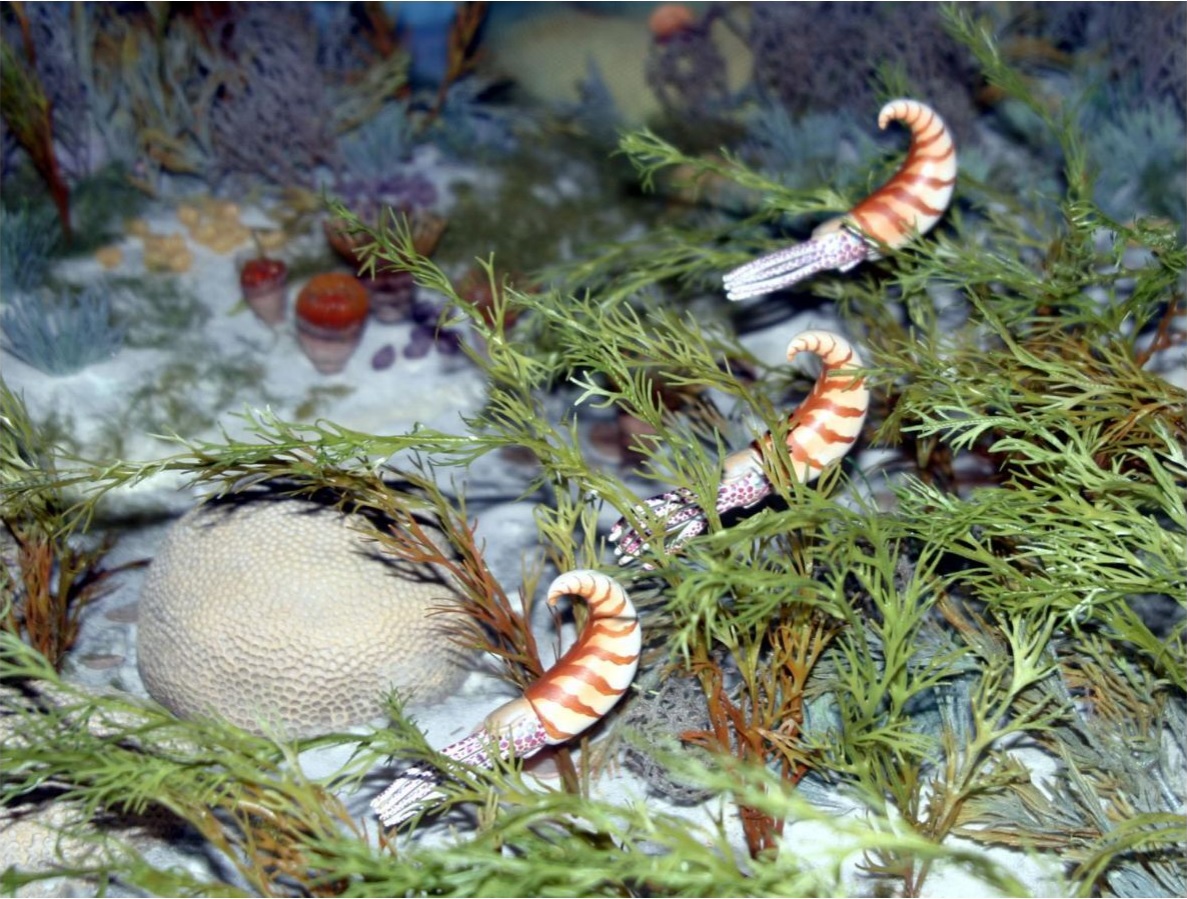
Diorama reconstruction of Beloitoceras oncocerids, from the Burpee Museum.


One factor that has drawn attention in recent years has been the arrangement of muscle scars on the shell. Large muscle attachment scars appear as raised annular elevations on the inside of the shell towards the rear end of the body chamber (in practice, they are more often observed in fossils as depressions on the internal mould). In the living nautilus, the muscles attached to these scars function in the retraction of the head (King & Evans 2019). Modern nautilus possess a pair of large lateral scars in an arrangement that has been labelled 'pleuromyarian'. However, many of the earliest cephalopods possessed a ring of numerous small scars, an arrangement referred to as 'oncomyarian'. Other cephalopods might have scars restricted to the dorsal ('dorsomyarian') or ventral ('ventromyarian') midline.

Primary types of muscle scar in nautiloids, from King & Evans (2019). 'D' and 'V' indicate dorsal and ventral, respectively, and arrows indicate direction of aperture.


Another feature that has been called out has been the structure of the connecting rings around the siphuncle. Shelled cephalopods, you will recall, have the shell divided into chambers separated by septa. Though the bulk of the animal is found in the final body chamber, a fleshy cord called the siphuncle runs back through the remaining chambers. In life, the siphuncle is used to control the levels of fluid in the chambers, which in turn controls the animal's buoyancy. The boundary between the siphuncle and the surrounding chamber is marked a toughened sheath, referred to as the connecting ring. In the modern nautilus, the connecting ring is comprised of two layers, an outer calcareous layer and an inner chitinous layer. In comparable fossils, the latter chitinous layer has decomposed after death so only the outer layer is preserved. However, some extinct cephalopod groups preserve evidence of calcification in the inner as well as the outer layer. Based on the distinction between these two siphuncle types, Mutvei (2015) supported dividing most of the nautiloids between two major lineages, the Nautilosiphonata (with a nautilus-type siphuncle) and the Calciosiphonata (with the internally calcified connecting rings).

A couple of years earlier, the same author (Mutvei 2013) had proposed recognition of a superorder Multiceratoidea for nautiloids that combined multiple muscle scars with a nautilus-type siphuncle. Examples of nautiloid orders with such a combination included the Ellesmeroceratida (small nautiloids with densely placed septa), the Oncoceratida (often short, squat nautiloids) and the Discosorida (similarly squat forms with complex bulging connecting rings). All of these were found in the earlier part of the Palaeozoic with the oncoceratids dieing off in the early Carboniferous. Mutvei (2013) also included the coiled Tarphyceratida and the egg-shaped Ascoceratida in this group. Later, King & Evans (2019) redefined this grouping as the Multiceratia, excluding the Tarphyceratida and Ascoceratida on the grounds that they had ventromyarian rather than oncomyarian muscle scars. Mutvei (2013) suggested that, rather than representing retractor muscles, these smaller repeated scars were associated with an outgrowth of the mantle, either as tentacles or a muscular 'skirt', that was used to capture micro-plankton.

Phylogeny of 'nautiloids' supported by King & Evans (2019). Though not shown on this diagram, the majority of authors have suggested that ammonoids and coleoids are descended from Orthoceratida.


King & Evans (2019) proposed a reclassification of the subclass Nautiloidea between five subclasses defined primarily by muscle structure. Apart from the earliest oncomyarian Plectronoceratia, most 'nautiloids' could be divided between two lineages. On one side were the dorsomyarian Orthoceratia (usually thought to include the ancestors of the ammonoids and coleoids). On the other, the oncomyarian Multiceratia would eventually give rise to the ventromyarian Tarphyceratia which in turn included the ancestors of the pleuromyarian Nautilida. Note that many of the reocognised subclasses (and orders) remain paraphyletic but we are at least approaching a more informative picture of cephalopod evolution than the earlier unceremonious dumping into 'Nautiloidea' (I should probably also remind you that, for various reasons, most invertebrate palaeontologists still don't regard strict monophyly as a taxonomic requirement in and of itself).

The usage of muscle scars and connecting rings as classificatory keys is handicapped by the difficulty of observing them. As internal structures, they each require careful preparation of a specimen to observe. And once you've gotten to a position where you can see them, it seems not to be particularly easy to tell just what you're looking at. As a result, muscle scarring and siphon structure remains undescribed for the majority of nautiloid species. Judging the structure of connecting rings seems to be particularly challenging and some have gone so far as to suggest that purported different structures may be the result of post-mortem taphonomic processes (King & Evans 2019). Nevertheless, what we do know suggests that such features remain reasonably consistent within each of the well-recognised nautiloid orders. And Mutvei's (2015) concept of Calciosiphonata vs Nautilosiphonata does largely line up with King & Evans' (2019) dorsomyarian vs oncomyarian-ventromyarian lineages. There are, of course, some notable exceptions. Whether these will cause the developing structure to collapse, or whether they indicate mistakes in interpretation, only continued research will tell.

REFERENCES

King, A. H., & D. H. Evans. 2019. High-level classification of the nautiloid cephalopods: a proposal for the revision of the Treatise Part K. Swiss Journal of Palaeontology 138: 65–85.

Mutvei, H. 2013. Characterization of nautiloid orders Ellesmerocerida, Oncocerida, Tarphycerida, Discosorida and Ascocerida: new superorder Multiceratoidea. GFF 135 (2): 171–183.

Mutvei, H. 2015. Characterization of two new superorders Nautilosiphonata and Calciosiphonata and a new order Cyrtocerinida of the subclass Nautiloidea; siphuncular structure in the Ordovician nautiloid Bathmoceras (Cephalopoda). GFF 137 (3): 164–174.

The Sordariales: In the Soil and Under the Skin

Microfungi are a very important factor in our lives. They play a key role in assuring that we are not literally up to our armpits in shit. Their hungry little hyphae break down ordure, cleaning up the planet and unlocking nutrients that will then be made available to other organisms. And among the most significant lineages of these largely unseen decomposers are the members of the order Sordariales.

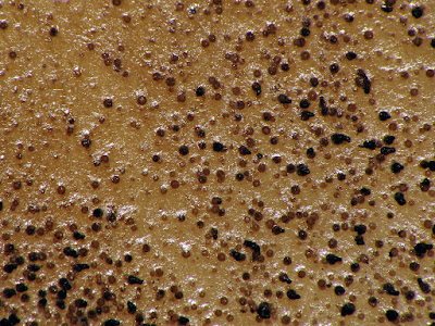
Lab culture of Sordaria fimicola, copyright BlueRidgeKitties.


Members of the Sordariales are, without exception, minute. Many species are coprophilous, growing on dung. Others may be found on rotting wood, or other decaying plant matter or soil. Fruiting bodies, when they appear, are flask-shaped perithecia protruding to a greater or lesser degree from the surface of their substrate. The walls of the perithecia are made up of large cells and have a membranous or coriaceous (leathery) texture. Within the fruiting body, the asci are single-walled and contain one- or two-celled ascospores that are often surrounded by a gelatinous sheath or bear various appendages. If the ascospores are two-celled, the cells are typically differentiated into an apical head and a basal tail (Kruys et al. 2015; Marin-Felix et al. 2020). Genera of Sordariales have historically been recognised on the basis of ascospore morphology but the advent of molecular data has indicated that such genera are highly polyphyletic. As a result, the Sordariales have seen (and are still seeing) a great deal of taxonomic reassessment. Miller & Huhndorf (2005) suggested that the structure of the fruiting body walls are more consistent with molecular phylogenies than ascospore morphology.

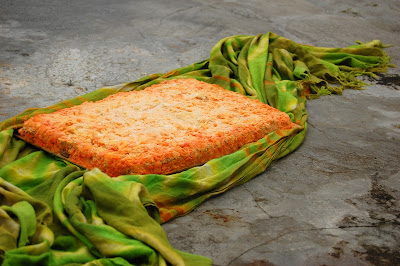
Cake of oncom-fermented beans, copyright Hariadhi.


Apart from their significant role as decomposers, most Sordariales have little direct impact on human economics. The mould Neurospora intermedia is used to make oncom, a fermented food similar to tempeh. A number of species of Sordariales such as Neurospora crassa and Sordaria fimicola have been widely used in genetic research, to the extent that they have been labelled the 'fruit flies of the fungal world'. Seriously, it's one of those expressions almost every publication seems obliged to crow-bar in somewhere. The analogy is made even more apropos by the fact that one of the most widely used species, Triangularia née Podospora anserina, has been made the subject of debate whether taxonomic considerations should be allowed to shake up the name of a popular model organism.

Molecular studies have also shown that the Sordariales encompass Madurella mycetomatis, a fungus causing subcutaneous inflammation in humans (van de Sande 2012). Seeing as sexual fruiting bodies are unknown in this species, and even asexual spore-producing structures are exceedingly rare, this organism would have previously been all but impossible to classify. Infection by M. mycetomatis is characterised by the production of granular swellings. It is most significant in central Africa but is also known from other tropical regions of the world. Madurella mycetomatis infects people via trauma such as animal bites and other wounds, and it has been isolated from soil and ant nests. In its normal state, M. mycetomatis is probably a quite innocent soil fungus. The trouble comes when it finds itself somewhere it shouldn't be.

REFERENCES

Kruys, Ã…., S. M. Huhndorf & A. N. Miller. 2015. Coprophilous contributions to the phylogeny of Lasiosphaeriaceae and allied taxa within Sordariales (Ascomycota, Fungi). Fungal Diversity 70: 101–113.

Marin-Felix, Y., A. N. Miller, J. F. Cano-Lira, J. Guarro, D. García, M. Stadler, S. M. Huhndorf & A. M. Stchigel. 2020. Re-evaluation of the order Sordariales: delimitation of Lasiosphaeriaceae s. str., and introduction of the new families Diplogelasinosporaceae, Naviculisporaceae, and Schizotheciaceae. Microorganisms 8: 1430.

Miller, A. N., & S. M. Huhndorf. 2005. Multi-gene phylogenies indicate ascomal wall morphology is a better predictor of phylogenetic relationships than ascospore morphology in the Sordariales (Ascomycota, Fungi). Molecular Phylogenetics and Evolution 35: 60–75.

van de Sande, W. W. J. 2012. Phylogenetic analysis of the complete mitochondrial genome of Madurella mycetomatis confirms its taxonomic position within the order Sordariales. PLoS One 7 (6): e38654.

The Cephalodiscids

Among the more obscure inhabitants of the world's oceans are the Cephalodiscidae, a family of small (only a few millimetres in length), largely sessile animals that mostly live in colonies within a shared domicile. Though rarely observed, cephalodiscids have received their fair share of attention due to being among the closest living relatives of the graptolites that once dominated the world's oceans during the early Palaeozoic era.

Preserved Cephalodiscus colony, copyright E. A. Lazo-Wasem.


Cephalodiscids are one of the two living branches of the pterobranchs (the other being the Rhabdopleuridae), which together with the acorn worms make up the phylum Hemichordata. Hemichordates are in turn one of the three living phyla of the deuterostomes, together with the echinoderms and chordates (to which, of course, we ourselves belong). Pterobranchs are filter feeders, using an arrangement of tentaculated arms arising just behind the head to collect particles from the water. In cephalodiscids, each individual usually possesses multiple pairs of arms in contrast to the single pair in rhabdopleurids (though at least one species of Cephalodiscus has small males with a single pair). The head carries a large glandular disc (hence the name of the family) that is used to secrete the horny tissue making up the external dwelling (referred to as the tubarium) in which a colony of Cephalodiscus lives. Both cephalodiscids and rhabdopleurids have a contractile stalk at the end of the body from which new individuals (zooids) are budded. However, whereas the zooids of rhabdopleurids (and presumably their extinct graptolite relatives) remain attached to each other throughout their life, cephalodiscid zooids split away from their parent by the time they mature. The majority of cephalodiscid species have distinct males and females though a small number may be hermaphrodites. Some species exhibit sexual dimorphism; males may be considerably smaller than females.

Individual zooid of Cephalodiscus dodecalophus, from Sedgwick et al. (1898).


About twenty species of living cephalodiscids are currently recognised. The majority of these have been included in a single genus Cephalodiscus, albeit divided between a number of subgenera. The single outlier, Atubaria heterolopha, was described in 1936 from a single dredge haul near Japan (Mitchell et al. 2013). No dwelling material was found in the haul so it was presumed this species does not construct a tubarium like other cephalodiscids. However, its zooids were otherwise little different from those of Cephalodiscus. The subgenera of Cephalodiscus are mostly distinguished by tubarium structure. In some species, each individual in the colony will have its own separate tube closed off at the base. In other species, tubes will open into a central chamber shared between multiple zooids (Maletz 2014). Openings of the tubarium may be surrounded by spines and the like, secreted by the zooids as they creep out from their domicile.

Recent studies have indicated that cephalodiscids represent the sister group to all other pterobranchs/graptolites, implying an history that may extend back to the Cambrian. However, the fossil record of cephalodiscids themselves is minimal. This is largely due to practical difficulties: because the soft-bodied zooids are not preserved, fossils can only be identified from the external tubarium structure alone. Unless the origin point of the tubarium is preserved and identifiable, there is little to distinguish a cephalodiscid tubarium from a benthic graptolite (graptolite colonies begin with a differentiated larval chamber called a sicula, cephalodiscids produce no such structure). A handful of fossil cephalodiscids have been identified, notably the early Devonian Eocephalodiscus, but as yet they tell us little about the evolution of this ancient lineage.

REFERENCES

Maletz, J. 2014. The classification of the Pterobranchia (Cephalodiscida and Graptolithina). Bulletin of Geosciences 89 (3): 477–540.

Mitchell, C. E., M. J. Melchin, C. B. Cameron & J. Maletz. 2013. Phylogenetic analysis reveals that Rhabdopleura is an extant graptolite. Lethaia 46: 34–56.

The Fate of Oligochiton

Chitons are one of the most distinctive and evolutionarily divergent groups of molluscs alive today. But compared to other groups of molluscs, the fossil record of chitons is rather sparse—or at least sparsely studied. It's not hard to see why. The multi-plated nature of the chiton shell means that it tends to fall apart after death, and the structure of the plates is such that critical features are easily abraded.

(Clockwise from top left) head, intermediate and tail valves of Lepidochitona lioplax, from Dell'Angelo et al. (2011).


Lepidochitona lioplax is one example of a fossil chiton. It was originally described from Oligocene rocks belonging to the Sooke Formation of southern Vancouver Island in British Columbia. Only four moderate-sized valves were initially identified: one head valve, one intermediate, and two tails (so at least two individuals were involved). The valves had a smooth outer surface without a strong distinction in appearance between the central and lateral areas. The insertion plates (lateral projections of the lower surface of the valves that in life anchor them into the surrounding girdle) were very short. The sutural laminae (anterior projections of the lower surface of the intermediate and tail valves that articulate with the valve in front) were low, wide, and divided in the middle by a broad shallow surface. Slits in the lateral insertion plates were numerous, with several in the tail valves and probably two or three on each side in the intermediate valves (Smith 1960). When first described, this species was thought distinct enough to belong in its own genus Oligochiton.

Oligochiton lioplax would then go little reported on until 2011 when Dell'Angelo et al. described an assemblage of chiton fossil from the latest Eocene or early Oligocene of the Lincoln Creek Formation in Washington State. Specimens of lioplax were relatively numerous in this collection and Dell'Angelo et al. were able to examine close to a hundred valves. Their observations would lead to something of a downgrade in the species status. Rather than deserving its own extinct genus, Dell'Angelo et al. felt that lioplax could be comfortably accommodated in the living genus Lepidochitona. Its smooth valves are unusual within Lepidochitona but not unique. The supposed multiple slits in the sides of the valves did not stand up to scrutiny. Instead, intermediate valves of L. lioplax bore only a single slit on each side, in line with other Lepidochitona species. The original inference of multiple slits was an error due to the original specimen being still partially embedded in the surrounding matrix.

Lepidochitona lioplax is one of the earliest known representatives of its genus but its exact significance is obscure. It has been suggested as a direct ancestor of the modern subgenus Spongioradsia but this, again, was based on the supposed slits in the intermediate valves that Dell'Angelo et al. refuted. To know how L. lioplax connects to the big picture of Lepidochitona evolution, we would probably need a better picture of Lepidochitona evolution overall.

REFERENCES

Dell'Angelo, B., A. Bonfitto & M. Taviani. 2011. Chitons (Polyplacophora) from Paleogene strata in western Washington State, U.S.A. Journal of Paleontology 85 (5): 936–954.

Smith, A. G. 1960. Amphineura. In: Moore, R. C. (ed.) Treatise on Invertebrate Paleontology pt I. Mollusca 1: Mollusca—General Features, Scaphopoda, Amphineura, Monoplacophora, Gastropoda—General Features, Archaeogastropoda and some (mainly Paleozoic) Caenogastropoda and Opisthobranchia pp. I41–I76. Geological Society of America, and University of Kansas Press.

In the Arms of Pseudisograptus

From the Ordovician to the early Devonian, the graptoloids were a major component of the oceanic fauna. These colonial floaters are among the characteristic fossils of the early Palaeozoic and have received a lot of attention due to their use in biostratigraphy. The evolution of graptoloids has been presented as a process of increasing simplification, of progressive reductions in colonial complexity and density. Like all illustrations of evolutionary trends, this is an overly simplistic representation of how things actually occurred but it's not entirely incorrect. The history of graptoloids was indeed marked by a number of significant transitions were particular growth forms overran their predecessors. One genus that may have played a significant role in the lead-up to one of these turnovers was Pseudisograptus.

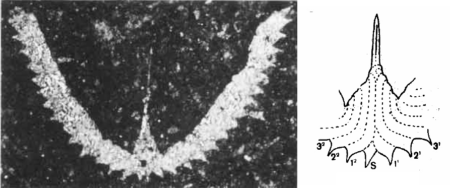
Pseudisograptus manubriatus koi, entire fossil and close-up diagram of initial thecae, from Cooper & Ni (1986).


The graptoloid genus Pseudisograptus has been collected from rocks in Australia, North America and eastern Asia dating to the latter part of the Floian stage of the early Ordovician, a bit over 470 million years ago (Cooper & Ni 1986). It is characterised by colonies growing in two branches (stipes) with the stipes spreading outwards and upwards like a pair of wings (indeed, one species of this genus luxuriates in the name of Pseudisograptus angel). In large specimens, the stipes reach about two centimetres in length and about three millimetres wide (from inner margin to the outer apex of the individual thecae). Pseudisograptus species are very similar to, and until 1972 where classified with, species of the related genus Isograptus. They differ, however, in the arrangement and growth of the earliest thecae in the colony. Whereas Isograptus stipes grow outwards immediately from the oldest theca, Pseudisograptus have the first few thecae on each stipes elongated and growing downwards before the stipes makes a later sharp turn upwards. As a result, between the two 'wings' of the stipes there is a more or less distinct triangle (referred to as the manubrium) formed from the bases of the early thecae. At the top of the manubrium is an upright thread, the nema. In a number of graptoloid fossils, an inflated structure has been identified at the top of the nema that probably functioned as a float. I don't know if such a structure has ever been identified in a Pseudisograptus fossil but I imagine it would quite easily be lost in the course of preservation.

Basal section of Cardiograptus amplus, from Fortey et al. (2005). Not a true biserial graptoloid but illustrative of the way biserial forms may have evolved from biramous ancestors.


Pseudisograptus' disappearance from the fossil record coincides with one of the aforementioned turnovers in graptoloid diversity, the appearance of the biserial graptoloids. These forms, which had two rows of thecae arising from a single central line, rapidly replaced most of the earlier branched forms. The rapid appearance of the biserial graptoloids has made their origins difficult to work out but current thinking is that they arose from a form similar to Pseudisograptus, in which the upward growth of the stipes became steep enough that they met in the middle along the nema. One interesting detail is that two lineages appear to have achieved biseriality at about the same time from closely related but separate ancestors. In the glossograptids, the conjoined stipes met each other side-by-side; in the diplograptids, they met back to back. Pseudisograptus has, at different times, been implicated in the ancestry of both of these groups. Cooper & Ni (1986) regarded Pseudisograptus as paraphyletic and including the direct ancestors of the glossograptids. In contrast, more recent studies by Fortey et al. (2005) and Maletz et al. (2009) have placed Pseudisograptus closer to the diplograptids. These studies have been more agnostic as to whether Pseudisograptus was a direct ancestor or a close relative. If the former is the case then, while the exact Pseudisograptus morphotype would disappear at the end of the Floian, their genetic lineage would continue strong for nearly ninety million more years.

REFERENCES

Cooper, R. A., & Ni Y. 1986. Taxonomy, phylogeny, and variability of Pseudisograptus Beavis. Palaeontology 29 (2): 313–363.

Fortey, R. A., Y. Zhang & C. Mellish. 2005. The relationships of biserial graptolites. Palaeontology 48 (6): 1241–1272.

Maletz, J., J. Carlucci & C. E. Mitchell. 2009. Graptoloid cladistics, taxonomy and phylogeny. Bulletin of Geosciences 84 (1): 7–19.

The Shells of Ducks and Swans

The freshwater environment has been a challenging one for bivalves. Though there is a reasonable diversity of freshwater bivalves around the world, they tend to be dominated by members of a select few lineages. One of the most successful groups of freshwater bivalves is the family Unionidae, and among the more widespread unionids are the freshwater mussels of the genus Anodonta.

Swan mussel Anodonta cygnea, copyright Gail Hampshire.


Anodonta species are found widely across northern Eurasia and North America, commonly referred to as 'mussels' in Eurasia and 'floaters' in North America. They are relatively large bivalves (one of the largest, the swan mussel Anodonta cygnea of Eurasia, can be up to about twenty centimetres across) with an irregularly elliptical shape and a relatively thin shell. One of their distinguishing features compared to other freshwater bivalves is the teeth of the hinge connecting the shell valves have been lost. Instead, the valves are primarily held together by the dorsal ligament (Moore 1969). Freshwater mussels are most commonly found in mud at the bottom of slow-moving or still waters, such as lakes or slow rivers.

One of the main hurdles to bivalve colonisation of fresh water has been the question of dispersal. In most marine bivalves, populations mostly disperse via their planktonic larvae. But because of the directed flow of water in rivers and the like, passive plankton fare less well in freshwater environments. If you just float along a stream, eventually you'll be washed out to sea. Anodonta species, like other unionids, solve the problem of getting back upstream through parasitic larvae called glochidia. Female Anodonta have the rear part of the gills modified into a pouch (or marsupium) in which the developing larvae are initially incubated. When they are released by their mother, the glochidia already possess a bivalved, sharp-edged shell. Released glochidia swim towards a suitable host in the form of a passing fish and use the valves of the shell to clamp onto a narrow appendage of the host's body such as its fins or gills. Eventually, a cyst forms around the attached glochid within with it develops until it is ready to emerge and attain maturity.

Winged floater Anodonta nuttalliana, a North American species, copyright Eric Wagner.


Freshwater molluscs have a history of being subject to taxonomic chicanery, through the Nouvelle École of late nineteenth-century France and other excesses of typological enthusiasm. Anodonta is no exception. The shells of freshwater mussels tend to be very plastic in morphology, their size, shape and appearance being strongly affected by their developmental environment. As a result, they include what were labelled by Riccardi et al. (2020) as "some of the most over-described species on the planet". The swan mussel A. cygnea alone has had somewhere in the region of 550 different species-group names applied to it at one time or another. Modern estimates of Anodonta diversity are considerably more conservative. Just four species are currently recognised from Eurasia (Riccardi et al. 2020) with the swan mussel and the duck mussel A. anatina being the most widespread (offhand, I don't know whether the mussels get their vernacular names because they're eaten by swans and ducks or because the shape of the shell is supposed to look like a swan or duck). Considering the travails of shell-based taxonomy, it is noteworthy that these species often cannot be distinguished with certainty without checking the soft tissue. North America is home to six or seven recognised species with diversity being higher to the west of the continent.

Nevertheless, there are still grounds for questioning the current taxonomy of Anodonta. Molecular studies of the genus by Chong et al. (2008), Bolotov et al. (2020) and Riccardi et al. (2020) have all suggested that Anodonta as currently recognised may be paraphyletic to closely related genera. In particular, there may be a divide between the Eurasian and North American lineages with the North American species closer to taxa found in eastern Asia. Anodonta has been a problem genus in the past and it sees no reason why it should allow itself to be reformed.

REFERENCES

Bolotov, I. N., A. V. Kondakov, E. S. Konopleva, I. V. Vikhrev, O. V. Aksenova, A. S. Aksenov, Y. V. Bespalaya, A. V. Borovskoy, P. P. Danilov, G. A. Dvoryankin, M. Y. Gofarov, M. B. Kabakov, O. K. Klishko, Y. S. Kolosova, A. A. Lyubas, A. P. Novoselov, D. M. Palatov, G. N. Savvinov, N. M. Solomonov, V. M. Spitsyn, S. E. Sokolova, A. A. Tomilova, E. Froufe, A. E. Bogan, M. Lopes-Lima, A. A. Makhrov & M. V. Vinarski. 2020. Integrative taxonomy, biogeography and conservation of freshwater mussels (Unionidae) in Russia. Scientific Reports 10: 3072.

Chong, J. P., J. C. B. Box, J. K. Howard, D. Wolf, T. L. Myers & K. E. Mock. 2008. Three deeply divided lineages of the freshwater mussel genus Anodonta in western North America. Conserv. Genet. 9: 1303–1309.

Moore, R. C. (ed.) 1969. Treatise on Invertebrate Paleontology pt N. Mollusca 6. Bivalvia vol. 1. The Geological Society of America, Inc. and The University of Kansas.

Riccardi, N., E. Froufe, A. E. Bogan, A. Zieritz, A. Teixeira, I. Vanetti, S. Varandas, S. Zaccara, K.-O. Nagel & M. Lopes-Lima. 2020. Phylogeny of European Anodontini (Bivalvia: Unionidae) with a redescription of Anodonta exulcerata. Zoological Journal of the Linnean Society 189: 745–761.

Gastrotrichs and their Tacky Little Tubes

When I was a student, I was taught that known animal diversity could be divided between somewhere in the region of a couple of dozen 'phyla'. These were the fundamental units of animal classification, the basic archetypes of animal morphology. Many of these were the major assemblages with which we all were familiar: chordates, arthropods, molluscs and the like. But many were the so-called 'lesser phyla', those taxonomic orphans that, whether small in size or small in number or both, tended to escape observation and study by the majority of people. One such 'minor phylum' was the collection of small worm-like animals known as the Gastrotricha.

Polymerurus nodicaudus, a paucitubulate gastrotrich, from Balsamo et al. (2015). Scale bar equals 100 µm.


Gastrotrichs are, in general, minute (Todaro et al. 2019). The largest reach about three-and-a-half millimetres in length, the smallest are about sixty microns, and there are probably many more at the lower range than the higher. They are dorsoventrally flattened with numerous cilia, and their cuticle may often be differentiated into a covering of scales or spines. Gastrotrichs are aquatic and are often referred to as part of the meiofauna, the assemblage of animals specialised for living within and crawling through the spaces between sand grains. That is indeed the preferred habitat for many species and gastrotrichs may be among the most abundant inhabitants of this milieu, edged out only by the nematodes and copepods. However, other species live above the sediment surface, crawling over the surface of aquatic vegetation or even floating among the plankton. Over 850 species are known to date, of which are a bit over 500 are marine (with all marine species being meiofaunal) and the remainder are found in fresh water. They feed on micro-organisms such as bacteria and algae, swallowing them by means of a muscular pharynx.

Gastrotrichs differ from other animals in a number of significant features. Among these is the differentiation of the outer cuticle into two distinct layers. The outermost of these layers, the epicuticle, covers the entire outer surface of the body, including coating the cilia. Gastrotrichs also possess characteristic tubular outgrowths ending in adhesive glands. Their relationships to other animals remain uncertain. Most authors now agree that they represent an early-diverging branch of the Lophotrochozoa, the animal superclade including such creatures as molluscs and annelids. It is possible that they are more closely related to flatworms than anything else but even then the relationship would hardly be close.

Pseudostomella etrusca, a macrodasyidan gastrotrich, from Todaro et al. (2011). Scale bar = 50 µm.


Historically, gastrotrichs have been divided between two orders, the Macrodasyida and Chaetonotida. This division was supported by structural features of the pharynx and the body wall but is also reflected in the distribution of the adhesive tubes. The Macrodasyida, which are usually vermiform, possess adhesive tubules at both the anterior and posterior ends of the body, as well as laterally. Macrodasyidans are always interstitial in habits and usually marine. The Chaetonotida, on the other hand, lack anterior tubules. Chaetonotidans were further divided between two major taxa. One of these was the isolated genus Neodasys which is vermiform and interstitial like a macrodasyidan, and possesses both lateral and posterior tubules. The remaining Chaetonotida were recognised as the suborder Paucitubulatina. As indicated by their name (meaning 'few tubules'), members of this suborder are characterised by the reduction in number of adhesive tubules, usually to a single pair at the end of the body (a few species have two pairs of tubules, others lack distinct tubules and have the adhesive glands opening directly on the main body). They are short, generally shaped more or less like a bowling pin, and are the most ecologically diverse major gastrotrich group, including both marine and freshwater forms.

A phylogenetic analysis of gastrotrichs by Kieneke et al. (2008), however, questioned the established classification of the group. Rather than affirming a basal division between Chaetonotida and Macrodasyida, their results placed Neodasys as the sister group of all other gastrotrichs. Such a division may be reflected in the nature of their adhesive tubules: Neodasys has tubules containing a single gland but Macrodasyida and Paucitubulatina have two glands per tubule (unfortunately, because of the lack of close outgroups, it's hard to know which tubule type was ancestral). Within the Macrodasyida + Paucitubulatina clade, the macrodasyidans were then paraphyletic to the paucitubulates. Interestingly, the sister group to the Paucitubulatina was a clade of the only two known freshwater macrodasyidans, Marinellina and Redudasys. The implication was that gastrotrichs may have made the move to fresh water on just one occasion (followed by a number of returns to the sea among paucitubulates). This is not an isolated case: a number of phylogenetic studies of micro-organisms have found deep divides between marine and freshwater lineages. It seems it's hard to adjust to a life less salty.

REFERENCES

Kieneke, A., O. Riemann & W. H. Ahlrichs. 2008. Novel implications for the basal internal relationships of Gastrotricha revealed by an analysis of morphological characters. Zoologica Scripta 37 (4): 429–460.

Todaro, M. A., J. A. Sibaja-Cordero, O. A. Segura-Bermúdez, G. Coto-Delgado, N. Goebel-Otárola, J. D. Barquero, M. Cullell-Delgado & M. Dal Zotto. 2019. An introduction to the study of Gastrotricha, with a taxonomic key to families and genera of the group. Diversity 11: 117.

Eurotiomycetes: Small but Significant Fungi

Mention the word 'fungi' and most people's thoughts will probably go to images of mushrooms or toadstools. A few may conjure up pictures of lichens. Nevertheless, the great majority of fungal species are microscopic and likely to pass unremarked by most observers. That does not, however, mean that they are of no consequence. Today's post involves one major group that, for all their visual insignificance, include some of the most significant fungal species for modern human society: the Eurotiomycetes.

Developmental stages of Aspergillus glaucus, with cleistothecia as figs 21–23, from Raper & Fennel (1965).


The class Eurotiomycetes has been recognised in recent years as including a diverse assemblage of fungi, associated with a wide range of morphologies and habitats, that are united as a clade by molecular analyses. Réblová et al. (2017) recognised five subclasses within the Eurotiomycetes of which the two largest (or at least the most studied) are the Eurotiomycetidae and the Chaetothyriomycetidae. The Eurotiomycetidae are, for the greater part, saprobes. They were largely recognised as a distinctive group even before the advent of molecular phylogenetic analysis owing to the production by sexually reproducing forms of a distinctive type of fruiting body, the cleistothecium. In cleistothecia, the fruiting body is completely enclosed with no openings to faciliatate the release of spores, which only escape when the fruiting body itself breaks down. Cleistothecia are most commonly produced by fungi that grow in enclosed locations such as underground (the Eurotiomycetidae are not the only group of fungi to produce cleistothecia though they are one of the most diverse). Within the cleistothecium, spores develop within globular asci with a single wall that breaks down shortly after maturity (Geiser et al. 2015).

Penicillium expansum on rotting pear, copyright H. J. Larsen.


For many people, though, the most familiar members of the Eurotiomycetidae are likely to be asexually reproducing forms. This is the clade containing the moulds of the genera Aspergillus and Penicillium. Even before a species of the latter achieved fame as the shource of the first known antibiotic, penicillin, members of these genera had a great impact on human lives. Species of Penicillium are the moulds used in the production of cheeses such as Roquefort and camembert. Species of Aspergillus are used to ferment soy beans and rice in the production of comestibles such as soy sauce and sake. On the flip side, a number of species of Eurotiomycetidae act as pathogens of mammals including humans, causing conditions such as respiratory illnesses or tinea, with the former being of particular concern in immunocompromised individuals. Eurotiomycetid moulds may also cause problems for food storage and the like, particularly as many species are capable of growing under remarkably hot and/or dry conditions. Some Aspergillus moulds produce dangerous toxins, capable of causing acute poisioning or cancer development.

Verrucaria maura, copyright Richard Droker.


The Chaetothyriomycetidae are less clearly defined morphologically than the Eurotiomycetidae but fruiting bodies are mostly produced as perithecia: flask-shaped structures with an apical pore through which spores are released. The asci within the perithecium usually possess a double wall. Like many eurotiomycetids, chaetothyriomycetids have a tendency to be associated with habitats where water availability is a concern such as in very dry and/or saline environments. A number of chaetothyriomycetid species form lichens. One genus, Verrucaria, is often found as a thin black lichen growing on rocks along the seashore. Some species grow within the cavities of myrmecophytes, plants that form mutualistic associations with ants (the plant provides food and/or accomodation for the ants and the ants help keep the plant clear of grazers or sap-suckers). The fungi are cultivated by the ants that use them for food.

The other three subclasses of the Eurotiomycetes are less well known and recognised as containing a single order each. The Sclerococcales were first recognised as such by Réblová et al. (2017) via molecular analysis. Fruiting bodies, where known, are apothecia (open bowls) bearing single-walled asci. Representatives are known from marine and terrestrial habitats, growing on wood or lichens, and some have been isolated from within the digestive tracts of bark beetles. The Coryneliaceae, living as parasites on podocarps, have been considered as morphologically intermediate between chaetothyriomycetids and eurotiomycetids. Molecular analysis positions them as sister to the latter (Wood et al. 2016). Finally, the Mycocaliciales live as parasites or commensals of other fungi, particularly lichens.

There are other representatives of the Eurotiomycetes that I haven't even had the time to gloss over, such as endophytes and ectomycorrhizal truffles. You may not know they're there but that doesn't mean they don't mean anything to you.

REFERENCES

Geiser, D. M., K. F. LoBuglio & C. Gueidan. 2015. Pezizomycotina: Eurotiomycetes. In: D. J. McLaughlin, & J. W. Spatafora (eds) The Mycota 2nd ed. vol. 7. Systematics and Evolution part B pp. 121–141. Springer-Verlag: Berlin.

Réblová, M., W. A. Untereiner, V. Å tÄ›pánek & W. Gams. 2017. Disentangling Phialophora section Catenulatae: disposition of taxa with pigmented conidiophores and recognition of a new subclass, Sclerococcomycetidae (Eurotiomycetes). Mycological Progress 16: 27–46.

Wood, A. R., U. Damm, E. J. van der Linde, J. Z. Groenewald, R. Cheewangkoon & P. W. Crous. 2016. Finding the missing link: resolving the Coryneliomycetidae within Eurotiomycetes. Persoonia 37: 37–56.

The Glyceriforms: Stabby Worms and Grabby Worms

Historically, the annelid worms have been considered a difficult group to classify. Whereas most of the recognised families have been fairly well established, higher taxa uniting these families have tended to be a bit on the vague side. Nevertheless, there are some supra-familial groups that can be considered well established, one such group being the Glyceriformia.

Specimen of Goniadidae (head to the right), from NOAA Fisheries.


The glyceriforms are two families of marine worms, the Glyceridae and Goniadidae. More than a hundred species are known in this clade (over forty glycerids and over sixty goniadids), found in habitats ranging from the intertidal to the abyssal. They range in size from about a centimetre in length to well over half a metre. The front end of the body tapers to a narrow, elongate conical point in front of the mouth, bearing two terminal pairs of small, slender appendages that may correspond to the antennae and palps of other worms. Eyes may be present or absent. The pharynx forms a remarkably elongate, eversible proboscis. In Glyceridae, the proboscis ends in a ring of four hook-shaped jaws, all similar to each other. In Goniadidae, the arrangement of jaws is more complex with the usual arrangement being small micrognaths on one side of the ring and larger macrognaths on the other. Glycerids usually have a transparent skin and an overall red or white colour reflecting the coloration of the internal fluids (red-coloured individuals are sometimes known as 'bloodworms', as are many other similarly coloured worm-like invertebrates). Goniadids have a more opaque cuticle and often have an iridescent sheen (Rouse & Pleijel 2001).

Glycera dibranchiata with everted proboscis, from the Yale Peabody Museum.


Glyceriforms most commonly live as burrowers in muddy or sandy substrates though some live on the surface of rocks. Most are carnivores of active invertebrates such as crustaceans or other worms; some may be detritivores. They may be vagile or they may construct permanent galleries of burrows with multiple entrance and exit openings in which they wait to lunge at anything foolish enough to pass nearby. In glycerids, the stabby jaws are associated with venom glands leading to ducts opening through pores on the jaw's underside. In some species, this venom is strong enough to cause a painful reaction in humans (though I haven't come across any references to long-term consequences). Goniadids lack venom glands and seem to rely on the physical use of their jaws to capture prey. As with many other marine worms, reproduction happens via pelagic epitokes. As a suitable time approaches (Prentiss, 2020, records goniadid epitokes emerging only during a full moon), the glyceriform worm undergoes a metamorphosis involving the break-down of the digestive system and enlargement of the parapodia. The transformed epitokes swim towards the surface where they release gametes through ruptures of the body wall, ending their life in a suicidal orgasm.

Close-up on proboscis of Glycera alba, copyright Hans Hillewaert.


Because of their hardened jaws, which are mostly constructed of protein but partially mineralised, glyceriforms have quite a good fossil record compared to many other worms (Böggemann 2006). Fossilised glyceriform jaws have been found as far back as the Triassic and are little different from those of modern glyceriforms. Body fossils are, unsurprisingly, much rarer but a worm from the Carboniferous Mazon Creek fauna, Pieckonia helenae, has been identified as a stem-group goniadid. The glyceriform body plan seems to have been a very successful one, remaining essentially unchanged over hundreds of millions of years.

REFERENCES

Böggemann, M. 2006. Worms that might be 300 million years old. Marine Biology Research 2: 130–135.

Prentiss, N. K. 2020. Nocturnally swarming Caribbean polychaetes of St. John, U.S. Virgin Islands, USA. Zoosymposia 19: 91–102.

Rouse, G. W., & F. Pleijel. 2001. Polychaetes. Oxford University Press.

Tricolia: Fluorescent Seashells

Tricolia pullus, copyright Ar rouz.


Search among patches of seaweed along the shores of Africa, Australia or warmer parts of Eurasia and you may be able to find represents of the marine gastropod genus Tricolia. Tricolia are small shells, less than a centimetre in height, with shiny shells that may be smooth or spirally ribbed. Most species have a moderately high spire and an ovate shape but some are lower and more globose (Knight et al. 1960). The shell may or may not have an umbilicus, and there is a calcareous, externally convex operculum. Tricolia belongs to the Phasianellidae, commonly known as pheasant shells, presumably in reference to the bold, intricate colour patterns of many species. Species of Tricolia and the closely related genus Eulithidium, which replaces it in the Americas, have shell pigments containing porphyrin that fluoresce under ultraviolet light (Vafiadis & Burn 2020). Over forty species of Tricolia are currently recognised with the highest diversity in southern Africa (Nangammbi et al. 2016). However, the taxonomy of the genus has historically been confused due to polymorphic species being named multiple times; it is possible that at least some of the apparent African diversity is an artefact of the genus being largely unrevised in that region. An analysis of some of the southern African taxa by Nangammbi et al. (2016) found that some 'species' could not be distinguished genetically. They were, nevertheless, distinct geographically and the authors suggested that they may be variants of a single species responding to different environments.

Variants of Tricolia kochii, copyright Brian du Preez.


Like other members of the Vetigastropoda (the clade containing most of what used to be called the 'archaeogastropods'), Tricolia species have a simple life cycle without an actively feeding planktonic larva. The basic mode of reproduction is by broadcast spawning with separate males and females releasing gametes into the water column. After fertilisation, a brief non-feeding planktonic phase is nourished by yolk from the egg before the larva settles. The brevity of this phase is reflected by the resultant form of the protoconch which accounts for less than an entire whorl. In the Indo-West Pacific species T. variabilis, the male is smaller than the female and sits directly on her, waiting to fertilise her eggs as they are laid as gelatinous capsules rather than freely broadcasted. A temperate Australian species, T. rosea, takes things a step further as the female broods embryos (up to nearly fifty at a time) within the cavity of the last shell whorl (Vafiadis & Burn 2020). How the eggs are actually fertilised remains unknown but all embryos within a brood are about the samesize and stage of development, indicating a single fertilisation event; perhaps males associate with females as in T. variabilis. After the young pheasant shells hatch or settle, they initially feed on diatoms and other microalgae until they eventually grow enough to move onto the seaweed fronds that will comprise their adult diet.

REFERENCES

Knight, J. B., L. R. Cox, A. M. Keen, R. L. Batten, E. L. Yochelson & R. Robertson. 1960. Gastropoda: systematic descriptions. In: R. C. Moore (ed.) Treatise on Invertebrate Paleontology pt I. Mollusca 1. Mollusca—general features, Scaphopoda, Amphineura, Monoplacophora, Gastropoda—general features, Archaeogastropoda and some (mainly Paleozoic) Caenogastropoda and Opisthobranchia pp. I171–I351. Geological Society of America: Boulder (Colorado), and University of Kansas Press: Lawrence (Kansas).

Nangammbi, T. C., D. G. Herbert & P. R. Teske. 2016. Molecular insights into species recognition within southern Africa's endemic Tricolia radiation (Vetigastropoda: Phasianellidae). Journal of Molluscan Studies 82: 97–103.

Vafiadis, P., & R. Burn. 2020. Internal embryonic brooding and development in the southern Australian micro-snail Tricolia rosea (Angas, 1867) (Vetigastropoda: Phasianellidae: Tricoliinae). Molluscan Research 40 (1): 60–76.

Key Limpets

On two occasions before, I've presented you with members of the Fissurellidae, the keyhole and slit limpets. It's time for a return visit to the fissurellids, in the form of the diverse keyhole limpet genus Diodora.

Various views of shell of Diodora italica, copyright H. Zell.


Species have been assigned to Diodora from coastal waters pretty much around the world except for the coolest regions. They are small to moderate-size limpets, the largest species growing about three centimetres in length and two centimetres in height. The shell opens through a 'keyhole' at the apex through which the animal ejects waste matter and water that has been passed over the gills. The internal margin of this keyhole is surrounded by a callus on the underside of the shell; a distinguishing feature of Diodora is that this callus is posteriorly truncate. The external ornament of the shell is cancellate (arranged in a criss-cross pattern) and the margin of the shell is internally crenulated (Moore 1960). Moore (1960) listed three subgenera of Diodora distinguished by features of the keyhole shape and position but Herbert (1989) notes that these subgenera are not clearly distinct. A phylogenetic analysis of the fissurellids by Cunha et al. (2019) did recognise a clade including the majority of Diodora species analysed. However, species from the eastern Pacific formed a disjunct clade that may prove to warrant recognition as a separate genus.

As far as is known, Diodora species have a long lifespan, surviving for some ten to twenty years. They do not have a planktonic larva; young Diodora hatch directly from the egg as benthic crawlers. For the most part, they are presumed to graze on algae in the manner of other fissurellids and limpets. However, the northeastern Australian species D. galeata has been found feeding on the soft tissues of coral (Stella 2012), a habit that went unrecognised until fairly recently owing to the animal's cryptic nature, hiding deep among the branches of the host. Whether other Diodora species might exhibit similar lifestyles would require further investigation.

REFERENCES

Cunha, T. J., S. Lemer, P. Bouchet, Y. Kano & G. Giribet. 2019. Putting keyhole limpets on the map: phylogeny and biogeography of the globally distributed marine family Fissurellidae (Vetigastropoda, Mollusca). Molecular Phylogenetics and Evolution 135: 249–269.

Herbert, D. G. 1989. A remarkable new species of Diodora/i> Gray, 1821 from south-east Africa (Mollusca: Gastropoda: Fissurellidae). Annals of the Natal Museum 30: 173–176.

Moore, R. C. (ed.) 1960. Treatise on Invertebrate Paleontology pt I. Mollusca 1. Mollusca—general features, Scaphopoda, Amphineura, Monoplacophora, Gastropoda—general features, Archaeogastropoda and some (mainly Paleozoic) Caenogastropoda and Opisthobranchia. Geological Society of America, Inc. and University of Kansas Press.

Stella, J. S. 2012. Evidence of corallivory by the keyhole limpet Diodora galeata. Coral Reefs 31: 579.

Donaldina: Palaeozoic Turrets

Within the last few decades, we've developed a reasonably good idea of what are the primary subdivisions of gastropods alive today. One such generally accepted lineage is the Heterobranchia (or, depending on the author, the Heterostropha), a group that includes (among others) the air-breathing pulmonates as well as the marine sea slugs and bubble shells. In the fossil record, the roots of this lineage extend well back into the Palaeozoic with early members recognisable by their distinctive mode of shell development. The larval shell, the protoconch, of these forms spirals in the opposite direction from the mature teleoconch, so the animal will start its life sinistral (spiralling left) and end it dextral (spiralling right; if you're having difficulty imagining how this works, the protoconch often ends up sitting upside down relative to the teleoconch). Among the earliest heterobranchs in the fossil record is the genus Donaldina.

Specimen of Donaldina (1.2 mm in height), with close-up of protoconch, from Bandel et al. (2002).


Fossils of Donaldina have been found around the world and the genus persisted for a long time. The earliest potential Donaldina have been described from the Early Devonian but their inclusion in the genus is uncertain (Bandel et al. 2002). The protoconch on these early forms is poorly preserved and it is uncertain whether they truly showed a heterobranch development. The genus was definitely present by the early Carboniferous and persisted into the lower Permian. This is an impressive length of time: the Carboniferous alone last for around sixty million years.

Donaldina was a genus of small, high-spired gastropods, less than a centimetre in height. Many early members of the Caenogastropoda, the likely sister group of the Heterobranchia, also had shells of this kind and it may have represented the ancestral form for the two lineages. The sinistral protoconch of Donaldina was almost planispiral (spiralling in a flat plane) and completed between one and two whorls. The multi-whorled, dextral teleoconch was characterised by an ornament of spiral cords, usually only on the lower half of the whorl.

So what were Donaldina doing with their time when alive? Modern high-spired gastropods occupy a range of lifestyles, including free-living grazers, burrowers, or sedentary forms that live as filter feeders or parasites of other animals (Signor 1982). The morphology of Donaldina suggests that it is unlikely to be a burrower. The whorls are individually rounded whereas those of habitual burrowers tend to be flattened so the shell moves more smoothly through the sediment. The ornamentation on the underside of the whorl would presumably also have presented resistance to burrowing. The shape of the aperture in Donaldina is more suggestive of a free roamer, as a sinus in the upper part of the outer margin would have allowed the animal to pull back into its shell while the plane of the aperture was held as flat as possible against the substrate to protect against predators. Overall, the lifestyle of Donaldina may not have been dissimilar to that of the modern mudsnails of Cerithium and similar genera, crawling about in search of algae and other tasty morsels.

REFERENCES

Bandel, K., A. Nützel & T. E. Yancey. 2002. Larval shells and shell microstructures of exceptionally well-preserved Late Carboniferous gastropods from the Buckhorn Asphalt Deposit (Oklahoma, USA). Senckenbergiana Lethaea 82 (2): 639–689.

Signor, P. W., III. 1982. Resolution of life habits using multiple morphologic criteria: shell form and life-mode in turritelliform gastropods. Paleobiology 8 (4): 378–388.

Air-breathing Limpets

For many people, the most familiar members of the gastropods are the terrestrial snails. Gastropods started their evolution as marine animals, breathing through gills, but members of one lineage would instead evolve their own version of a lung, a large hollow in the mantle cavity opening through a hole alongside the head called the pneumostome. Possession of this lung cavity would enable slugs and snails to thrive in the terrestrial environment but the structure had originally evolved in a marine context, and even today one may find marine lung-bearers occupying habitats along the coast. One such group is the siphon limpets or 'false limpets'* of the Siphonarioidea.

*You know, normally I don't overly concern myself with vernacular names. They are not regulated and not obliged to follow reason. But even so, the name 'false limpet' makes me grit my teeth. The name is presumably inspired by the fact that siphonariids are not direct relatives of the 'true' limpets of the Patellogastropoda. But the limpet morphotype, where the typical spiral gastropod shell is reduced to a simple cap, has evolved on multiple occasions. As well as the siphonariids and patellogastropods, there are the keyhole limpets of the Fissurellidae, the freshwater limpets of the Ancylini, and many others, all consistently referred to as 'limpets'. The name refers to a morphology, not to a clade, and by that measure the siphonariids are no more 'false' than any other limpets.

Flat siphon limpets Siphonaria atra, copyright Ria Tan.


Living siphonarioids are placed within a single family, the Siphonariidae, whose members with their cap-shaped, often radially ribbed shells are found in littoral environments in temperate and tropical regions of the world. A second family, the Acroreiidae, is recognised from the Cretaceous and early Tertiary; the inclusion of these smooth, thin shells in the Siphonarioidea is somewhat tentative (classification of limpets in the fossil record is always a challenge because their simple shell form renders them light on distinguishing characters). Siphonariids are readily distinguished from other living limpets by the presence of a groove on the underside of the right side of the shell marking the position of the pneumostome. In dorsal view, this groove is often indicated by an asymmetry in the outline of the shell with one side produced. The pneumostome is also associated with a broad gap in the ring of muscle holding the shell in place; the ring is more complete in other limpets. Seemingly as a result of this lower extent of muscle, siphonariids cling to their home rocks with less tenacity than other limpets and are mostly restricted to more sheltered locations (Simone & Seabra 2017). On the other hand, they do have a more flexible foot than their competitors, allowing them to potentially move more quickly. Like other limpets, siphonariids are grazers, scraping microalgae as they crawl about. Siphonariids have a weaker radula than patellogastropods and so scrape somewhat less forcefully; when members of the two clades occupy the same habitats, patellogastropods are generally the more abundant. The majority of siphonariids (where known) have planktonic larvae but some species are known to be direct developers.

Siphonaria lessonii, copyright Mikelzubi.


Obviously, the marine but lung-possessing siphonariids are potentially of great interest in understanding how the gastropod lung evolved. Many earlier researchers thought that the siphonariids may have evolved from terrestrial ancestors who had returned to the seashore but this is no longer thought likely to be the case. In most lunged gastropods, gas exchange is effected in the mantle cavity via dense blood vessels in the cavity wall but in siphonariids a gill structure is present within the lung (this lung-gill combination makes the siphonariids particularly well suited for moving freely both above and below the water surface). The gill of siphonariids is quite similar to that of the sacoglossans, a group of herbivorous sea-slugs. Though it was long presumed that the lung-bearing gastropods belonged to a single clade, more recent molecular phylogenies have confused the issue (Kocot et al. 2013). The sacoglossans are likely to be close to the ancestry of lunged gastropods as a whole, but it is possible that siphonariids are more closely related to the sacoglossans than the other lung-bearers. It remains an open question whether the siphonariid combination of lung and gill represents an intermediate stage towards the vascular lung of the terrestrial forms, or whether siphonariids and other lung-bearers each evolved their pneumostome from close but distinct ancestors.

REFERENCES

Bouchet, P., J.-P. Rocroi, B. Hausdorf, A. Kaim, Y. Kano, A. Nützel, P. Parkhaev, M. Schrödl & E. E. Strong. 2017. Revised classification, nomenclator and typification of gastropod and monoplacophoran families. Malacologia 61 (1–2): 1–526.

Simone, L. R. L., & M. I. G. L. Seabra. 2017. Shell and body structure of the plesiomorphic pulmonate marine limpet Siphonaria pectinata (Linnaeus, 1758) from Portugal (Gastropoda: Heterobranchia: Siphonariidae). Folia Malacologia 25 (3): 147–164.

Crystal Butterflies of the Sea

All chains of life in the open ocean are ultimately dependent on plankton. Photosynthetic micro-plankton convert the energy of sunlight into their own stores that are in turn commandered by animal plankton through consumption and digestion. Both animal and photosynthetic plankton provide food sources for larger animals, both plankton and nekton, and even deeper dwelling organisms may take their sustenance from the rain of planktonic corpses settling from above. A wide range of animal lineages may be identified among oceanic plankton, one of the most prominent being the gastropod group known as the Thecosomata.

An orthoconch thecosomatan, Clio pyramidata, copyright Russ Hopcroft.


The Thecosomata are small gastropods, rarely exceeding a couple of centimetres in size at their largest. Many marine molluscs will spend at least part of their lives as planktonic larvae but relatively few mollusc groups have taken the route that the Thecosomata have, remaining part of the plankton through their entire life cycle. They maintain their position in the plankton by means of broad expansions of the foot on either side of the mouth, known as parapodia. The appearance and movement of these parapodia have given the Thecosomata the vernacular name of 'sea butterflies'. They also inspired the name Pteropoda ('wing-foot'), used for a clade that unites the Thecosomata with another group of planktonic gastropods, the Gymnosomata. Historically, many authors have questioned the association of the pteropods, in part because of the differing dispositions of the parapodia in the two component groups (Gymnosomata, commonly known as 'sea angels', have the wings of the parapodia located further back on the body rather than around the mouth). Nevertheless, more recent studies have corroborated pteropod monophyly (Klussmann-Kolb & Dinapoli 2006). The Thecosomata themselves fall between two major sublineages, known as the Euthecosomata and Cymbulioidea (or Pseudothecosomata). Members of the Euthecosomata have well-divided parapodia and the viscera are contained within a delicate, translucent, calcareous shell. In some euthecosomes such as the genus Limacina, this shell is coiled like that of other gastropods, but in others the shell has become straight and bilaterally symmetrical, being conical or globular with lateral projections. Recent phylogenetic analysis suggests that the straight-shelled sea butterflies may form a single monophyletic lineage known as the Orthoconcha* (Corse et al. 2013). In the Cymbulioidea, the parapodia are fused around the front of the animal to form a single swimming plate. Of the three families of cymbulioids, the Peraclidae have a calcareous shell as in the euthecosomes. The Cymbuliidae shed the larval calcareous shell over the course of their development and replace it with a pseudoconch, a hardened gelatinous, slipper-shaped structure that is still secreted by the mantle. In the third family, the Desmopteridae, the shell has been lost entirely.

*In the early days of invertebrate palaeontology, pteropod affinities were suggested for a number of groups of early Palaeozoic conical shells of uncertain affinities, such as the hyoliths and tentaculitoids. It is worth noting that, at the time, the pteropods themselves were often thought to represent a distinct molluscan class independent of the gastropods. Such proposals have long since fallen by the wayside. Not only is there nothing to connect such Palaeozoic forms with modern pteropods but the most superficial of resemblances in overall shape to certain Orthoconcha, but all indications now are that the Orthoconcha themselves did not evolve until some time in the Cenozoic (Corse et al. 2013), leaving a gap of some hundreds of millions of years between them and their erstwhile forebears.

Cymbulia peronii, copyright Vincent Maran.


Live sea butterflies feed on a wide range of organisms, including both micro-algae and other planktonic animals. The ancestral radula is reduced or lost and prey is captured by means of a mucous web, globular in euthecosomes and a funnel-shaped sheet in cymbulioids (Gilmer & Harbison 1986). This web may be absolutely gigantic relative to the animal itself, reaching up to two meters in diameter. While the web is extended, the sea butterfly does not swim actively but hangs suspended in the water column below, drawing food into the mouth by means of tracts of cilia on lobes of the foot. A further mucous array may trail away from the animal containing faecal particles and/or particles rejected as food; this may keep such particles from re-entering the feeding web. Should the animal be disturbed, the mucous web is rapidly ingested or abandoned before swimming away. Sea butterflies have commonly been referred to as suspension feeders but Gilmer & Harbison (1986) noted that a case could potentially be made for considering them as predators. Though micro-algae make up a large proportion of thecosome gut contents, the mucous web allows them to also capture active prey such as copepods that might have otherwise eluded them. It is possible that such prey is in fact more important overall for satisfying the sea butterfly's nutritional requirements. A number of sea butterflies possess brightly coloured mantle appendages that may lure active prey; the faecal trails may also assist in this way.

Limacina helicina, copyright Russ Hopcroft.


Sadly, any discussion of Thecosomata is forced to end on a tragic note. Recent increases in atmospheric carbon dioxide have lead to oceanic waters becoming more acidic than previously, which in turn reduces the concentration of dissolved carbonate. Because carbonate is a vital component of molluscan shells, ocean acidification compromises shell production. Studies of recent thecosome samples show that their shells have become thinner and more porous as acidification increases (Roger et al. 2012). If this trend continues, we may reach a point where shell secretion becomes impossible for these animals, leading to tragic consequences both for the thecosomes themselves and for the countless other organisms ecologically dependent on them. In recent years, concern has been expressed that ecological degredation may mean that we can no longer see butterflies flying in our gardens; their marine analogues are no less vulnerable.

REFERENCES

Corse, E., J. Rampal, C. Cuoc, N. Pech, Y. Perez & A. Gilles. 2013. Phylogenetic analysis of Thecosomata Blainville, 1824 (holoplanktonic Opisthobranchia) using morphological and molecular data. PLoS One 8 (4): e59439.

Gilmer, R. W., & G. R. Harbison. 1986. Morphology and field behavior of pteropod molluscs: feeding methods in the families Cavoliniidae, Limacinidae and Peraclididae (Gastropoda: Thecosomata). Marine Biology 91: 47–57.

Klussmann-Kolb, A., & A. Dinapoli. 2006. Systematic position of the pelagic Thecosomata and Gymnosomata within Opisthobranchia (Mollusca, Gastropoda)—revival of the Pteropoda. Journal of Zoological Systematics and Evolutionary Research 44 (2): 118–129.

Roger, L. M., A. J. Richardson, A. D. McKinnon, B. Knott, R. Matear & C. Scadding. 2012. Comparison of the shell structure of two tropical Thecosomata (Creseis acicula and Diacavolinia longirostris) from 1963 to 2009: potential implications of declining aragonite saturation. ICES Journal of Marine Science 69 (3): 465–474.