Field of Science

Showing posts with label Hypsogastropoda. Show all posts

Colus and Co.

The neogastropods have long been a challenge taxonomically. They are extremely diverse, encompassing a large number of species with a wide range of lifestyles, but they also exhibit exhibit regular patterns of convergence and/or conservatism between different lineages. Perhaps the most challenging group of all has been the whelks, commonly recognised as the superfamily Buccinoidea, a massive radiation of over 3300 known species. Whelks are particularly diverse in colder regions of the world's oceans, including amongst their number there the members of the family Colidae.

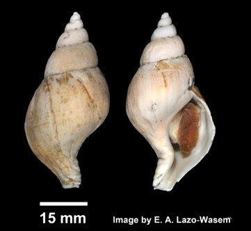
Hairy colus Colus pubescens, copyright E. A. Lazo-Wasem.

Colus has been used as the basis of a family group name at many levels of whelk classification, whether it be Colidae, Colinae or Colini. The gastropod classification laid out by Bouchet et al. (2017) recognised 'Colini' as a diverse tribe within the main whelk family Buccinidae, including a range of cold-water taxa. However, a more recent phylogenetic analysis of the buccinoids by Kantor et al. (2021) found Bouchet et al.'s concept of Colini to be polyphyletic, placing the type genus Colus outside what the called the 'core Buccinoidea'. As such, they raised Colidae to the status of a separate family and restricted it to just two genera, Colus and Turrisipho.

In this restricted form, the Colidae are thin-shelled, medium-sized to large whelks with the largest having shells up to twenty centimetres in length. The shells are fusiform to ovate in shape with a more or less elongate siphonal canal and covered by a brown periostracum. Axial sculpture is absent; spiral sculpture is expressed as more or less prominent cords. The aperture is closed with a operculum bearing a terminal nucleus. The animal has a more or less long proboscis. The radula bears three teeth per row; the middle tooth has a more or less square base and one to three cusps, with the middle cusp the largest, whereas the lateral teeth bear three hooked cusps with the outermost cusp significantly larger than the other two. None of these features, it should be noted, is entirely unique to the Colidae (Kantor et al. 2021).

Turrisipho dalli, from BoldSystems.


Members of the Colidae are found in the Arctic and northern Atlantic Oceans, from subtidal to bathyal depths. Because they are not targeted commercially, the life habits of colids have not been well studied. However, what we do know indicates that they are likely predators on other invertebrates (Kosyan 2007). The long proboscis of most species is probably used to pull infaunal animals such as amphipods and bivalves out of their burrows. Colids have well-developed salivary glands and it is possible that these may produce toxins as found in other neogastropods. They do not have anything like the elaborate venom delivery setups like those found in the conoids, but even a little dose of toxic saliva helps to subdue a struggling crustacean.

REFERENCES

Bouchet, P., J.-P. Rocroi, B. Hausdorf, A. Kaim, Y. Kano, A. Nützel, P. Parkhaev, M. Schrödl & E. E. Strong. 2017. Revised classification, nomenclator and typification of gastropod and monoplacophoran families. Malacologia 61 (1–2): 1–526.

Kantor, Y. I., A. E. Fedosov, A. R. Kosyan, N. Puillandre, P. A. Sorokin, Y. Kano, R. Clark & P. Bouchet. In press 2021. Molecular phylogeny and revised classification of the Buccinoidea (Neogastropoda). Zoological Journal of the Linnean Society.

Kosyan, A. R. 2007. Morphological features, ecology, and distribution of poorly studied molluscan genera of the Colinae subfamily (Gastropoda, Buccinidae) from the far eastern seas of Russia. Oceanology 47 (4): 531–536.

Crossing the Busycon

I must admit that when I think about the biodiversity hotspots of the world, the eastern seabord of the United States would not be among the first regions to come to mind. But for this post, I'm looking at a dramatic and eye-catching radiation of molluscs for which this is their centre of distribution. I speak of the giant whelks of the Busyconidae.

Left-handed whelk or lightning whelk Sinistrofulgur sinistrum, copyright Andrea Westmoreland.


Busyconid whelks first appeared in the waters of eastern North America during the early Oligocene, about 32 million years ago, in what was then the Mississippi Sea and is now the Mississippi River Basin. As the oceans receded from the Mississippi, they spread into the Gulf of Mexico and are now found between Massachusetts in the north and the Yucatan Peninsula in the south. Except for an introduced population of the channeled whelk Busycotypus canaliculatus that has become established in San Francisco Bay in California since the 1930s, the family has never been found elsewhere. These are remarkably large snails: smaller examples are still more than five centimetres in length, and the largest of all get close to a foot (Petuch et al. 2015). Mature shells have a large body whorl, generally higher than the visible spire, with a long siphonal canal. SCulpture of the shell, if present, is dominated by spiral elements, and the shoulder of the whorls may be marked by prominent carinae and/or spines. As is standard for neogastropods, the classification of this group has shifted around a bit over the years, whether treated as their own family or as a subfamily Busyconinae of the related families Buccinidae or Melongenidae. In a recent review of the busyconids, Petuch et al. (2015) recognised fifteen living species in six genera. The number of fossil species that has been described is significantly larger (over one hundred); not surprisingly, these large solid shells have an excellent fossil record. However, it is worth noting that some of the living species may be remarkably variable in shell morphology and I don't know whether fossil representatives have been subject to the same systematic scrutiny.

Knobbed whelk Busycon carica, copyright Matt Tillett.


All busyconids are predators on bivalves, particularly on burrowing clams. In general, the whelk envelops its victim in its muscular foot and then uses the edge of the shell lip to open the clam's shell, allowing the whelk to insert its radula and rasp out the clam's flesh. The preferred method of opening the shell depends on the species of whelk and may be classed as 'wedging' and 'chipping'. 'Wedging' is the most straightforward method and believed to be the more primitive; wedgers insert the shell lip into the gap between valves and directly force them apart and/or prevent the clam shell from closing. 'Chipping' is more involved and performed by members of the genera Busycon and Sinistrofulgur. In this method, the edge of the whelk shell is rhythmically pounded against the commissure between the clam shell valves, progressively wearing at the valve margins until enough of an opening has been made to insert the radula. The process may take multiple hours of patient hammering. Chipping requires more power and a heavier shell than wedging (chipping whelks may damage their own shell as well as the prey's) but also allows the whelk to attack thicker-shelled clams.

Though each species of busyconid will generally use one or the other method of opening prey, there are borderline examples. Larger individuals of Busycotypus canaliculatus, usually a wedger, may adopt a process like chipping though their attacks on the prey shell are usually less systematic than true chippers. And while I haven't found anywhere that says as much, I suspect that young chippers may spend the earlier parts of their life as wedgers untill they have developed the shell strength for chipping. Dietl (2004) suggested that chipping behaviour may have originated twice among busyconids, based on the fossil evidence of its traces left on clam shells. The modern chippers appear to derive from a single origin in the later Pliocene. However, evidence of an earlier and now seemingly extinct chipping lineage was also found in shells from the late Miocene. These earlier chippers seemingly did not belong to any of the modern chipping genera which are not known from the Miocene deposits in which chipped clams were found. Instead, Dietl proposed that the culprit was a large Busycotypus.

Channeled whelk Busycotypus canaliculatus laying a string of egg cases, copyright Eric Heupel.


Busyconid whelks have long been of significance to people living in areas where they are found. Not only are the shells eye-catching and ornamental objects in themselves, the animals are also harvested for food (though their meat is often sold under misleading names such as 'conch' or 'clam strips'). Archaeological examples have been found of busycon shells being used for tools; Petuch et al. (2015) illustrate an example of a left-handed whelk Sinistrofulgur sinistrum shell with holes drilled into it that would have allowed it to be attached to a stick and used as a shovel. These animals are truly an icon of North America's eastern seaboard.

REFERENCES

Dietl, G. P. 2004. Origins and circumstances of adaptive divergence in whelk feeding behavior. Palaeogeography, Palaeoclimatology, Palaeoecology 208: 279–291.

Petuch, E. J., R. F. Myers & D. P. Berschauer. 2015. The Living and Fossil Busycon Whelks: Iconic Mollusks of Eastern North America. San Diego Shell Club, Inc.

Murderous Cones

The cone shells of the family Conidae have long been the subject of extreme interest from collectors. Their architectural form, polished surface and intricate patterning make it hard to argue that they are things of beauty, indeed. Not surprisingly, this long-standing aesthetic interest has also made them the subject of much taxonomic interest—some for the better, some arguably for the worse. For today's post, I've selected a particular subgroup of the cone shells: the species of the subgenus Textilia.

Bubble cone Conus bullatus, copyright H. Zell.


To describe the generic taxonomy of cone shells as 'messy' is something of an understatement. Part of the problem is that cone shells are another one of those groups in which a high level of species diversity contrasts with a low level of morphological disparity. Though species are readily distinguishable on the basis of superficial features such as colour patterning, they generally hew pretty closely to a particular overall morphotype. This can make it difficult to associate particular species into evolutionary groups. For many authors, the problem has been solved (or at least satisfyingly swept under the rug) by treating all cone shells as belonging to a single genus Conus. But with over 800 known species of conid, many showing intriguing variations in biology and natural history, many have yearned for a more informative system. Those who would divide, however, have disagreed significantly on how many divisions there should be. At the most disassociative end on the scale, one recent system divided the cone shells between no less than 113 genera, separated into five families. A more conservative approach was taken by Puillandre et al. (2014) who recognised four genera of cone shells (in a single family) with the larger genera encompassing multiple subgenera. Textilia was treated by PUillandre et al. as a subgenus within Conus, which remains the largest genus in the family by a considerable margin.

Pallisade cone Conus cervus, copyright James St. John.


Ten species of cone shell were included in Textilia by Puillandre et al. (2014). The species are found in the Indo-west Pacific, between south-east Africa and Hawaii. They are medium- to large-sized cone shells with the largest species, the pallisade cone Conus cervus reaching close to 12 cm in length. The smallest, the Timor cone C. timorensis, is at least 13 mm long. Textilia species have smooth, inflated shells and flared lips on the aperture (Old 1973). Only one species of Textilia, the bubble cone C. bullatus, can be considered well known. Not only is it found over almost the subgenus' entire range (other species are more localised), it is the only species found in shallower waters, being most common from slightly subtidally to 50 m (Hu et al. 2011). All other Textilia species are restricted to deeper waters. Just to confuse matters slightly, the textile cone C. textile is not a member of subgenus Textilia but another subgenus Cylinder.

Video of cone shells capturing fish, from here. The first individual is a striated cone Conus striatus (subgenus Pionoconus), the second is a bubble cone Conus bullatus.


Textilia forms part of a clade of cone shells with a diet composed primarily of fish. A slow-moving gastropod is obviously ill-suited to taking down a fast-moving fish by brute strength alone so cone shells make use of a quite different tactic: lethal poisons. The venom of a cone shell can be exceedingly powerful, enough so that multiple species have been known to cause severe injury or fatality to humans unwise enough to handle them live (cone shells may use their venom for defense as well as for attack). The teeth of the cone shell's radula have been modified into elongate, hollow needles. While most of the teeth are retained in a sac at the rear of the buccal cavity, only a single tooth is in use at any one time. When a suitable prey animal comes within reach, the snail's proboscis is stealthily extended towards it. The active tooth is then fired along the proboscis into the target, injecting a complete payload of toxins. Among Textilia, Conus bullatus is the only species whose toxic characteristics and capabilities have been studied as yet, but it is probably representative of the subgenus as a whole. As with other fish-hunting cone shells, the injected venom carries a mixture of toxic peptides that can be divided between two functional groups (Hu et al. 2011). These have been referred to as the "lightning-strike cabal" and the "motor cabal". The peptides of the lightning-strike cabal are the first to take effect, causing a rapid (almost instantaneous) tetanic immobilisation of the prey. After this, the motor cabal of peptides act to block neuromuscular transmission, preventing the prey from recovering from its freeze. And all this in a matter of milliseconds: as of 2011, at least, C. bullatus had the fastest immobilisation capacities of any fish-hunting cone shell. As beautiful as they are, cone shells are a force to be feared.

REFERENCES

Hu, H., P. K. Bandyopadhyay, B. M. Olivera & M. Yandell. 2011. Characterization of the Conus bullatus genome and its venom-duct transcriptome. BMC Genomics 12: 60.

Old, W. E., Jr. 1973. A new species of Conus from Indonesian waters. Veliger 16 (1): 58–60.

Puillandre, N., T. F. Duda, C. Meyer, B. M. Olivera & P. Bouchet. 2014. One, four or 100 genera? A new classification of the cone snails. Journal of Molluscan Studies 81: 1–23.

Moles, Tortoises, Calves and Cowries

The cowries of the family Cypraeidae are one of the most readily recognisable groups of tropical and subtropical shells. Their distinctive shape (with no spire and a long narrow aperture running the length of the shell) and highly polished appearance are guaranteed to catch the eye (to the extent that one species, the money cowry Monetaria moneta, famously has a history of being used as a form of currency in many regions around the Indian Ocean). Though there are a large number of cowry species found around the world, they tend to be similar enough to each other that, until relatively recently, many authors would place all within a single genus Cypraea. This approach has fallen out of fashion in more recent years and, indeed, the current favoured approach divides the family between several subfamilies. One such subgroup is the subfamily Luriinae.

Live mole cowry Talparia talpa, copyright Juuyoh Tanaka.


In a phylogenetic analysis of the cowries, Meyer (2003) recognised the Luriinae as including two tribes, the Luriini and Austrocypraeini. This concept of Luriinae was essentially based on molecular phylogenetic analysis though it was also corroborated by radular morphology (with a reduced shaft on all teeth). The underside of the shell in luriines is mostly smooth with the 'teeth' being restricted to alongside the aperture. As in other cowries, the mantle is widely extended and mostly covers the shell in life (this is how cowry shells stay so shiny). In most luriines, the mantle is covered by warty papillae. In species of the genus Luria these warts are obsolete (Schilder 1939) but they are particularly prominent in the Indo-west Pacific mole cowry Talparia talpa. Members of the Luriinae vary greatly in size: the Pacific Annepona mariae is only a centimetre or two in length but the tortoise cowry Chelycypraea testudinaria of the Indian and western Pacific Oceans grows to ten centimetres or more. Species of Luriini have shells that are banded in coloration, with three or four broad dark bands divided by narrower light bands. The Austrocypraeini are most commonly marked with brown speckles or blotches on a pale background; these blotches may be irregular as in Chelycypraea testudinaria or more regularly rounded as in Annepona mariae. The calf cowry Lyncina vitellus of the Indo-Pacific is marked with white spots on a brown background, and some species or forms of Austrocypraeini may have coloration patterns more like the banded arrangement of Luriini.

Lynx cowry Lyncina lynx, copyright Patrick Randall.


My impression is that species of Luriinae tend to be mostly nocturnal, sheltering in crevices in coral reefs during the day before emerging to feed at dusk (the name of the aforementioned mole cowry is, I suspect, more likely to refer to its appearance in some way than to any actual burrowing habit). Though I haven't (though a cursory search, at least) found any reference to species of Luriinae in particular being endangered, a number of cowries in general have been threatened by overcollecting for their shells. Certainly, luriines would be subject to the broad range of threats that currently hang over coral reefs and their inhabitants anywhere in the world.

REFERENCES

Meyer, C. P. 2003. Molecular systematics of cowries (Gastropoda: Cypraeidae) and diversification patterns in the tropics. Biological Journal of the Linnean Society 79: 401-459.

Schilder, F. A. 1939. Die Genera der Cypraeacea. Archiv für Molluskenkunde 71 (5–6): 165–201.

Slippers on the Coast

The 'limpet' form is something that has evolved numerous times among gastropods, as various lineages of marine snail converted to a more or less unwhorled shell and low profile. In many cases, the evolution of the limpet form is also associated with high energy environments, the ability to nestle against rocks helping the gastropod maintain its grip against the surge of the waves. In the modern world, the most diverse and familiar lineage of limpets is that including the common limpets of the genus Patella and their relatives, but there also many independent lineages to be found. One of these is the slipper limpets of the genus Crepidula.

Various views of shell of Crepidula onyx, copyright H. Zell.


Slipper limpets get their vernacular name from the shape of their shell, whose more or less oval shape together with a jutting internal horizontal shelf (the septum) at one end gives the overall impression of a carpet slipper. About forty species (including fossils) of Crepidula are currently recognised worldwide. Species recognition has historically been difficult owing to their simple form and tendency to vary according to the environment in which they mature, but Hoagland (1977) identified a number of key distinguishing features such as disposition and shape of the muscle scars, features of the septum, and conformation of the apical beak of the shell. In contrast to the grazing common limpets, slipper limpets are filter feeders using their gill to capture micro-algae from the water column. They are protandric hermaphrodites, beginning their life as males but maturing into females as they grow. Eggs are brooded under the shell when first produced; in some species, the eggs are subsequently released to hatch into planktonic larvae whereas other species produce fewer eggs but retain them until the young have developed to the crawling stage. For instance, two species found on the east coast of North America that are very similar in adult appearance and have been confused historically differ in that Crepidula ustulatulina, found around Florida and the Gulf of Mexico, produces free-living larvae whereas the more northerly C. convexa does not.

Mating stack of Crepidula fornicata, copyright Dendroica cerulea.


The most renowned species of slipper limpet is the northern Atlantic Crepidula fornicata. This species was originally native to the eastern coast of North America but was accidentally imported to Europe in the late 1800s in association with oysters being transported as stock for farming (Blanchard 1997). In the subsequent years, C. fornicata has become increasingly widespread on the shores of Europe, and is often a significant fouling pest for oyster farms. It has also been introduced to even further flung locations such as Japan and Washington State. Crepidula fornicata is famed for its habit of forming high mating stacks with several smaller males living permanently on the dorsal surface of larger females. If the female of a stack dies, the largest male may develop into a female. Not all Crepidula species form such stacks: in some, just two or three individuals may form a temporary cluster when mating.

Historically, Crepidula has been distinguished from other genera in the limpet family Calyptraeidae by their posterior shell apex and flat septum (other calyptraeid genera may have a cone-shaped shell and/or cup-shaped septum). However, a molecular analysis of the family by Collin (2003) found that species of Crepidula sensu Hoagland (1977) did not form a single clade within Calyptraeidae, and the genus' prior members are now divided between at least four genera. While these genera may be distinguishable using features of the soft anatomy, they are almost indistinguishable from the shells alone.

REFERENCES

Blanchard, M. 1997. Spread of the slipper limpet Crepidula fornicata (L. 1758) in Europe. Current state and consequences. Scientia Marina 61 (Suppl. 2): 109–118.

Collin, R. 2003. Phylogenetic relationship among calyptraeid gastropods and their implications for the biogeography of marine speciation. Systematic Biology 52 (5): 618–640.

Hoagland, K. E. 1977. Systematic review of fossil and recent Crepidula and discussion of evolution of the Calyptraeidae. Malacologia 16 (2): 353–420.

Hypsogastropods: Gastropods on High

Historically, the classification of molluscs has been a challenging prospect. Early researchers focused almost entirely on the shell which provided a somewhat limited range of characters with a definite possibility for convergence. Over time, more attention came to be paid to features of the soft anatomy but that required access to freshly collected material that might be difficult or impossible to obtain. As such, it has only been in the last few decades that a well-structured classification for many molluscan groups has begun to develop, and even now many significant uncertainties remain.

Common periwinkles Littorina littorea, a pretty typical hypsogastropod, copyright Fritz Geller-Grimm.


Until maybe the late 1990s, gastropods were primarily classified using a heavily grade-based system that was established in the 1930s. Gastropods were divided between three subclasses: the torted, gill-breathing prosobranchs, the untorted opisthobranchs, and the lung-breathing pulmonates. Prosobranchs were in turn divided into three main groups whose names directly reflected the 'level' of evolution at which they were supposed to sit: the archaeogastropods, the mesogastropods and the neogastropods. Many of these subdivisions were implicitly assumed to be ancestral to others. As the philosophical underpinnings of biological classification came to favour recognition of monophyletic taxa, it was obvious that such a system had to change. The prosobranchs and archaeogastropods both faded away as formal taxa. A major clade uniting the neogastropods and most of the mesogastropods came to be recognised as the caenogastropods. And while many questions still remain about relationships within the caenogastropods, most recent analyses have agreed in supporting a clade that was dubbed the Hypsogastropoda by Ponder & Lindberg (1997).

False cowrie Dentiovula dosruosa, copyright Nick Hobgood.


The prefix 'hypso-' means 'high' and was chosen because this clade corresponded to a group that had previously been known as the 'higher' caenogastropods (including the neogastropods and a fair chunk of the 'mesogastropods'). Hypsogastropods include many of the best known marine gastropods, such as whelks, periwinkles, moon snails, cones, cowries, conches and doubtless a ton of other things beginning with C (they also include freshwater and terrestrial forms but these are mostly minute and lack the public image of their marine relatives). They are ecologically diverse, including grazers, detritivores, filter feeders, predators and even parasites. The violet snails of the genus Janthina are planktonic, using a raft of bubbles to float on the water's surface so they can feed on Portuguese men-of-war. The similarly pelagic heteropods of the superfamily Pterotracheoidea have the foot extended and flattened to form a fin for active swimming.

Paraspermatozoon of violet snail Janthina, from Buckland-Nicks (1998). The arrow indicates the much smaller euspermatozoa attached to the tail.


Among the characters originally cited by Ponder & Lindberg (1997) as uniting the hypsogastropods were features of the spermatozoa. Most hypsogastropods have vermiform paraspermatozoa, sterile sperm cells that are released by the male together with the functioning euspermatozoa. The function of the paraspermatozoa seems to warrant further study. In some cases they may actively assist in the transport of the euspermatozoa; for instance, in violet snails a large number of euspermatozoa will be attached to a single super-sized paraspermatozoon able to swim harder and faster than any of the smaller cells could do on their own. In others, however, the two sperm cell types are not directly associated. It is possible that the paraspermatozoa act as a nuptial gift, providing nutrients to the female as a reward for mating, or that they somehow function to suppress sperm cells from any other males the female might made with (Buckland-Nicks 1998). Other synapomorphies of the clade include an external penis located behind the right cephalic tentacle, and statocysts (balance organs) each containing a single large statolith (Simone 2011).

Relationships within the Hypsogastropoda remain more poorly supported. Most researchers have agreed that the traditionally recognised neogastropods represent a clade united by numerous features, many of them related to the digestive system. The 'mesogastropods' included in the Hypsogastropoda mostly possess a taenioglossan radula with seven teeth in each row. In neogastropods, the number of teeth becomes more varied and the teeth themselves become modified so that the lateral teeth are strongly distinct in form from the central tooth. Some of these neogastropod modifications have been discussed in earlier posts on this site. A number of recent analyses have further associated the neogastropods with 'mesogastropod' taxa such as cowries and tun shells that they resemble in possessing an inhalent siphon forming a groove at the front of the shell (Simone 2011). A number of the remaining 'mesogastropods', such as the periwinkles of the Littorinidae and the Rissoidae, have been united by molecular analyses into a group that has been labelled the 'asiphonate clade' or the 'GC group' (the latter name chosen by Colgan et al., 2007, in reference to a particular genetic sequence motif). This clade is less universally recovered, however, and the scope for further investigation certainly remains.

REFERENCES

Buckland-Nicks, J. 1998. Prosobranch parasperm: sterile germ cells that promote paternity? Micron 29 (4): 267–280.

Colgan, D. J., W. F. Ponder, E. Beacham & J. Macaranas. 2007. Molecular phylogenetics of Caenogastropoda (Gastropoda: Mollusca). Molecular Phylogenetics and Evolution 42: 717–737.

Ponder, W. F., & D. R. Lindberg. 1997. Towards a phylogeny of gastropod molluscs: an analysis using morphological characters. Zoological Journal of the Linnean Society 119: 83–265.

Simone, L. R. L. 2011. Phylogeny of the Caenogastropoda (Mollusca), based on comparative morphology. Arquivos de Zoologia 42 (4): 161–323.

Alvania

It's a general rule with organisms that species diversity increases as size decreases (at least down to about the millimetre range, below which things get a bit more complicated). That's certainly the case with molluscs, whose range clearly favours the tiny.

Alvania cimex, copyright Alboran Shells.


Alvania is a cosmopolitan genus of marine gastropods, found in most parts of the world except the Antarctic and sub-Antarctic (Ponder 1984). The average Alvania species is less than five millimetres in total length, and other members of the family they belong to, the Rissoidae, are similarly wee. The shell of Alvania species varies from elongate-conical to more squatly conical in shape, and generally has a sculpture of both axial and spiral ridges. In some species the axial and spiral ribs are both similarly prominent; in others, the spiral ridges are more strongly developed.

Rissoids may be found crawling on seaweed or sheltered amongst stones or other rubble. Alvania species seem to be more likely to be found in the latter habitat than the former. Alvania have a smaller mucous gland on the rear of the foot than species of Rissoa, a related genus that is more likely to be found on the weeds. The mucus produced by this gland assists rissoids in clinging to their substrate or the surface film, and its reduction in Alvania is presumably connected to their preference for the low life. Rissoids are grazers on microalgae or deposit feeders; those species found on seaweeds will feed on diatoms and the like growing over the seaweed rather than on the seaweed itself. Among European species, A. punctura is known to selectively pick out diatoms and dinoflagellates from among detritus when feeding whereas A. jeffreysi may be less discriminating in what it swallows.

Alvania subcalathus, copyright H. Zell.


The greater number of Alvania species are planktotrophic as larvae, and as described in some of my previous posts on turrids, their shells have protoconches to match. Nevertheless, the genus also includes some direct-developing species with fewer protoconch spirals. The Mediterranean species A. cimex and A. mammillata are almost indistinguishable when mature except by features of the shell apex, which is broader with fewer spirals to the protoconch in the latter (Verduin 1986). If A. mammillata is a direct developer while A. cimex has a planktotrophic larva, it would tally up with the situation elsewhere seen among turrids.

REFERENCES

Ponder, W. F. 1984. A review of the genera of the Rissoidae (Mollusca: Mesogastropoda: Rissoacea). Records of the Australian Museum Supplement 4: 1–221.

Verduin, A. 1986. Alvania cimex (L.) s.l. (Gastropoda, Prosobranchia), an aggregate species. Basteria 50: 25–32.

Narona decaptyx: A Fossil Vampire

13.5 mm long specimen of Narona decaptyx, from Landau et al. (2012).


Narona decaptyx was described by Brown & Pilsbry (1911) from a single small, fusiform fossil shell, 11 mm in length, from the Gatun Formation of Panama, north of Panama City. They regarded the formation as probably Oligocene in age but Landau et al. (2012) later referred N. decaptyx to the upper Miocene. Until it's redescription by the latter, this species was only known from Brown & Pilsbry's original holotype; Landau et al. described further material from the Bocas del Toro region to the west of the original locality. Both the sites from which N. decaptyx are known are on the Caribbean coast of Panama.

Narona decaptyx is a member of the Cancellariidae, the nutmeg snails. Cancellariids are one of the smaller families of the great neogastropod radiation, the group that also includes such forms as whelks and cone shells. They generally have a more or less developed sculpture of criss-crossing spiral and axial ribs; the latticed pattern this produces is formally referred to as 'cancellate' and provides the source of the family's name. The most distinctive feature of Cancellariidae is their radula, a slender ribbon of long, flexible teeth arranged in a single row. How this radula functioned was long a mystery. Dissections of the gut of cancellariids failed to find any trace of solid food, and it was suggested they may be adapted to some form of suctorial feeding. Some authors suggested that cancellariids might feed by slurping up micro-organisms. Then, in the 1980s, one species of cancellariid Cancellaria cooperi was observed feeding on sleeping electric rays. The snail would cut incisions in the ray's skin, presumably with its radula, before inserting its proboscis to slurp up the fish's blood (O'Sullivan et al. 1987). Other cancellariids have been observed feeding on fluids from other invertebrates such as benthic molluscs or their egg masses.

Cancellaria cooperi feeding on an electric ray, copyright Clinton Bauder.


The genus Narona to which N. decaptyx belongs is now restricted to the north Pacific. In this respect, it is not unique. Cancellariids are poorly represented in the modern Caribbean fauna with only six species known from the sea's shallow waters but they were much more diverse there in N. decaptyx's time. However, following the rise of the isthmus of Panama, many cancellariid taxa once found widely in the tropical Americas became extinct for whatever reason on the eastern side of the divide. This happened regularly enough that the term 'paciphile' has been coined for referring to such taxa. A number of distinct waves of paciphile extinctions have been identified in the Caribbean cancellariid fossil record and they have been used to identify distinct chronological zones. Narona decaptyx became extinct as part of the GNPMU (Gatunian Neogene Paciphilic Molluscan Unit) 1 period. Other Narona species persisted in the Caribbean and Gulf of Mexico for longer, surviving into the Pliocene, but eventually they too succumbed to whatever dampened this family's prospects in the region.

REFERENCES

Brown, A. P., & H. A. Pilsbry. 1911. Fauna of the Gatun Formation, Isthmus of Panama. Proceedings of the Academy of Natural Sciences of Philadelphia 63 (2): 336–373.

Landau, B., R. E. Petit & C. M. da Silva. 2012. The family Cancellariidae (Mollusca: Gastropoda) in the Neogene of the Bocas del Toro region, Panama, with the description of seven new species. Journal of Paleontology 86 (2): 311–339.

O'Sullivan, J. B., R. R. McConnaughey & M. E. Huber. 1987. A blood-sucking snail: the Cooper's nutmeg, Cancellaria cooperi Gabb, parasitizes the California electric ray, Torpedo californica Ayres. Biological Bulletin 172 (3): 362–366.

Turridae

Shell of Turris crispa crispa, copyright H. Zell.


At this point, I've made numerous references on this site to the gastropod family Turridae, discussing its members and non-members and alluding to its sordid history. So maybe I should set out the basics of the story properly.

The Conoidea are a diverse group of marine predatory gastropods with over 4000 known living species. They are best known for the production by many species of venom used to paralyse their prey, in some species being potent enought to threaten humans. In the majority of conoideans, this venom is delivered via a tooth that becomes detached from the radula and is held at the end of the retractable proboscis. Until relatively recently, Conoidea were commonly divided between three families. Two of these families, the Conidae (cone shells) and Terebridae (awl shells) were well defined and constrained. The third family was the Turridae, including by far the greater number of species but not really defined within Conoidea beyond 'the rest'. Many of 'the rest' were small, many were restricted to deep water, many were poorly known. Different systems were proposed over the years in an attempt to break the turrid mass into more manageable units but each system differed significantly from the next and no one system became universally accepted. Some authors would focus on the protoconch as their guide to classification, others would focus on the radula, others might call out features of the operculum. One author commented in 1922 that turrids were "considered by those who meddle with them to be more perplexing than any other molluscan family", and this complaint was still being upheld by Kilburn (1983) over sixty years later.

Though it had long been accepted that the 'turrids' probably did not represent an evolutionarily coherent group, it wasn't really until the advent of molecular phylogenies that things started falling into place. Puillandre et al. (2011) identified two main lineages within the Conoidea, leading to the dissolution of the original Turridae into no less than 13 families in order to maintain the already-established Conidae and Terebridae. Turridae in the strict sense was restricted to a much smaller clade of a bit over a dozen genera, sister to the Terebridae (Bouchet et al. 2011).

In contrast to the bewilderment of the original turrid array, Turridae sensu Bouchet et al. is a morphologically quite coherent group. They are more or less fusiform (spindle-shaped) shells, often with a narrow, high spire and relatively weak sculpture. Indeed, but for the fact that most tend to have a long siphonal canal at the base of the shell, they often bear a distinct resemblance to their sister group, the terebrids. The majority of turrids have a multispiral protoconch, indicating an extended, planktonic-feeding larval stage in development, but there are some species with a paucispiral protoconch indicative of direct development.

Radula of Xenuroturris legitima, from Kantor & Puillandre (2012); ct = central tooth.


The radula of turrids usually comprises three apparent teeth in each row. The central tooth is actually formed from three teeth (the original pointed central tooth and two plate-like lateral teeth) fused together; in some species the division between these teeth remains visible whereas in others the central tooth disappears entirely. The main business part of the radula is the single pair of marginal teeth which, as in other conoideans, are enlarged and modified for venom delivery. They have a distinctive 'duplex' form; in older publications, this was referred to as a 'wishbone' form because the tooth appears under light microscopy to be divided between two branches. After the advent of electron microscopy, it was discovered that these two 'branches' in fact represent the thickened margins of an undivided tooth. The larger of the two margins is mostly attached to the radular membrane with only the tip of the tooth being free; the smaller margin is held free of the radula. The thinner part of the tooth between the two margins forms a gutter along which venom can flow. However, the radula is placed in such a position that it cannot be protruded through the mouth in the manner of grazing gastropods. As with other conoideans, prey (in this case probably worms) is despatched through the use of a detached marginal tooth transferred to the end of the proboscis. However, whereas other conoideans such as cone shells may have the tooth functioning like a hypodermic syringe for delivering prey, turrids use their tooth to slash at the prey like a switchblade, with venom passively entering through the resulting cuts. The proboscis is then used to draw the prey back into the mouth, where the radula is used to grasp and swallow it, sucking the unlucky worm down the gullet like spaghetti.

REFERENCES

Bouchet, P., Y. I. Kantor, A. Sysoev & N. Puillandre. 2011. A new operational classification of the Conoidea (Gastropoda). Journal of Molluscan Studies 77: 273–308.

Kantor, Y. I., & N. Puillandre. 2012. Evolution of the radular apparatus in Conoidea (Gastropoda: Neogastropoda) as inferred from a molecular phylogeny. Malacologia 55 (1): 55–90.

Kilburn, R. N. 1983. Turridae (Mollusca: Gastropoda) of southern Africa and Mozambique. Part 1. Subfamily Turrinae. Annals of the Natal Museum 25 (2): 549–585.

Puillandre, N., Y. I. Kantor, A. Sysoev, A. Couloux, C. Meyer, T. Rawlings, J. A. Todd & P. Bouchet. 2011. The dragon tamed? A molecular phylogeny of the Conoidea (Gastropoda). Journal of Molluscan Studies 77: 259–272.

Conus jaspideus or Conasprella jaspidea, Take Your Pick

Live Conasprella jaspidea, copyright Anne DuPont.


Cone shells are one of the classic varieties of tropical sea shells, perhaps only rivalled in their familiarity with the general public by cowries and conches. Over 800 species of the family Conidae have been described from around the world. The specimen above represents one of these species, going by the name of Conasprella jaspidea or Conus jaspideus. The alternatives reflect the conflict between those who would treat all cone shells as belonging to a single genus Conus, or those who would divide them between multiple genera (Conasprella jaspidea is the name used for this species by Puillandre et al., 2014). One 2009 classification went so far as to divide the cone shells between 89 genera in five separate families, which does seem perhaps a little excessive. Among other features, Conasprella species differ from Conus sensu stricto in having a higher spire to the shell.

The type specimen of Conasprella jaspidea, copyright MHNG.


Conasprella jaspidea is found in coastal sections of the western Atlantic between Florida and the area of Rio de Janeiro. It is a medium-sized shell, reaching about three centimetres in length. Whorls of the spire are marked by distinct shoulders, and the body whorl is ornamented by spiral cords. The colour of the shell is white, orange or brown with darker brownish or violet spots. Shells of C. jaspidea may vary in texture from granular to smooth. These variants were initially recognised as distinct species or subspecies Conus jaspideus and C. verrucosus but, not only can both forms be found intermixed within a single population, the difference between them may be simply a question of the degree of wear a shell has been exposed to (Santos Gomes 2011).

Like other cone shells, Conasprella jaspidea is venomous with the radula bearing a single functional tooth modified into a short of hypodermic needle for injecting venom. Species of Conasprella are vermivorous (that is, they feed on worms). Feeding by a live individual of C. jaspidea was observed in an aquarium by Santos Gomes (2011). Photographs therein show the individual ingesting a polychaete worm that was perhaps not too much shorter in length than the cone shell itself; the process of feeding (from the initial strike with the radula to completion of ingestion) took about eighteen minutes from start to finish.

REFERENCES

Puillandre, N., T. F. Duda, C. Meyer, B.M. Olivera & P. Bouchet. 2014. One, four or 100 genera? A new classification of the cone snails. Journal of Molluscan Studies 81: 1–23.

Santos Gomes, R. dos. 2011. Conus jaspideus (Mollusca: Neogastropoda: Conoidea) on the Brazilian coast. Journal of the Marine Biological Association of the United Kingdom 91 (2): 531–538.

Cochlespira

Shell of Cochlespira radiata, photographed by Jan Delsing.


This beauty is a member of the genus Cochlespira, another one of the conoid shells previously classed as 'turrids' (it now belongs in the family Cochlespiridae since the disassembly of Turridae in the broad sense). Cochlespira species can be relatively large as conoids go, reaching lengths of up to five centimetres. They are found in deep waters in various parts of the world, with a fossil record going back to the Eocene (Powell 1966; Powell treated the western Atlantic species as a separate genus Ancistrosyrinx, but this and the Indo-Pacific Cochlespira have since been synonymised). One of the genus' more distinctive features is a little hard to miss: that eye-catching keel around the outside of the whorls, ornamented with serrations or spines.

As with other deep-water conoids, our knowledge of how Cochlespira species live their lives seems to be pretty limited. The radula has a broad-based central tooth with a single median cusp, and a pair of marginal teeth that are elongate but not as slender as those of many other conoids (Powell 1966). The rhynchodeal walls in the foregut are muscular and the proboscis is long. The venom glands are well-developed but join the oesophagus at about its midlength rather than in the buccal mass (Simone 1999). The arrangement looks to my admittedly inexpert eyes like it might be suited for sucking up invertebrate prey, perhaps something that might be expected to be relatively slow-moving or soft-bodied.

REFERENCES

Powell, A. W. B. 1966. The molluscan families Speightiidae and Turridae. An evaluation of the valid taxa, both recent and fossil, with lists of characteristic species. Bulletin of the Auckland Institute and Museum 5: 1-184.

Simone, L. R. L. 1999. The anatomy of Cochlespira Conrad (Gastropoda, Conoidea, Turridae) with a description of a new species from the southeastern coast of Brazil. Revista Brasileira de Zoologia 16 (1): 103–115.

Zemacies: A Toothless Wonder

The Recent species Zemacies queenslandica, photographed by Jan Delsing.


What makes an organism a 'living fossil'? The phrase is one that has been thrown about a bit over the years but whose actual definition can be ambiguous. Many people use it to refer to a species that is supposedly little changed from its distant ancestors. An alternative interpretation, however, and I suspect the phrase's original inspiration, would be something that was first discovered as a fossil, only to be found alive at a later date. The coelacanth, of course, would be the textbook example of such a case (without necessarily being a good example). But you might be surprised at some of the animals that could be called a 'living fossil' under this definition. The white-tailed deer would be one, as would the bush dog of South America. And so would the subject of today's post.

Zemacies is a genus of conoid gastropods known from the south-west Pacific. It is relatively large as conoids go, with some growing over three inches in length. Shells are a slender, fusiform shape, often with prominent nodules on the whorls. The first known species, Z. elatior, was described from the Miocene of New Zealand. Over time, additional species were described from New Zealand and Australia, extending the age range of the genus from the Palaeocene to the Pliocene (Powell 1969). It wasn't until 2001, however, that living species of Zemacies were recognised in the deep sea around New Caledonia and Queensland.

Figure from Fedosov & Kantor (2008) showing appearance of pyriform organ in foregut of Zemacies excelsa (left), with cross-section of organ to show internal structure (right). Abbreviations: bl, bulb-like structure; gt, glandular tissue; ms, muscles; sgp, semicircular glandular pad; tf, tall folds underlain by glandular tissue; tn, tentacles.


The discovery of living specimens (or, at least, living until the time of their collection) led to the revelation that Zemacies was a very intriguing genus, in a way that would have probably never been guessed from fossil material alone. As has been described previously, one of the great innovations of conoids was the modification of the radula into a system for the injection of paralysing toxins. When researchers investigated the soft anatomy of Zemacies excelsa, however, they discovered that it turned away from this trend. Zemacies has lost both the radula and its associated poison glands, as well as the associated proboscis. In their place, one side of the foregut has grown a pear-shaped outgrowth, referred to as the pyriform organ, that is covered with glandular tentacles (Fedosov & Kantor 2008). This structure is so unusual that its initial discovery lead to the proposal of a new subfamily to separate Zemacies from all other conoids (it has since been placed by Bouchet et al., 2011, as a distinctive member of the family Borsoniidae).

Unfortunately, the bathyal habitat of living Zemacies means that we as yet have no idea of its preferred prey and consequently little idea of how the pyriform organ is used when feeding. The internal cavity of the pyriform organ contains an array of longitudinal muscles, suggesting that it can be extended out the front of the animal in place of the usual proboscis. The tentacles may function to grasp or adhere to the prey, and/or the glandular tissue underlying them may secrete toxins or digestive enzymes, while the action of the pyriform organ against the foregut wall during withdrawal of the prey may serve to crush it (I wonder what the efficacy of this arrangement may be against something with a strong but not calcified cuticle, such as a deep-water crustacean). A similar foregut structure (also associated with loss of the radula and proboscis) has been identified in another conoid genus, Horaiclavus, though phylogenetic analysis of the Conoidea indicates that the two genera almost certainly evolved these structures independently. Horaiclavus also differs from Zemacies in that the muscular foregut outgrowth lacks any associated glandular tissue and is presumably entirely mechanical in its action (Fedosov & Kantor 2008). Perhaps one day someone will finally observe one of these deep-sea genera in their native habitat and provide us with a solution to the mystery of their life styles.

REFERENCES

Bouchet, P., Y. I. Kantor, A. Sysoev & N. Puillandre. 2011. A new operational classification of the Conoidea (Gastropoda). Journal of Molluscan Studies 77: 273–308.

Fedosov, A., &. Y. Kantor. 2008. Toxoglossan gastropods of the subfamily Crassispirinae (Turridae) lacking a radula, and discussion of the status of the subfamily Zemaciinae. Journal of Molluscan Studies 74: 27–35.

Powell, A. W. B. 1969. The family Turridae in the Indo-Pacific. Part 2. The subfamily Turriculinae. Indo-Pacific Mollusca 2 (10): 215–415.

Deep Pleurotomella

The type species of Pleurotomella, P. packardi, copyright Forum Natura Mediterraneo.


'Turrid' time again! Though the disassembly of the enormous mass that was the old gastropod family Turridae (now several families of the superfamily Conoidea) has left the subject of today's post, the genus Pleurotomella, as a member of the Raphitomidae rather than the Turridae. Pleurotomella is a widespread genus, with species found in deeper parts of ocean basins around the world. As with many deep-water animals, we know relatively little about their lifestyles, though they are undoubtedly predators like other conoids. Like other conoids, Pleurotomella species have a radula with the teeth modified into hypodermic syringes for the injection of toxins. At least some species (including the type) are blind (Bouchet & Warén 1980) and I can imagine that they attack relatively sedentary prey such as worms.

Taxonomically speaking, Pleurotomella has one of those histories that can make a grown taxonomist just want to sit down and cry. I've already mentioned this horrible genus in my earlier post on Asperdaphne as a player in one of those scenarios where a misunderstood type species leads a genus to jettison almost all of the species previously associated with it and pick up a whole bunch of new ones that it never held before. An inordinate number of deep-water 'turrid' species seem to have been dumped into Pleurotomella at some time or other, many of which are probably only remotely related to the true Pleurotomella. However, since Bouchet & Warén (1980) redescribed the type species Pleurotomella packardi as part of a revision of north-east Atlantic 'turrids', we have much better grounds for the genus' recognition (Beu 2011). Species of Pleurotomella have strongly inflated whorls that are evenly rounded except for a concave 'ramp' below the suture between whorls. The shell contracts rapidly to a narrow base, and has prominent, sharp and often curved axial ridges.

Multispiral (left) and paucispiral (right) protoconches of Mangelia species, from Bouchet (1990). Scale bars = 200 µm.


Again as was the case in the Asperdaphne post, a notable factor in the taxonomic complications of Pleurotomella has been matters relating to the protoconch, the larval shell that remains perched throughout development at the tip of the post-larval shell, the teleoconch. Because the features of the protoconch such as ornamentation may often differ from those of the teleoconch, it can often be of significance in gastropod taxonomy. A lead proponent of the importance of the protoconch in 'turrid' taxonomy was the New Zealand malacologist A. W. B. Powell who produced an influentiall classification of turrids between the 1940s and 1960s. Nevertheless, Powell did note an interesting phenomenon: the common existence of 'genus pairs' that were all but indistinguishable in teleoconch morphology but very distinct in their protoconches. Because Powell regarded the teleoconch as phylogenetically less significant than the protoconch (in accord with Ernst Haeckel's old dictum that ontogeny should recapitulate phylogeny), he concluded that these 'genus pairs' must represent separate lineages converging on a single adult morphology.

More recent authors agree that, in this, Powell was wrong (Bouchet 1990). As noteworthy a source of taxonomic characters it may be, protoconch development is subject to selective and evolutionary pressures just as much as teleoconch development. The most regular difference between Powell's 'genus pairs' is that one would have a conical protoconch with a number of whorls (say three or four, referred to as multispiral) whereas the other would have a stubby round protoconch with at most about one-and-a-half whorls (paucispiral). This difference in protoconch morphology reflects a difference in how the larval shell is fed. In the original development path for gastropods, eggs hatch out to planktic larvae that feed themselves on other plankton (planktotrophy) before eventually settling and developing to maturity. However, many conoids (and other gastropods) have evolved eggs that have a large yolk; the developing embryos obtain their energy from the reserves in the yolk (lecithotrophy) and bypass the planktic stage, hatching directly as benthic crawlers. Because planktotrophs need their larval shell for longer than lecithotrophs, it becomes more developed; planktotrophs are multispiral, lecithotrophs are paucispiral. Powell's 'genus pairs' did not represent separate lineages evolving similar adult lifestyles, but members of the same lineage tackling early development different ways. As such, and because of the possibility that the change between planktotrophic and lecithotrophic development may have occured multiple times within a single group, most recent authors would not automatically recognise multispiral and paucispiral species as separate genera. Pleurotomella species mostly have multispiral protoconches, but some (including P. packardi and a number of Pacific species) have paucispiral ones.

Which is not to say that protoconch morphology has become irrelevant. Bouchet & Warén (1980) did maintain the genus Neopleurotomoides as separate from Pleurotomella on the basis of protoconch morphology, despite these two genera having very similar teleoconches. In this case, the difference is not just the number of spirals in the protoconch, but its ornamentation. Pleurotomella species with a multispiral protoconch have a cancellate (cross-hatch) pattern of ridges covering it, but Neopleurotomoides has a sparser ornament of one or two spiral keels crossed by axial ribs. The distinction between the two genera remains problemematic: species with a paucispiral protoconch (which is usually more or less unornamented) cannot be readily assigned to either genus, and there are many 'Pleurotomella' species for which the protoconch remains undescribed. But the take-away lesson, as so often in taxonomy, is this: no source of characters should be ignored, but nor should it be fetishised.

REFERENCES

Beu, A. G. 2011. Marine Mollusca of isotope stages of the last 2 million years in New Zealand. Part 4. Gastropoda (Ptenoglossa, Neogastropoda, Heterobranchia). Journal of the Royal Society of New Zealand 41 (1): 1–153.

Bouchet, P. 1990. Turrid genera and mode of development: the use and abuse of protoconch morphology. Malacologia 32 (1): 69–77.

Bouchet, P., & A. Warén. 1980. Revision of the north-east Atlantic bathyal and abyssal Turridae (Mollusca, Gastropoda). Journal of Molluscan Studies, Supplement 8: 1–119.

Syntomodrillia

Syntomodrillia cybele, copyright Korina Sangiouloglou.


Time, I think, for another visit to the often overlooked hotbed of gastropod diversity that is the 'turrids'. As alluded to here and here, these are the less differentiated members of the cone shell superfamily Conoidea, treated in the past as a single family Turridae but now classified into several different families.

Syntomodrillia is a genus in the conoid family Drilliidae. These are small shells, with recent species no more than a centimetre in length (Woodring 1970). Recent species of Syntomodrillia are found only in the American tropics, mostly in the Caribbean and the Gulf of Mexico, with a single species S. cybele (the one shown above) at the Galapagos Islands. The fossil record, however, may indicate a broader range for Syntomodrillia in the past, as Powell (1966) assigned species to this genus from the Oligocene to the Pliocene of Australasia and Okinawa. Syntomodrillia is similar in appearance to another drilliid genus, the somewhat magnificently named Splendrillia, and has been treated by some authors as a subgenus of the latter. Among the features distinguishing the two is the appearance of the longitudinal ribs running down the shell: in Syntomodrillia, the ribs completely cross each whorl, but in Splendrillia they are interrupted on the shoulder. The protoconch (larval shell) also differs between the two, with that of Syntomodrillia being slender with two whorls, whereas that of Splendrillia is broadly rounded and paucispiral (Powell 1966). This may indicate that the larval stage of Syntomodrillia is slightly longer and/or more active than that of Splendrillia.

Radula of Splendrillia, from Kantor & Puillandre (2012); mt = marginal teeth.


As described in an earlier post, the Conoidea have alternatively been known as the 'Toxoglossa' because many conoids have the radula modified for the injection of toxins (taken to the utmost in the cone shells, which may be capable of killing humans). The median and lateral teeth of the radula are reduced or lost, and the marginal teeth turn into disposable syringes. The Drilliidae, however, have not gone down this path: they retain a radula with well-developed saw-like lateral teeth. Though records of drilliid diet are decidedly sparse, they probably hunt soft-bodied prey by actively grabbing and tearing it, in contrast to the more refined eating habits of other conoids.

REFERENCES
Kantor, Y. I., & N. Puillandre. 2012. Evolution of the radular apparatus in Conoidea (Gastropoda: Neogastropoda) as inferred from a molecular phylogeny. Malacologia 55 (1): 55–90.

Powell, A. W. B. 1966. The molluscan families Speightiidae and Turridae: an evalution of the valid taxa, both Recent and fossil, with lists of characteristic species. Bulletin of the Auckland Institute and Museum 5: 1–184, 23 pls.

Woodring, W. P. 1970. Geology and paleontology of Canal Zone and adjoining parts of Panama: description of Tertiary mollusks (gastropods: Eulimidae, Marginellidae to Helminthoglyptidae). Geological Survey Professional Paper 306-D: 299–452, pls 48–66.