Field of Science

Arranging Nautiloids

For years, the higher taxonomy of cephalopods was expressed as a division between three subclasses: the Nautiloidea, the Ammonoidea and the Coleoidea. Coleoids were the clade of cephalopods that had lost the external shell, ammonoids were a Mesozoic lineage with complex septa dividing the chambers of the shell, and nautiloids were... the rest. From the tiny, possibly benthic, curved cones of the Cambrian where the class began, to gigantic straight-shelled monsters of the later Palaeozoic, to the modern chambered nautilus, all were lumped together as 'nautiloids'. The nautiloid subclass was explicitly understood to include the ancestors of the others but recognition of more phylogenetically coherent subgroups has been hampered by poor understanding about how the various nautiloid lineages were interrelated. And part of the problem in this regard has been uncertainty about just what features of their fossils we should be paying attention to.

Diorama reconstruction of Beloitoceras oncocerids, from the Burpee Museum.


One factor that has drawn attention in recent years has been the arrangement of muscle scars on the shell. Large muscle attachment scars appear as raised annular elevations on the inside of the shell towards the rear end of the body chamber (in practice, they are more often observed in fossils as depressions on the internal mould). In the living nautilus, the muscles attached to these scars function in the retraction of the head (King & Evans 2019). Modern nautilus possess a pair of large lateral scars in an arrangement that has been labelled 'pleuromyarian'. However, many of the earliest cephalopods possessed a ring of numerous small scars, an arrangement referred to as 'oncomyarian'. Other cephalopods might have scars restricted to the dorsal ('dorsomyarian') or ventral ('ventromyarian') midline.

Primary types of muscle scar in nautiloids, from King & Evans (2019). 'D' and 'V' indicate dorsal and ventral, respectively, and arrows indicate direction of aperture.


Another feature that has been called out has been the structure of the connecting rings around the siphuncle. Shelled cephalopods, you will recall, have the shell divided into chambers separated by septa. Though the bulk of the animal is found in the final body chamber, a fleshy cord called the siphuncle runs back through the remaining chambers. In life, the siphuncle is used to control the levels of fluid in the chambers, which in turn controls the animal's buoyancy. The boundary between the siphuncle and the surrounding chamber is marked a toughened sheath, referred to as the connecting ring. In the modern nautilus, the connecting ring is comprised of two layers, an outer calcareous layer and an inner chitinous layer. In comparable fossils, the latter chitinous layer has decomposed after death so only the outer layer is preserved. However, some extinct cephalopod groups preserve evidence of calcification in the inner as well as the outer layer. Based on the distinction between these two siphuncle types, Mutvei (2015) supported dividing most of the nautiloids between two major lineages, the Nautilosiphonata (with a nautilus-type siphuncle) and the Calciosiphonata (with the internally calcified connecting rings).

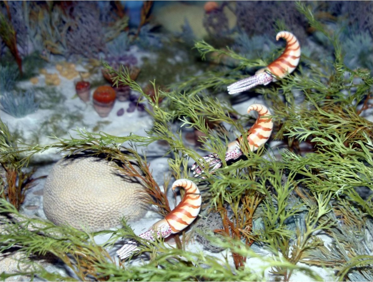
A couple of years earlier, the same author (Mutvei 2013) had proposed recognition of a superorder Multiceratoidea for nautiloids that combined multiple muscle scars with a nautilus-type siphuncle. Examples of nautiloid orders with such a combination included the Ellesmeroceratida (small nautiloids with densely placed septa), the Oncoceratida (often short, squat nautiloids) and the Discosorida (similarly squat forms with complex bulging connecting rings). All of these were found in the earlier part of the Palaeozoic with the oncoceratids dieing off in the early Carboniferous. Mutvei (2013) also included the coiled Tarphyceratida and the egg-shaped Ascoceratida in this group. Later, King & Evans (2019) redefined this grouping as the Multiceratia, excluding the Tarphyceratida and Ascoceratida on the grounds that they had ventromyarian rather than oncomyarian muscle scars. Mutvei (2013) suggested that, rather than representing retractor muscles, these smaller repeated scars were associated with an outgrowth of the mantle, either as tentacles or a muscular 'skirt', that was used to capture micro-plankton.

Phylogeny of 'nautiloids' supported by King & Evans (2019). Though not shown on this diagram, the majority of authors have suggested that ammonoids and coleoids are descended from Orthoceratida.


King & Evans (2019) proposed a reclassification of the subclass Nautiloidea between five subclasses defined primarily by muscle structure. Apart from the earliest oncomyarian Plectronoceratia, most 'nautiloids' could be divided between two lineages. On one side were the dorsomyarian Orthoceratia (usually thought to include the ancestors of the ammonoids and coleoids). On the other, the oncomyarian Multiceratia would eventually give rise to the ventromyarian Tarphyceratia which in turn included the ancestors of the pleuromyarian Nautilida. Note that many of the reocognised subclasses (and orders) remain paraphyletic but we are at least approaching a more informative picture of cephalopod evolution than the earlier unceremonious dumping into 'Nautiloidea' (I should probably also remind you that, for various reasons, most invertebrate palaeontologists still don't regard strict monophyly as a taxonomic requirement in and of itself).

The usage of muscle scars and connecting rings as classificatory keys is handicapped by the difficulty of observing them. As internal structures, they each require careful preparation of a specimen to observe. And once you've gotten to a position where you can see them, it seems not to be particularly easy to tell just what you're looking at. As a result, muscle scarring and siphon structure remains undescribed for the majority of nautiloid species. Judging the structure of connecting rings seems to be particularly challenging and some have gone so far as to suggest that purported different structures may be the result of post-mortem taphonomic processes (King & Evans 2019). Nevertheless, what we do know suggests that such features remain reasonably consistent within each of the well-recognised nautiloid orders. And Mutvei's (2015) concept of Calciosiphonata vs Nautilosiphonata does largely line up with King & Evans' (2019) dorsomyarian vs oncomyarian-ventromyarian lineages. There are, of course, some notable exceptions. Whether these will cause the developing structure to collapse, or whether they indicate mistakes in interpretation, only continued research will tell.

REFERENCES

King, A. H., & D. H. Evans. 2019. High-level classification of the nautiloid cephalopods: a proposal for the revision of the Treatise Part K. Swiss Journal of Palaeontology 138: 65–85.

Mutvei, H. 2013. Characterization of nautiloid orders Ellesmerocerida, Oncocerida, Tarphycerida, Discosorida and Ascocerida: new superorder Multiceratoidea. GFF 135 (2): 171–183.

Mutvei, H. 2015. Characterization of two new superorders Nautilosiphonata and Calciosiphonata and a new order Cyrtocerinida of the subclass Nautiloidea; siphuncular structure in the Ordovician nautiloid Bathmoceras (Cephalopoda). GFF 137 (3): 164–174.

A Brief Spotlight on Scopariines

The moths of the Pyraloidea are perhaps one of the more under-appreciated sectors of lepidopteran diversity. With many thousands of species, they comprise a significant proportion of the order in terms of both taxonomic and ecological diversity. Nevertheless, with most species being small and dull in coloration, many Lepidoptera enthusiasts will tend to lump them in the too-hard basket for study. One subgroup of the pyraloids to which this issue definitely applies is the subfamily Scopariinae.

Scoparia spelaea, copyright Donald Hobern.


Close to 600 species of Scopariinae are known from around the world with the highest diversity found on tropical mountains and islands (Léger et al. 2019). They are mostly a mottled greyish in coloration, blending in among the rocks and tree trunks on which they settle during the day. Like other pyraloids, they have large palps that extend in front of the head; pyraloids as a whole are sometimes referred to as 'snout moths' in reference to the appearance this gives them. Forewing venation is characterised by clear separation of vein R2 from R3+4 and absence of CuP (Nielsen & Common 1991).

Meadow grey Scoparia pyralella, copyright Hectonichus.


The majority of scopariine species feed as larvae on mosses, living concealed within a slight silk web. A smaller number feed on dicotyledons or lichens. One New Zealand species, the sod webworm Eudonia sabulosella, has been known to cause economic damage to pasture during sporadic outbreaks. Other species generally do not cause significant impact to humans.

Eudonia lacustrata, copyright Tony Morris.


Identification of scopariines is notoriously difficult with many species closely approximating each other in pattern or exhibiting confounding intra-specific variation. The two largest genera Scoparia and Eudonia can only be reliably separated by examination of the genitalia. Two genera, the Indo-Australian Micraglossa and the Neotropical Gibeauxia, are distinguished by the presence of shiny golden scales on head, thorax and abdomen. With such significant challenges to their study, it would not be surprising if 600 species should turn out to be a marked under-estimate of their true diversity.

REFERENCES

Léger, T., B. Landry & M. Nuss. 2019. Phylogeny, character evolution and tribal classification in Crambinae and Scopariinae (Lepidoptera, Crambidae). Systematic Entomology 44: 757–776.

Nielsen, E. S., & I. F. B. Common. 1991. Lepidoptera (moths and butterflies). In: CSIRO. The Insects of Australia: A textbook for students and research workers 2nd ed. vol. 2 pp. 817–915. Melbourne University Press: Carlton (Victoria).

Psalidothrips

Many of you may know thrips as small insects that infest buds and young shoots of garden plants, stymieing growth and causing malformed development. However, there is also a wide diversity of thrips species that feed on fungi, inhabiting leaf litter and other fallen vegetation. In tropical and subtropical regions of the world, one of the more numerous genera of such fungus-feeders is Psalidothrips.

Winged female (left) and wingless male of Psalidothrips comosus, from Zhao et al. (2018).


Close to fifty species of Psalidothrips have been described from various locations around the world (Wang et al. 2019). They are most commonly found among leaf litter and are believed to feed on fungal hyphae. Most Psalidothrips are relatively small, pale thrips, yellowish or light brown in coloration. As members of the family Phlaeothripidae, the last segment of the abdomen is modified into a tube ending in a ring of setae; in Psalidothrips, this tube is commonly short and the terminal setae are often longer than the tube.

As is common among thrips, the recognition of Psalidothrips and its constituent species is often complicated by within-species variation. Many species are known as both winged and wingless forms (Wang et al., 2019, note that Australian species seem particularly prone to winglessness). Wingless forms often show reductions in the sclerotisation of the thorax. It is difficult to name a single feature of the genus that does not find exception in some species or other. Most species are weakly sculpted. For the most part, the maxillary stylets are short and sit low and far apart in the head when retracted. The mouth-cone is similarly short and rounded. The head is often fairly short with rounded cheeks that do not bear strong setae. Setae on the anterior margin of the pronotum are often reduced. The wings, if present, are often more or less constricted at about mid-length. Many phlaeothripids possess a series of large setae on the abdomen that hold the wings in place when folded back; in individuals of Psalidothrips with such setae (obviously, they tend to disappear in wingless individuals), they are often relatively few in number and simply curved.

Many of these features are related to the thrips' litter-dwelling habits. The short mouthparts, for instance, presumably reflect how these thrips are gleaning fungi from the surface of leaves without needing to pierce the leaf's cuticle. As such, it will be interesting to see how the genus holds out as our understanding of thrips phylogeny improves. Is this a true evolutionarily coherent assemblage, or disparate travellers who are following a fashion?

REFERENCE

Wang, J., L. A. Mound & D. J. Tree. 2019. Leaf-litter thrips of the genus Psalidothrips (Thysanoptera, Phlaeothripidae) from Australia, with fifteen new species. Zootaxa 4686 (1): 53–73.

In Honour of Amblyseius

At this point in time, the Phytoseiidae are one of the most intensely studied families of mites. They are the only group of mesostigmatan mites to have significantly diversified among the foliar environment (on and around plant leaves) where they are mostly predators on other small invertebrates. The taxonomic history of phytoseiids is storied and complex but one taxon that has been consistently recognised as a major part of the family is the genus Amblyseius.

Swirski mite Amblyseius swirskii, from here.


When reviewed by Chant & McMurtry in 2004, Amblyseius was a sizeable assemblage of close to 350 known species (I quite expect that number to have expanded by now). Species of Amblyseius are lightly sclerotised, mostly pale in colour, and usually have a smooth shield covering most of the dorsum. The genus is characterised by the presence of eighteen or nineteen pairs of setae on the dorsum of the idiosoma (the central body) with three sublateral pairs being particularly long: one about the level of the third pair of legs (referred to as the s4 pair) and the other two towards the rear of the body. Except for a few pairs forward of the s4 setae, the remaining dorsal setae are all minute.

The primary focus of human interest in phytoseiids has been their role as predators of crop pests. I described some of the ways in which phytoseiids have been commercially utilised in an earlier post. Species used in this way include several Amblyseius though matters are complicated slightly by changes in taxonomy (for instance, one species which has been widely traded as Amblyseius cucumeris is now placed in the genus Neoseiulus). One of the most widely used of the commercial phytoseiids in recent years has been Amblyseius swirskii, commonly known as the Swirski mite (E. Swirski being an acarologist after whom the species was named). This species was first described in 1962 from almond trees in Israel and subsequently identified from a wide range of plant and crop species. Its history in pest control has been described in detail by Calvo et al. (2015).

The Swirski mite feeds on a range of prey, including mite, thrips and whitefly species, as well as on pollen and micro-fungi. It was first promoted as a commercial control for silverleaf whitefly Bemisia tabaci in the early 2000s. However, it did not get taken up in a big way until media publicity about pesticide residues on capsicum crops in Spain led to a crash in demand. Farmers in that country were forced to look for alternative means of pest control and found great success with A. swirskii (previous attempts to use the cooler-clime preferring Neoseiulus cucumeris in Spain had not been promising). Since then, the Swirski mite has been adopted in numerous countries for use on a range of crops to control various pests such as western flower thrips Frankliniella occidentalis. Because of its ability to grow and thrive on non-insect foods, including artificial diets, this mite is easily cultured commercially. It may also be released on crops before pest infestations develop, building up numbers on a diet of pollen until suitable prey presents itself. For the same reason, Swirski mite populations do not crash before pest control is complete. Overall, a remarkable success and a prime example of the value of Amblyseius species to mankind.

REFERENCES

Calvo, F. J., M. Knapp, Y. M. van Houten, H. Hoogerbrugge & J. E. Belda. 2015. Amblyseius swirskii: what made this predatory mite such a successful biocontrol agent? Experimental and Applied Acarology 65: 419–433.

Chant, D. A., & J. A. McMurtry. 2004. A review of the subfamily Amblyseiinae Muma (Acari: Phytoseiidae): part III. The tribe Amblyseiini Wainstein, subtribe Amblyseiina n. subtribe. International Journal of Acarology 30 (3): 171–228.

By the Light of the Pony

Light-emitting organs have evolved in many different species of marine fish. For the greater part, they are associated with inhabitants of the deep sea, the twilight and midnight zones beyond the reach of celestial light. Light production by species found in shallow waters is much less common. Nevertheless, one particularly notable radiation of near-surface glowers is the ponyfishes of the family Leiognathidae.

Leiognathus equulus, copyright Sahat Ratmuangkhwang.


Ponyfishes are small, mostly silvery fishes found in coastal and brackish waters in tropical regions of the Indo-West Pacific. The largest ponyfishes grow to about 25 cm in length but most species are much smaller (Woodland et al. 2002). They live in large schools that forage near the surface at night, descending close to the bottom sediment during the day. Why these animals are referred to as 'ponyfishes', I have no idea (perhaps the head is meant to look a bit pony-like?) An alternative vernacular name of 'slipmouth' makes a lot more sense as these fish have highly extensible jaws that can be used to snipe prey out of the water. A groove along the top of the skull allows for reception of a long, mobile premaxilla, supporting the mouth as an elongate tube when extended. Most ponyfishes are planktivores with simple, minute teeth in the jaw and the mouth extending horizontally. Species of the genus Deveximentum have the mouth tilted obliquely at rest so that it stretches upwards when extended. Members of the genus Gazza are piscivores when mature, feeding on other fish, and possess a pair of large caniniform teeth in each of the upper and lower jaws to hold their prey (James 1975).

Ponyishes are also notable for their elaborate light-producing organs. In most bioluminescent fishes, the photophores sit on or close to the skin surface but in leiognathids it is an internal outgrowth of the gut. A cavity around the end of the oesophagus houses colonies of bioluminescent bacteria, usually the species Photobacterium leiognathi. This light organ sits alongside or projects into the gas bladder which has a reflective internal coating. In many species, patches of scale-less, translucent skin allow the transmitted light to shine forth brightly. Muscular 'shutters' associated with the light organ allow the fish to control light transmission more directly (Woodland et al. 2002).

Photopectoralis bindus, copyright D. G. R. Wiadnya.


In a review of ponyfish taxonomy by James (1975), no mention was made of the light-emitting organ or many of its associated structures (though reference was made to the absence of scales on certain parts of the body). With the exceptions of the distinctive genera Gazza and Deveximentum, ponyfishes were assigned to a broad genus Leiognathus. Since then, variations in the structure of the light organ have been recognised as taxonomically significant, allowing the recognition of several genera divided between two subfamilies Leiognathinae and Gazzinae (Chakrabarty et al. 2011). Leiognathinae is defined by plesiomorphic characters and is likely to be paraphyletic to Gazzinae (Sparks & Chakrabarty 2015).

Because of the nocturnal habits of ponyfish and the delicacy of the light-emitting structures, our understanding of how light production functions in Leiognathidae remains somewhat limited. In Leiognathinae and females of Gazzinae, the light organ is relatively small and the external body surface lacks translucent patches. For the most part, light is expressed in these individuals as a uniform ventral glow that probably functions as counter-illumination (the light from the venter prevents the fish from appearing as a silhouette against light from the water surface to predators swimming below). Alternatively, light may be flashed to warn school-mates of danger. In males of Gazzinae, conversely, the light organ is enlarged relative to females and associated with translucent 'windows'. The shape of the organ and the arrangement of the 'windows' is a primary factor in distinguishing genera. Rhythmic flashing of light has been observed in males of many gazzine species and is probably characteristic of the group as a whole. Woodland et al. (2002) observed a school of several hundred Eubleekeria splendens flashing their lights synchronously shortly after nightfall. The exact function of such displays is uncertain, whether in courtship displays, co-ordinating school movements, attracting prey or dissuading predators. The sexually dimorphic nature of the light organ system, together with its species-specific expression, might seem to favour the first of these options but it should be noted that they are not all mutually exclusive.

Despite their small size, ponyfishes are often significant food fish for people living in areas where they are found. Thanks to their schooling behaviour, they are often a major component of dredge catches. In the Philippines, they are used for making bagoong, a fermented fish paste. In other places, they may be cooked whole after cleaning. The glow, sadly, does not survive the process.

REFERENCES

Chakrabarty, P., M. P. Davis, W. L. Smith, R. Berquist, K. M. Gledhill, L. R. Frank & J. S. Sparks. 2011. Evolution of the light organ system in ponyfishes (Teleostei: Leiognathidae). Journal of Morphology 272: 704–721.

James, P. S. B. R. 1975. A systematic review of the fishes of the family Leiognathidae. J. Mar. Biol. Ass. India 17 (1): 138–172.

Sparks, J. S., & P. Chakrabarty. 2015. Description of a new genus of ponyfishes (Teleostei: Leiognathidae), with a review of the current generic-level composition of the family. Zootaxa 3947 (2): 181–190.

Woodland, D. J., A. S. Cabanban, V. M. Taylor & R. J. Taylor. 2002. A synchronized rhythmic flashing light display by schooling Leiognathus splendens (Leiognathidae: Perciformes). Marine and Freshwater Research 53: 159–162.