Field of Science

Showing posts with label Echinodermata. Show all posts

The Pelecocrinidae

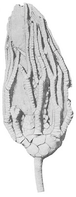
The latter part of the Palaeozoic represented a peak in crinoid diversity. More families of crinoid have been recognised from the Carboniferous and Permian than any period before or since. Among the various families of the Late Palaeozoic were representatives of the Pelecocrinidae.

Pelecocrinus insignis, from Moore & Teichert (1978).


The fossil record of the pelecocrinids was long-lasting but patchy. They are known from the early Carboniferous of North America (Pelecocrinus) and Great Britain (Forthocrinus), the late Carboniferous of North America (Exoriocrinus), and the late Permian of Italy (Tetrabrachiocrinus) and Indonesia (Drepaocrinus, Malaiocrinus). These locations largely correspond to what would have been a distribution along the northern coast of the Palaeotethys Ocean, possibly becoming extinct in the west as the gap between North America and the southern continent of Gondwana closed to form the Pangean supercontinent. Pelecocrinids seem to be so far unknown from the southern continents.

In life, pelecocrinids were characterised by a high crown arising from a low, bowl-shaped cup. The stem could be round or pentagonal. The base of the cup was flattened or shallowly concave, so the infrabasals (the lowest circle of plates above the stem) were barely or not visible where the cup to be observed from the side. The articulations between the upper plates of the cup and the bases of the arms were angled downwards and outwards with the articular facets being somewhat narrower than the plates they sat on. The arms themselves had a wedge-shaped cross-section and divided into equal branches two or more times along their length. Each arm bore one or two rows of pinnules. The anal sac, as described for Pelecocrinus, was relatively short and slender and summited by heavy, spinose plates.

The structure of the arm articulations and pinnules indicates that the arms would have been subject to muscular control with individual arms being able to be moved in more than one plane. This arrangement became increasingly common among crinoids from the Carboniferous onwards, allowing them to function in more high-current habitats. The position of the arms and pinnules could be adjusted to optimise filtration from the water column, while the current provided lift to the crown so it did not need to be mechanically supported by the stem alone. The effectiveness of this arrangement is attested to by the long history of the pelecocrinids. Nevertheless, the end-Permian extinction was to end their lineage along with that of so many of their contemporaries.

REFERENCE

Moore, R. C., & C. Teichert (eds) 1978. Treatise on Invertebrate Paleontology pt T. Echinodermata 2. Crinoidea vol. 2. The Geological Society of America, Inc.: Boulder (Colorado), and The University of Kansas: Lawrence (Kansas).

Taxocrinus

Below is an example of Taxocrinus, a genus of fossil crinoids known from the later Devonian and earlier Carboniferous of Europe and North America. It is a relatively plesiomorphic representative of the flexible crinoids, one of the major crinoid lineages of the Palaeozoic era.

Taxocrinus colletti, copyright James St. John.


Flexible crinoids are characterised by arms that lack pinnules, the small side-branches found on the arms of most other crinoids. As a result, the preserved arms have a somewhat tentacle-like appearance, and are commonly preserved coiled in over the oral surface of the central cup. In Taxocrinus, the arms were regularly and isotomously bifurcated: that is, they divided between two branches of more or less equal size. The central cup itself in flexible crinoids was (somewhat counter-intuitively) quite inflexible, with the plates of the aboral surface firmly jointed together. The oral surface bore a more flexible covering of small plates, and an anal tube (visible near the midline of the fossil above) directed waste away from the mouth. The stem was round in cross section and lacked lateral cirri (Moore 1978).

Flexible crinoids were around for a very long time but it is rare for them to be found in abundance. As such, they were probably specialised for particular habitats that were either uncommon or less likely to be preserved. It has been suggested that, because their pinnule-less arms would have been poorly suited for filtering particles from strong currents, flexible crinoids may have inhabited calm, low-energy waters (Breimer 1978) (though I do wonder if enlarged tube feet may have partially filled the role of pinnules; is it possible to estimate the size of the tube feet from the preserved skeleton?) Crinoids living in such habitats will often hold the arms in a bowl arrangement so they may capture particles settling from higher in the water column. In the case of the flexible crinoids, moving the arms in and out may have created local water movements to further draw such particles in.

Though Taxocrinus itself would disappear in the mid-Carboniferous, flexible crinoids as a whole would persist to the end of the Permian. In more derived forms, the branching of the arms was often unequal, with the smaller branches effectively replacing the missing pinnules. In the end, though, the specialised flexibles were yet another casualty of the end-Permian cataclysm that so shook the composition of life on this planet.

REFERENCES

Breimer, A. 1978. Autecology. In: Moore, R. C., & C. Teichert (eds) Treatise on Invertebrate Paleontology pt T. Echinodermata 2 vol. 1 pp. T331–T343. The Geological Society of America, Inc.: Boulder (Colorado), and The University of Kansas: Lawrence (Kansas).

Moore, R. C. 1978. Flexibilia. In: Moore, R. C., & C. Teichert (eds) Treatise on Invertebrate Paleontology pt T. Echinodermata 2 vol. 2 pp. T759–T812. The Geological Society of America, Inc.: Boulder (Colorado), and The University of Kansas: Lawrence (Kansas).

Echinoids: Regularly Irregular

In manufacturing, one of the most desired qualities is regularity. Success is achieved by ensuring that each unit matches the last, that its qualities remain predictable and reliable. In evolution, by contrast, the opposite is often true: embracing irregularity may allow a lineage to expand in directions not previously available. For evidence, just look at the success of the irregular echinoids.

Echinoneus cyclostomus, one of the few living holectypoid urchins, copyright Philippe Bourjon.
.

The Echinoidea, sea urchins, are commonly divided between regular and irregular forms. In regular echinoids, representing the ancestral type for the class, the mouth and anus are positioned at opposite points on the test. The mouth sits squarely in the centre of the animal's underside (the oral surface) while the anus sits at the centre of the upper (aboral) surface. The five ambulacra, the lines of small plates in the test from which the tube feet emerge, are more or less evenly arranged around the superficially radially symmetrical test. Irregular echinoids, in contrast, have the anus more or less displaced from the midpoint of the test. In the earliest irregular echinoids, this displacement might be relatively slight: the periproct (the membrane through which the anus opens, usually covered in echinoids with an array of small plates) was still found at the centre of the aboral surface but was enlarged and/or stretched towards one end of the test (Saucède et al. 2007). In more derived forms, the periproct has moved more significantly, potentially being found on the side of the test or even on the oral surface near the mouth.

Front view of heart urchin Spatangus purpureus, copyright Roberto Pillon.


This displacement of the anus indicates a directionality to the test that isn't found in regular echinoids. A number of other changes have associated it in the evolution of echinoids, such as reduction of the size of the spines covering the test and an increased directionality in their axes of movement. The mouth may also become displaced towards the front of the test, and the test as a whole may become more bilateral in its overall shape. The jaws become modified or, in a couple of groups, lost entirely. All these alterations add up to indicate a distinct change in lifestyle between regular and irregular echinoids. Whereas regular echinoids roam the surface of sea bottom, using their powerful jaws to graze directly on algae or scavenge on animal carcasses, irregular echinoids are deposit feeders that tend to live at least partially buried in the sidement. They may swallow large amounts of sediment and digest organic matter mixed therein, or gather up organic particles with their tube feet and/or by means of mucous strands transported in ciliary grooves. Burrowing is achieved by movement of the spines or by using the tube feet to pass sand grains above the aboral surface. In the shallow-burrowing heart urchin Spatangus purpureus, an array of longer spines on the aboral surface are used to keep a funnel open between the buried urchin and the surface, allowing water to carry oxygen to it. Echinocardium cordatum, which burrows as deep as 18 cm beneath the substrate surface, maintains an opening to the surface by means of elongate tube feet (Durham 1966).

One of the most irregular of irregular echinoids, the deep-sea Pourtalesia miranda, from Oliver (2016). The enlarged insert shows a symbiotic bivalve Syssitomya pourtalesiana.


The change in lifestyle was certainly a successful one: nearly 60% of living echinoids are irregular. The earliest irregular echinoids appeared in the early Jurassic, with recent analyses agreeing that they represent a monophyletic group (Saucède et al. 2007; Kroh & Smith 2010). Nevertheless, a certain degree of parallelism in adaptations appears to have been occurred. Living irregular echinoids can be divided between two clades: one is relictual, containing only two genera in the order Holectypoida, whereas the remaining species belong to the larger clade Microstomata. The earliest known members of the holectypoid lineage retained strong jaws even after they evolved the ability to burrow in sediment. In contrast, the earliest known member of the Microstomata retained large spines, indicating a non-burrowing lifestyle, but already possessed the adaptations for a particulate diet (Saucède et al. 2007). With time, both lineages developed the feature that they lacked, adding them together for a winning combination.

REFERENCES

Durham, J. W. 1966. Echinoids—ecology and paleoecology. In: Moore, R. C. (ed.) Treatise on Invertebrate Paleontology pt U. Echinodermata 3 vol. 2 pp. U257–U265. The Geological Society of America, Inc., and The University of Kansas Press.

Kroh, A., & A. B. Smith. 2010. The phylogeny and classification of post-Palaeozoic echinoids. Journal of Systematic Palaeontology 8 (2): 147–212.

Saucède, T., R. Mooi & B. David. 2007. Phylogeny and origin of Jurassic irregular echinoids (Echinodermata: Echinoidea). Geological Magazine 144 (2): 333–359.

The Pisocrinidae: Babyface Crinoids

One question that I haven't yet found an answer to is why the Palaeozoic marine fauna seems to have included so many filter feeders. Cystoids, blastoids, graptoloids... so many of the distinctive taxa occupying this niche would be gone by the period's end, without leaving any clear analogues behind them. What was the cause underlying this abundance? Is it simply a misapprehension caused by the filtering effect of history, with the modern fauna containing fewer major lineages but no fewer actual species? Is it the distorting lens that causes us to tend to assign a higher 'rank' to those lineages arising earlier in time, whatever their practical levels of disparacy? Or was there actually something different about what could be found in Palaeozoic seawater?

Reconstructions of short-armed and long-armed species of Pisocrinus, from Rozhnov (2007).


The Pisocrinidae are one of those distinctive Palaeozoic marine groups, known from around the world during the Silurian and Devonian. As crinoids, they were perhaps not as immediately unfamiliar to the modern eye as some of the other taxa that could be found at that time, but they were certainly different from any modern crinoid. The majority of the crinoids that have ever lived can be assigned to one of two main clades. One, the cladid lineage, includes all the crinoids alive today. Pisocrinids belong to the other major lineage, the disparids, which were prominent for most of the Palaeozoic era but failed to make it past the end of the Permian. Disparids differed from cladids in that their calyx included a single circlet of plates (the inferradials) beneath the circlet of the radials (the large plates making up the main body of the calyx) whereas cladids (at least to begin with) had two such circlets. Many disparid sublineages showed a tendency towards reduction and/or simplification of the calyx. In pisocrinids, most of the calyx was made up of just three plates: two large radials (representing the A and D rays of the basic crinoid calyx) and a greatly enlarged B inferradial. The B, C and E radials were all reduced in size. The arms of pisocrinids mostly lacked lateral pinnules and were undivided; one genus, Cicerocrinus, had bifurcating arms bearing lateral ramules (Moore et al. 1978). The length of the arms varied considerably between species: in some they were quite short and broad, in others they were remarkably long. Because their derived morphology made it difficult to compare pisocrinids to related families, their origins have been regarded as mysterious. Rozhnov (2007) suggested a derivation from an earlier, more typical crinoid family, the Homocrinidae, via paedomorphosis, possibly as a result of the evolution of a longer larval period in the life cycle (he specifically suggested that this extended larval phase may have allowed the ancestors of pisocrinids to spread across the Iapetus Ocean between the then-existing continents of Laurentia and Baltica). A direct pisocrinid-homocrinid connection was not supported in the phylogenetic analysis of disparids by Ausich (2018) but Rozhnov's overall model of pisocrinid paedomorphosis remains a possibility.

Assemblage of Triacrinus, from here.


During the Silurian, pisocrinids were among the most abundant, if not the most abundant, groups of crinoids. They were found in a variety of habitats but were particularly abundant around reefs in deeper waters. At first glance, the non-pinnulate arms of pisocrinids appear poorly suited for filter feeding, and one might be inclined to propose a more tentacular method of obtaining food items. However, such a method would seem unlikely for the short-armed species, whose arms would have been almost entirely inflexible. Even the long-armed species sometimes had arms made up of relatively long segments whose flexibility may have been limited. An alternative possibility, I suppose, is that in life pisocrinids may have had long tube feet that took the place of the missing pinnules. Meanwhile, the absence of the pinnules meant that the arms could be lain tightly alongside each other when the crown was closed. Earlier authors presumed that, because of their preference for deeper waters, pisocrinids were rheophobic (that is, they were found in places where the water lacked a noticeable current). However, Ausich (1977) proposed that they were low-energy rheophilic, seeking locations where a moderate but steady current prevailed. The current would provide a steady supply of organic particles that could be captured by the crown, and the ability to close the arms tight would protect the oral region during occasional bouts of rougher conditions.

REFERENCES

Ausich, W. I. 1977. The functional morphology and evolution of Pisocrinus (Crinoidea: Silurian). Journal of Paleontology 51 (4): 672–686.

Ausich, W. I. (in press, 2018) Morphological paradox of disparid crinoids (Echinodermata): phylogenetic analysis of a Paleozoic clade. Swiss Journal of Palaeontology.

Moore, R. C., N. G. Lane, H. L. Strimple, J. Sprinkle & R. O. Fay. 1978. Inadunata. In: Moore, R. C., & C. Teichert (eds) Treatise on Invertebrate Paleontology pt T. Echinodermata 2. Crinoidea vol. 2 pp. T520–T759. The Geological Society of America, Inc.: Boulder (Colorado), and The University of Kansas: Lawrence (Kansas).

Rozhnov, S. V. 2007. Changes in the Early Palaeozoic geography as a possible factor of echinoderm higher taxa formation: delayed larval development to cross the Iapetus Ocean. Palaeogeography, Palaeoclimatology, Palaeoecology 245: 306–316.

Brittle Stars, Brittle Taxa

Amphiura arcystata brittle stars extending their arms above the sediment, copyright James Watanabe.


The brittle stars are something of the poor cousin among echinoderm classes. Their tendency to relatively small size and cryptic habitats means that they do not attract the level of attention given to starfish, sea urchins or sea cucumbers. Despite this, they are perhaps the most diverse of the living echinoderm classes, with more recognised species around today than any other.

It should therefore come as no surprise that the internal classification of brittle stars remains decidedly up in the air. The basic framework of the surrent system was established over a hundred years ago by Matsumoto (1915) and changes to this arrangement since have been fairly cosmetic. However, a significant challenge to Matsumoto's system has been arisen following the input of molecular data to the mix: many of Matsumoto's higher groupings have not been supported by moleculat analyses. Perhaps the nail in the Matsumoto system's coffin has come from a recent publication by Thuy & Stöhr (2016) who found that a formal analysis of morphological data also failed to support the pre-existing classification. At this point in time, we know that a new classification of brittle stars is needed but we don't yet know what form it will take.

Excavated specimen of Amphiuridae, copyright Arthur Anker. The radial plates are visible as a pair of bars alongside the base of each arm; I don't think that the genital plates are visible externally.


Perhaps one of Matsumoto's groupings that will survive the transition is the Gnathophiurina. Notable features of this group include a ball-and-socket articulation between the radial shields (large plates that sit on the aboral side of the central body on either side of the insertion of each arm) and the genital plates (sitting below and alongside the radial shields), with the socket in the radial shield and the ball on the genital plate. The genital plates are also firmly fixed to the basal vertebra of each arm. I haven't been able to find what the functional significance of this arrangement is, such as whether it renders the body more flexible that in other groups where the radial-genital plate articulation is more fixed. At least one of the families of Gnathophiurina, the Amphiuridae, includes species that commonly live in burrows with the tips of their arms extended into the water column, using their tube feet to capture food particles (Stöhr et al. 2012). In contrast, some Ophiotrichidae are epizoic, living entwined around black corals and the like. The Gnathophiurina as a whole seem to be most diverse in relatively shallow waters.

Matsumoto's (1915) original concept of the Gnathophiurida included species that are now classified into four families, the Amphiuridae, Ophiotrichidae, Amphilepididae and Ophiactidae, and recent analyses have returned results not inconsistent with this association. In Thuy & Stöhr's (2016) morphological analysis, Gnathophiurina species all belong to, and make up the bulk of, their clade IIIc. In the molecular analysis presented by Hunter et al. (2016), the families belong to two separate clades but the branch separating them is very weakly supported. Further research is needed, of course, but it may turn out that Matsumoto was on to something when he focused on that ball-and-socket joint.

REFERENCES

Hunter, R. L., L. M. Brown, C. A. Hill, Z. A. Kroeger & S. E. Rose. 2016. Additional insights into phylogenetic relationships of the Class Ophiuroidea (Echinodermata) from rRNA gene sequences. Journal of Zoological Systematics and Evolutionary Research 54 (4): 269–275.

Matsumoto, H. 1915. A new classification of the Ophiuroidea: with descriptions of new genera and species. Proceedings of the Academy of Natural Sciences of Philadelphia 67 (1): 43–92.

Stöhr, S. T. D. O'Hara & B. Thuy. 2012. Global diversity of brittle stars (Echinodermata: Ophiuroidea). PLoS One 7 (3): e31940.

Thuy, B., & S. Stöhr. 2016. A new morphological phylogeny of the Ophiuroidea (Echinodermata) accords with molecular evidence and renders microfossils accessible for cladistics. PLoS One 11 (5): e0156140.

The Erisocrinoidea: Shallow Crinoids

Articulated calyx of Erisocrinus typus, copyright Richard Paselk.


The close of the Permian period saw the largest mass extinction ever recorded. It has been estimated that about 95% of all marine species were wiped out. Many prominent Palaeozoic lineages disappeared entirely; others were reduced to a mere remnant of their former selves.

One of the casualties of the end-Permian extinction was the crinoid group known as the Erisocrinoidea (or Erisocrinacea in older texts). These were a diverse group of crinoids divided between several families, recorded from the Carboniferous and Permian periods. One species, Erisocrinus typus, is known from a large number of well-preserved, articulated specimens from the mid-Late Carboniferous of the United States and is one of the best representatives of the Palaeozoic cladid crinoids. Erisocrinoids are characterised by a low cup, dominated by the ring of radial plates. The base of cup was often recessed, meaning that the basal and infrabasal plate rings were often partially or entirely obscured in outer view. Most significantly, the array of anal plates found in other crinoids was reduced to a single plate or even lost. The insertion points of the arms bear signs of strong muscular articulation, indicating that these were animals of higher-energy environments requiring more exertion to maintain an ideal feeding position. The anal sac, where it is preserved, was only weakly plated and would have been reasonably soft in life (Moore et al. 1978).

In other respects, though, the erisocrinoids could be somewhat disparate. Many, such as the type family Erisocrinidae and the families Protencrinidae and Catacrinidae, have biserial arms in which the arm's skeleton is comprised of paired rows of plates. In other families, such as the Graphiocrinidae and Diphuicrinidae, the arms were uniserial, with only a single row of plates. Webster & Maples (2006) noted that, even though all erisocrinoids shared the character of a reduced anal plate array, the exact position in the cup of the anal plate or its remnant differed between families. They therefore suggested that the erisocrinoids might not be a monophyletic group, but members of a number of different lineages that had converged on a similar morphology and presumably lifestyle.

This was not an entirely novel suggestion. Even while recognising a single superfamily Erisocrinacea, Moore et al. (1978) had suggested connections between individual erisocrinoid families and families placed in other superfamilies. The integrity of the Erisocrinoidea had also been questioned in relation to Encrinus, a genus from the Middle Triassic that had been included with the erisocrinoids on the basis of its combination of biserial arms and lack of an anal plate. If this assignment was correct, erisocrinoids would have survived the end-Permian extinction: the only crinoid lineage to do so other than the Articulata, the clade including the living sea lilies and feather stars. Articulates retain uniserial arms, a more plesiomorphic characteristic. However, while investigating the evolutionary origins of the articulates, Simms & Sevastopulo (1993) pointed out that Encrinus shared derived features with articulates that were absent in erisocrinoids. For instance, while Encrinus and the erisocrinoids both had each of the basic five echinoderm arms branching to form a total array of ten arms, in Encrinus they branched from the second primibrachial plate as in articulates, instead of from the first as in erisocrinoids. Rather than being a late-surviving erisocrinoid, Encrinus was an early side-branch of the articulates, and as far as is known only a single crinoid lineage survived the Permian.

REFERENCES

Moore, R. C., N. G. Lane, H. L. Strimple, J. Sprinkle & R. O. Fay. 1978. Inadunata. In: Moore, R. C., & C. Teichert (eds.) Treatise on Invertebrate Paleontology pt T. Echinodermata 2. Crinoidea vol. 2 pp. T520–T759. The Geological Society of America, Inc.: Boulder (Colorado), and The University of Kansas: Lawrence (Kansas).

Simms, M. J., & G. D. Sevastopulo. 1993. The origin of articulate crinoids. Palaeontology 36 (1): 91–109.

Webster, G. D., & C. G. Maples. 2006. Cladid crinoid (Echinodermata) anal conditions: a terminology problem and proposed solution. Palaeontology 49 (1): 187–212.

Bits of Cucumber in the Fossil Record

Aggregation of Eocaudina septaforminalis sclerites from Boczarowski (2001). Scale bar = 200 µm.


Echinoderms are a dream group of animals for invertebrate palaeontologists (that's palaeontologists who study invertebrates, not palaeontologists who are invertebrates). Their calcified skeletons mean that their fossil record is extensive and detailed. When most of the body is covered in plates, looking at the fossil can give you an instant idea of what the animal looked like when alive. But as with all things in biology, there are notable exceptions. The sea cucumbers are one group of echinoderms that has significantly reduced the original skeleton, sacrificing a hard outer skeleton for increased flexibility. Instead of solid plates, the sea cucumber skeleton is made up of many minute sclerites embedded in the skin. And while this may be all well and good for the sea cucumber, it is not so convenient for the palaeontologists. In the fossil record, these minute sclerites become separated, and one separated sclerite does not tell you much about the appearance of the sea cucumber as a whole.

As a result, palaeontologists looking at sea cucumber remains have found themselves presented with a conundrum. The classification of modern sea cucumbers is largely based on features of the soft body that are usually not preserved in the fossil record, making comparison of living and fossil cucumbers difficult. Also, the skeleton of a single sea cucumber may include different forms of sclerite, performing different roles. If two different types of sclerite are found close together in the fossil record, did they come from a single sea cucumber or from two different sea cucumbers that died close together? To bypass these barriers, palaeontologists have often used what are referred to as 'parataxa'. A single type of sclerite is treated as a single 'parataxon', with the recognition that there may not be a perfect correlation between the parataxon and the theoretical taxon that it originally came from.

Individual calclamnine sclerite, Priscocaudina crucensis, from Boczarowski (2001).


The Calclamnidae has been recognised as one such 'parafamily' of sea cucumber sclerites. As defined by Frizzell & Exline (1966), the Calclamnidae grouped together rounded or polygonal sclerites that are perforated with holes like a sieve, and that don't have any sort of stalk or other ventral protrusion. This is a very common sort of sclerite for echinoderms: 'calclamnid' sclerites have been identified as far back as the Ordovician (Boczarowski 2001), and sclerites of this sort are still found in sea cucumbers today (just to confuse matters, the skeletons of some brittle stars also include very similar sclerites, raising the spectre of misidentification). Boczarowski (2001) recognised two subfamilies of Calclamnidae: in one, the Eocaudininae, the perforations of the sclerite are all more or less even in size, while in the other, Calclamninae, the pores towards the centre of the plate are larger and arranged in a cross-shape. The eocaudinines include the earliest calclamnid plates, with the calclamnines appearing during the Devonian.

Recognition of parataxa is a convenient tool for keeping records of things like biostratigraphy without getting bogged down, but what sort of sea cucumber did calclamnids actually come from? The calclamnids resemble sclerites found in the group of modern sea cucumbers called the Dendrochirotacea, so they have often been classified with this group. However, a number of features of the dendrochirotaceans, including perforated calclamnid-like sclerites, have been suggested to be primitive for sea cucumbers, so similarities between calclamnids and dendrochirotaceans may represent shared ancestral features rather than true affinities. Haude (1992) commented on a number of cases of sclerites found preserved in assemblages that he believed represented original life associations, including some containing calclamnids. One of these contained sclerites that Haude identified as similar to Calclamna germanica, the type species of the family, in association with large hook-shaped sclerites. Hooks are not characteristic of dendrochirotaceans, but of Apodacea, a different group of sea cucumbers characterised by the loss of tube feet (with the hooks working to provide mobility in their place). Haude suggested the possibility that Calclamna might represent a stem-group apodacean that retained some primitive sclerite features. In other fossil groups such as conodonts, the identification of preserved assemblages has allowed palaeontologists to progress beyond the use of parataxa and integrate more recognition of evolutionary relationships. Hopefully we get the same opportunity with sea cucumbers.

REFERENCES

Boczarowski, A. 2001. Isolated sclerites of Devonian non-pelmatozoan echinoderms. Palaeontologia Polonica 59: 1-219.

Frizzell, D. L., & H. Exline. 1966. Holothuroidea—fossil record. In: Moore, R. C. (ed.) Treatise on invertebrate Paleontology pt U. Echinodermata 3 vol. 2, pp. U646-U672. The Geological Society of America, Inc., and The University of Kansas Press.

Haude, R. 1992. Fossil holothurians: sclerite aggregates as 'good' species. In: Scalera-Liaci, L., & C. Canicatti (eds) Echinoderm Research 1991, pp. 29-33. Balkema: Rotterdam.

Paracomatula: Feather Star, or Feather Star Wannabe?

Fossilised accumulation of Paracomatula helvetica, from here.


Earlier posts on this site have discussed examples of the feather stars, the most successful representatives in the modern environment of the crinoids. Originally a group whose members lived permanently attached to the substrate by a stalk, at some point crinoids diversified to a more mobile (or least shiftable) lifestyle, discovering the joys of travel. This was not, it should be noted, an entirely direct process. Many stalked crinoids are also mobile, able to detach themselves from their substrate and crawl to a new position. Other crinoids than the feather stars lost their stalks. And at least one group within the feather stars, the Mesozoic Thiolliericrinidae, reverted back to retaining as adults the larval stalk that most feather stars lose in the course of development.

Nevertheless, the feather stars had definitely made their appearance by the early Jurassic. One of the earliest taxa that has been assigned to the feather stars is Paracomatula, which is known from the very late Triassic to the middle Jurassic (Hess 2013). Paracomatula would have largely resembled a modern feather star in appearance, but had one significant difference. In modern feather stars, the base of the central cup is formed by a large conical plate known as the centrodorsal. In Paracomatula, however, the centrodorsal is replaced by a stack of five narrow plates. These correspond to much-shortened versions of the columnals that make up the stalked in other crinoids, and Rasmussen (1978) and other authors suggested that Paracomatula's separate columnals became fused to form the centrodorsal of the feather stars proper.

However, not all authors have accepted this interpretation. Hess (2013) has argued that details of the development of modern feather stars from a stalked juvenile to a free-living adult indicate that the centrodorsal is derived from the enlargement of a single columnal rather than the fusion of a series. In the earliest definitive feather star, the Jurassic Palaeocomaster, the cirri (tentacle-like appendages) on the centrodorsal are arranged in a haphazard fashion consistent with their development on a single expanding plate, while Paracomatula has a more orderly array of one ring of cirri per columnal without the development of supernumerary cirri. Hess therefore argues that Paracomatula species were not the forebears of feather stars, but their rivals: a closely related group that was independently experimenting with a stalk-free way of life.

REFERENCES

Hess, H. (in press, 2013) Origin and radiation of the comatulids (Crinoidea) in the Jurassic. Swiss Journal of Palaeontology.

Rasmussen, H. W. 1978. Articulata. In: Moore, R. C., & C. Teichert (eds) Treatise on Invertebrate Paleontology pt T. Echinodermata 2: Crinoidea, vo. 3, pp. T813-T927. The Geological Society of America, Inc., and The University of Kansas Press.

The Antedoninae: (Relatively) Big-Bellied Feather Stars

Several individuals of Antedon bifida attached to a kelp stipe, photographed by Bernard Picton.


The feather stars and other crinoids are both the most divergent and least known of the modern echinoderms. This is the second post here at Catalogue of Organisms on modern feather stars; an earlier post gave a brief overview of some of the details of the feather star lifestyle and anatomy. The main subject of the earlier post was the family Charitometridae; this post will focus on a different group, the Antedoninae. Yes, Virginia, there are different varieties of feather star.

The Antedonidae and related families differ from most other feather stars in that the internal cavity of the centrodorsal, the plate that forms the base of the calyx (central cup) of the feather star, is relatively large compared to the centrodorsal's diameter (in life, this cavity has organs nestled in it). When the antedonids were reviewed by the American echinodermatologist Austin H. Clark (Clark & Clark 1957*), he regarded this difference as significant enough to treat the antedonids and related families as a separate group, the Macrophreata, from other families in the Oligophreata with only a small centrodorsal cavity. However, later researchers have downplayed the significance of this distinction (e.g. Wienberg Rasmussen 1978), and even the monophyly of the Antedonidae has been questioned. Well-developed muscular articulations in the upper part of the calyx also indicate that antedonids are generally stronger swimmers than other feather stars (Meyer 1972). Clark & Clark (1967) divided the antedonids between six subfamilies, but Ailsa Clark commented that the distinctions between subfamilies were not always clear.

*Austin Clark's epic revision of the living crinoids was left incomplete after his death in 1954, until it was taken up by the British researcher Ailsa Clark (no relation, as far as I've found). The section of Clark's monograph on the 'Macrophreata' therefore made its debut under both researcher's names.

Specimen of Dorometra photographed by Lyle Vail and Anne Hoggett. The page linked notes that this species swims actively when disturbed, before 'holding their arms above the disk to form a shuttlecock shape and then plummeting towards the bottom'.


The Antedoninae generally differ from other antedonids in having rather short cirri (though the Philippine species Eumetra chamberlaini has exceptionally long cirri, up to about a third of the length of its arms). The cirri are also rounded dorsally, without dorsal spines or ridges, and most species lack ventral spines except one on the penultimate segment of the cirrus that opposes the terminal claw. The centrodorsal is low and rounded in the majority of species, though it may become raised and closer to conical. Antedonines include some of the shallowest-living of recent crinoids, with some species even found in tide pools; the deepest-living antedonines are known from 932 m. Clark and Clark (1967) recognised ten genera within the Antedoninae; a fossil genus Palaeantedon (from the Eocene to Quaternary) was listed in addition to the Recent genera by Wienberg Rasmussen (1978), and an eleventh Recent genus Ctenantedon was described by Meyer (1972).

The rosy feather star Antedon bifida and the Mediterranean feather star A. mediterranea are among the best-studied of all feather stars, primarily due to both being found in shallow waters around Europe. However, the greater diversity of antedonines is known from the Indo-Pacific. Apart from species of Antedon, the only Recent antedonine known from the Atlantic is the Caribbean Ctenantedon kinziei (the fossil species of Palaeantedon are also Atlantic). Characters used to distinguish genera include features of the pinnules, the slender side-branches of the arms. Antedon species, for instance, usually have the second and third pinnules on each arm similar in size to each other, and both distinctly shorter than the first pinnule. Ctenantedon kinziei is unusual in having a comb of 'teeth' developed in the distal part of the proximal pinnules. The function of these teeth is not entirely certain, though Meyer (1972) noted that he had observed the oral pinnules of comasterid feather stars (which also bear similar teeth) moving in and out from the central disk in a manner that suggested they were being used to remove undigested food and other waste material.

REFERENCES

Clark, A. H., & A. M. Clark. 1967. A monograph of the living crinoids. Volume 1. The comatulids. Part 5—suborders Oligophreata (concluded) and Macrophreata. Smithsonian Institution, United States National Museum, Bulletin 82.

Meyer, D. L. 1972. Ctenantedon, a new antedonid crinoid convergent with comasterids. Bulletin of Marine Science 22 (1): 53-66.

Wienberg Rasmussen, H. 1978. Articulata. In Treatise on Invertebrate Paleontology pt. T. Echinodermata 2. Crinoidea (R. C. Moore & C. Teichert, eds) vol. 3 pp. T813-T927. The Geological Society of America, Inc.: Boulder (Colorado), and The University of Kansas: Lawrence (Kansas).

Hemiaster: An Echinoid with Heart

The Upper Cretaceous Hemiaster (Hemiaster) bufo, in (a) aboral, (b) oral, (c) lateral and (d) posterior view. From Fischer (1966).


For today's post subject, I've drawn the echinoid genus Hemiaster. Hemiaster is a member of the group of echinoids known as heart urchins, in reference to their overall shape when viewed from above. Species of Hemiaster are also fairly deep, so their overall shape when viewed from the side is somewhat reminiscent of a hoof. Heart urchins mostly live burrowed into sediment (mud, in the case of Hemiaster). One notable feature compared to other echinoids is that they have lost the Aristotle's lantern, the 'jaw' structure found in regular echinoids. Heart urchins are detritivores feeding on organic matter either buried in sediment or deposited on the surface of their substrate. The specific habits of living Hemiaster species seem to be poorly known, due to their living in deep-water habitats, but an Atlantic specimen of H. expergitus has been found living in a 12 cm deep burrow with a narrow funnel opening to the surface (Gage 1987).

In order to maintain their burrows, heart urchins have exceedingly long and well-developed tube feet, the openings for which in the test are visible as a petal-shaped pattern (and I must expose my ignorance, here: before I started looking up stuff for this post, I had always assumed that the petaloid pattern on heart urchins was on the underside. It is, in fact, on the aboral side). Also characteristic of heart urchins are fascioles, bands of closely-crowded tiny spines covered with cilia, that are believed to function in respiration by increasing water flow over themselves (a necessary process when the respiratorily available surface of the animal has been mostly buried by mud) (Fischer 1966). In Hemiaster, the only fasciole present runs around the space occupied by the petaloids; other heart urchins may have different patterns of fascioles on different parts of the body.

Cretaceous Hemiaster whitei, from here.


Fossils attributed to Hemiaster date back as far as the Cretaceous, and it appears to be better known as a fossil than a living animal. This is not entirely unusual for echinoderms: I have a vague recollection of Chris Mah, who works on living echinoderms, complaining about this very point, but I can't recall exactly where/when he did so (sorry, Chris!). Still, in some justification, Hemiaster was more diverse in the past than it is now: of the seven subgenera recognised in Hemiaster by Fischer (1966), only the nominotypical subgenus survives to the present, and none of the others postdates the Palaeocene.

REFERENCES

Fischer, A. G. 1966. Spatangoids. In: Moore, R. C. (ed.) Treatise on invertebrate Paleontology pt U. Echinodermata 3 vol. 2, pp. U543-U628. The Geological Society of America, Inc., and The University of Kansas Press.

Gage, J. D. 1987. Growth of the deep-sea irregular sea urchins Echinosigra phiale and Hemiaster expergitus in the Rockall Trough (N.E. Atlantic Ocean). Marine Biology 96: 19-30.

Callocystitids: Ambulacra Advancement and Rhomb Reduction

The Upper Silurian callocystitid Staurocystis quadrifasciata, from Museum Victoria.


The Palaeozoic echinoderms included many distinctive groups that have no close relatives among the modern fauna: blastoids, cornutes, solutes, ctenocystoids... to name just a few. From the Ordovician to the Devonian, this diverse fauna also included a hodge-podge assemblage known as cystoids. Cystoids are a grouping of mostly stalked echinoderms in which certain plates in the theca are perforated by regular arrangements of pores that probably functioned in respiration. Cystoids were not always regularly pentamerous like other echinoderms, and some were notably asymmetrical. The ambulacra were recumbent on the theca, and the feeding appendages were brachioles rather than arms (for the difference between brachioles found in many fossil echinoderms and arms found in crinoids, see the post on blastoids). Cystoids would have been filter-feeders and were probably largely sedentary. Cystoids include some very disparate forms, and many researchers have suggested that they may represent a polyphyletic assemblage. Various authors have suggested cystoid ancestry for other echinoderm groups, such as blastoids or crinoids, but this remains controversial.

The Upper Silurian Schizocystis armata, from Kesling (1967). The two pore rhombs of this species are visible just above the center and at the lower right of the theca.


The Callocystitidae were a family of cystoids that persisted over most of the total cystoid time range. Callocystitids belonged to the major cystoid subgroup called the Rhombifera, in which the diagnostic pore groups were arranged as paired assemblies, commonly called pore rhombs, that spanned the border between two thecal plates (as opposed to the remaining cystoids, the Diploporita, in which pore assemblies each occupied a single plate). Broadhead & Strimple (1978) diagnosed the Callocystitidae based on the arrangement and position of the pore rhombs, together with their possession of a relatively small periproct (the circle of plates that indicates the position of the anus) and the number of radial plates in the theca. All callocystitids possessed a stalk, often divided into a flexible proximal section and a more rigid distal section. Broadhead & Strimple (1978) recognised four subfamilies of callosystitids, but one of these, the Apiocystitinae was explicitly suggested to be paraphyletic to the Callocystitinae and Staurocystinae. This was supported by the numerical phylogenetic analysis of Sumrall & Brett (2002), who furthermore suggested that the Callocystitinae was polyphyletic.

Theca of the Upper Silurian apiocystitine Lovenicystis angelini, from Kesling (1967).


The fourth of Broadhead & Strimple's subfamilies, the Scoliocystinae, was suggested to lie outside the clade formed by the other three; Sumrall & Brett's analysis only included Scoliocystis, but does not contradict this. Scoliocystines have the ambulacra relatively short, restricted to the summit of the theca, and would have had only a small number of brachioles. The most extreme example was the Lower Silurian Osculocystis, which had only a single extremely long brachiole (Paul & Donovan 2011). Another scoliocystine, Schizocystis, had one side of the theca relatively flat and the pore rhombs reduced in number and restricted to the other side, and may have lain on its side in life rather than standing upright.

Reconstruction of Pseudocrinites together with a number of individuals of the discosorid Phragmoceras by Alison Carey.


The remaining three subfamilies had more extensive ambulacra, extending right down to the base of the theca in some species. Apiocystitines and callocystitines had four or five ambulacra, usually branched in callocystitines and unbranched in apiocystitines, that did not strongly protrude above the surface of the theca and had widely spaced brachioles. The more distinctive Staurocystinae had two to four stongly protruding ambulacra that carried tightly packed brachioles. In the staurocystine Pseudocrinites, the theca was discus-shaped with its two ambulacra running around the outer rim of the disc (Kesling 1967).

REFERENCES

Broadhead, T. W., & H. L. Strimple. 1978. Systematics and distribution of the Callocystitidae (Echinodermata, Rhombifera). Journal of Paleontology 52 (1): 164-177.

Kesling, R. V. 1967. Cystoids. In Treatise on Invertebrate Paleontology pt. S. Echinodermata 1. General characters. Homalozoa-Crinozoa (except Crinoidea) (R. C. Moore, ed.) vol. 1 pp. S85-S267. The Geological Society of America, Inc., and The University of Kansas: Lawrence (Kansas).

Paul, C. R. C., & S. K. Donovan. 2011. A review of the British Silurian cystoids. Geological Journal 46: 434-450.

Sumrall, C. D., & C. E. Brett. 2002. A revision of Novacystis hawkesi Paul and Bolton 1991 (Middle Silurian: Glyptocystitida, Echinodermata) and the phylogeny of early callocystitids. Journal of Paleontology 76 (4): 733-740.

Name the Bug: Polyplacus kilmeri


Close-up of plates of Polyplacus kilmeri. Figure from Wilbur (2006).


Even among the generally bizarre world of Palaeozoic echinoderms, helicoplacoids stand out as particularly wierd (see this page by Chris Mah for an overview of their wierdness). But if there was to be such a thing as a helicoplacoid family reunion then there would be one family member that even the other helicoplacoids would be looking sideways at and muttering that they were a little odd; that member would be Polyplacus.

The Helicoplacoidea were a short-lived group of animals from very early in the Cambrian period. Their overall body shape was similar to a football, or a spindle, or a sort of armour-plated turd. The ambulacral (feeding groove) arrangement was essentially Y-shaped with two upper branches and one lower branch, but this 'Y' was then wrapped around the body in a left-handed spiral. One of the upper branches stopped further away from the uppermost point than the other while the lower branch stopped some distance short of the body's lower point. Most authors regard helicoplacoids as having been sessile in life with the ambulacrum-free lower part buried into soft sediment to hold the animal upright (like a rugby ball sitting in a kickstand). The body wall was made up of a large number of small plates held together by soft tissue; the plates were not anchored to each other directly, so the animal would have been able to expand or contract as it required. However, because of this lack of direct articulation, the plates in even well-preserved fossils have invariably shifted somewhat relative to each other so that any fine-scale features, such as were the body openings were located, are obscured. A few different reconstructions of helicoplacoid anatomy have been suggested, none of which (it must be said) make a huge deal of sense. For instance, Sprinkle & Wilbur (2005) (among others) locate the mouth at the junction of the three ambulacral branches; this is the most reasonable position in comparison to the anatomy of other echinoderms but implies a lateral position for the mouth in the living animal when pretty much all other sessile animals have their mouths positioned dorsally. In contrast, Durham (1993) suggested that the mouth might be located at the upper apex which seems more sensible from a functional perspective, but implies a branching and reversal of direction in the ambulacrum that is completely unlike anything seen in any other echinoderm (and, I can't help suspecting, may be developmentally impossible).


The type specimen of Polyplacus kilmeri as figured in Durham (1967). The entire specimen is five centimetres long.


Durham (1993) recognised nine species of helicoplacoid in four genera but Wilbur (2006) recently reduced the number of species to three, regarding the diagnostic features of the remaining 'species' as due to ontogeny and/or the degree of expansion of the specimen when preserved. Two of those species, Helicoplacus gilberti and Waucobdella nelsoni, have the whorls of the ambulacra (with biserial floor-plates and flanking cover plates) divided by distinct interambulacral zones, similar to the arrangement in other echinoderms. In Polyplacus kilmeri, however, while the overall arrangement in plates is spiral as in other helicoplacoids, there are no distinguishable ambulacra. Or, to put it another way, the skeleton appears to be all ambulacra, as the interambulacral zones have been replaced by arrays of small plates identical to the ambulacra of Helicoplacus gilberti (Wilbur, 2006). The true ambulacra of Polyplacus kilmeri have not yet been identified on either of the two specimens of this species known (Wilbur, 2006, seems to allude to the possibility that Polyplacus may be a pathological monstrosity rather than a true species but unfortunately there is simple not enough material available to establish this).

The phylogenetic position of helicoplacoids relative to other echinoderms remains highly debatable. Many authors have suggested a very basal position for helicoplacoids on the basis of their overall distinctiveness and early appearance in the fossil record, suggesting that they represent a trimerous stage in echinoderm evolution that preceeded the pentamerous stage more characteristic of the phylum. Others (e.g., Sprinkle & Wilbur, 2005) regard helicoplacoid trimery as derived rather than ancestral, perhaps from the pentamerous edrioasteroids. The suggestion of Smith (1988) that helicoplacoids might even be para- or polyphyletic, with Polyplacus closer to other echinoderms than to Helicoplacus, is based on a very speculative interpretation of Polyplacus and seems highly unlikely. The unique spiral morphology of helicoplacoids seems unlikely to have arisen twice, nor does it seem likely to have given rise to more orthodox echinoderms.

REFERENCES

Durham, J. W. 1967. Notes on the Helicoplacoidea and early echinoderms. Journal of Paleontology 41 (1): 97-102.

Durham, J. W. 1993. Observations on the early Cambrian helicoplacoid echinoderms. Journal of Paleontology 67 (4): 590-604.

Smith, A. B. 1988. Patterns of diversification and extinction in early Palaeozoic echinoderms. Palaeontology 31 (3): 799-828.

Sprinkle, J., & B. C. Wilbur. 2005. Deconstructing helicoplacoids: reinterpreting the most enigmatic Cambrian echinoderms. Geological Journal 40: 281-293.

Wilbur, B. C. 2006. Reduction in the number of Early Cambrian helicoplacoid species. Palaeoworld 15 (3-4): 283-293.

A Beginner's Guide to Blastoids


No, not that. These (photo by DanielCD):


Blastoids are small but reasonably common Palaeozoic (Silurian to Permian) fossils. The name means, roughly, 'bud-like' and refers to the common resemblance of the fossils to flower buds. However, blastoids were not plants but echinoderms, animals of the same phylum as modern crinoids, starfish and sea urchins.


Pentremites, the best-known blastoid. Photo from here.


Most blastoid fossils are less than an inch in diameter, and all have a very clear pentaradial arrangement (the following account is primarily based on Beaver et al., 1967). The theca (the main body) is very solidly built from a very regular arrangement of plates, with five ambulacra (feeding grooves) running down the sides (the ambulacra are what house the tube feet in living echinoderms). At the top of the fossil where the ambulacra meet is a central opening that in life would have led to the mouth, with a number of other openings around it. The largest of these was the anus while the smaller openings are known as the spiracles.


The spiracles are connected to the hydrospires, a series of folds of the internal body wall underlying the ambulacra (reconstruction above from Schmidtling & Marshall, 2010) that are generally believed to have function in respiration. Presumably, water was drawn into the hydrospires on either side of the ambulacra and expelled through the spiracles. Two orders of blastoids are distinguished based on the arrangement of the hydrospires and spiracles. In members of the order Fissiculata, the hydrospires open directly to the outside world through a series of slits in the thecal plates; the spiracles are often small or slit-like and may not be readily distinguishable from the hydrospire slits (as far as I can see, anyway). In members of the order Spiraculata, such as Pentremites, the hydrospire slits were internal and entry to the hydrospires was through minute pores on either side of the ambulacra while the spiracles were much larger and more distinct. The remaining internal anatomy (such as the mode of reproduction) remains largely unknown.


In life the theca was only a small (but significant) part of the whole. A slender stem (up to about 25 cm long) attached the blastoid to the substrate while on either side of each ambulacrum was a row of arm-like structures known as brachioles. The brachioles would have captured food particles in the water, transporting the particles down a groove on the underside to the ambulacrum below. The stem and brachioles were comparatively delicate and rarely preserved but the positions of the brachioles can still be otherwise distinguished by the presence of attachment sockets alongside the ambulacra.

Brachioles are also found in a number of other extinct echinoderm groups (such as cystoids and eocrinoids) but are not found in any living echinoderms. Despite a certain superficial resemblance, brachioles are not comparable to the arms of living crinoids. In recent years, it has been proposed that the echinoderm exoskeleton can be divided on morphological and developmental grounds into two distinct components, the axial skeleton which grows through the alternating addition of plates at the distal points and the extraxial skeleton which can grow through the addition of new plates in between pre-existing ones (David et al., 2000). The axial skeleton makes up the ambulacra and associated structures while the extraxial skeleton makes up the remainder of the body wall. Crinoid arms, which carry the ambulacra along their axis and contain radial extensions of the internal coelom, are made up of both axial and extraxial components. Blastoid brachioles, which sit alongside the ambulacra and do not contain coelomic extensions, are entirely axial*. While many authors have suggested that crinoids may be derived from brachiolar echinoderms (particularly cystoids, some of which have a similar arrangement of thecal plates to early crinoids - e.g. Ausich, 1998), proponents of the extraxial-axial division regard the similarities between the groups as convergent; indeed, the phylogeny proposed by David et al. (2000) would place brachiolar echinoderms such as blastoids entirely outside the echinoderm crown group.

*If I understand things correctly, deriving crinoid arms from blastoid brachioles would be a little like deriving human arms from lemur fingernails.

REFERENCES

Ausich, W. I. 1998. Early phylogeny and subclass division of the Crinoidea (phylum Echinodermata). Journal of Paleontology 72 (3): 499-510.

Beaver, H. H., R. O. Fay, D. B. Macurda Jr, R. C. Moore & J. Wanner. 1967. Blastoids. In Treatise on Invertebrate Paleontology pt. S. Echinodermata 1. General Characters. Homalozoa-Crinozoa (except Crinoidea) (R. C. Moore, ed.) pp. S297-S455. The Geological Society of America, Inc. and The University of Kansas.

David, B., B. Lefebvre, R. Mooi & R. Parsley. 2000. Are homalozoans echinoderms? An answer from the extraxial-axial theory. Paleobiology 26 (4): 529-555.

Schmidtling, R. C., II & C. R. Marshall. 2010. Three dimensional structure and fluid flow through the hydrospires of the blastoid echinoderm, Pentremites rusticus. Journal of Paleontology 84 (1): 109-117.

Crinoids of the Open Seas


Lateral view of calyx of Saccocoma tenella from Brodacki (2006) showing the emergence angle of the arms.

Living crinoids can be divided morphologically between the stalked sea lilies and the stemless feather stars but, as described in an earlier post, the feather stars are not really entirely stemless. Rather, the column has been reduced to a single plate that still functions as the point of attachment for the cirri, the small tentacle-like appendages that the feather star uses to hang onto the substrate or move about. There were two groups of Mesozoic crinoids that went a step further, completely losing both column and cirri.

The Uintacrinida (of the late Cretaceous) and the Roveacrinida (throughout the Mesozoic) were both subgroups of the Articulata, the clade that includes all living crinoids, but they are very distinct from each other and probably lost their stalks independently. Milsom et al. (1994) placed the Uintacrinida as a derived subgroup of the feather stars while the relationships of the Roveacrinida remain mysterious. Because of the lack of any means of attachment to the substrate, both have been regarded as pelagic; as I'll explain below, this seems likely for the roveacrinidans but not for the uintacrinidans.


Ventral reconstruction of Saccocoma from Milsom (1994) showing the arrangement of lateral plates in the proximal part and long branches in the distal part of the arms.

The roveacrinidans were absolutely tiny animals with the central cup only a couple of millimetres across and the total armspan up to a few centimetres. In the best-known example, Saccocoma, broad wing-like plates were attached to either side of the proximal part of the slender multi-branched arms while the skeleton as a whole was very thin and light. In an influential interpretation of Saccocoma, Otto Jaekel referred to the lateral plates on the arms as "Schwimmplatten" and suggested that they were used to propel the animal through the water. However, Brodacki (2006) pointed out that the mobility of the proximal part of the arms would not have been sufficient for the plates to be used in swimming. Instead, the distal branched parts of the arms would have provided the swimming force while the Schwimmplatten would have provided extra friction to reduce the rate of sinking. Because roveacrinidans would have been heavier than the surrounding water even with their lightened plates, they must have been active (and fairly continuous) swimmers rather than passive floaters. Swimming was probably done by slowly coiling the distal part of the arms inwards then rapidly straightening them outwards so the animal flicked itself through the water. An alternative suggestion (Milsom, 1994) that Saccocoma was benthic on soft mud with the "Schwimmplatten" protecting the animal from being buried is contradicted by the fact that the arms would have emerged from the top of the theca at an angle of 45° rather than being flat. Also, Saccocoma plates are commonly found in coprolites whose mode of deposition indicates that they were produced by pelagic animals (Hess, 1999a).


Fossil assemblage of the very aptly named Uintacrinus socialis, from Hess 1999b.

In contrast to the minute, light roveacrinidans, the two uintacrinidan genera Uintacrinus and Marsupites were very large crinoids with sack-like, flexible thecas up to 75 mm in diameter and arms up to a metre or more in length. Unlike roveacrinidans, uintacrinidan plates are not reduced but remain robust and heavy. The proximal parts of the arms were integrated into the theca which would have limited their ability to spread outwards as in Saccocoma. Orientation of preserved specimens (and Uintacrinus can sometimes be preserved in extraordinarily dense concentrations) indicates that the habitual life position of uintacrinidans was with the mouth upwards, contradicting suggestions that the theca could have contained some sort of buoyancy organ. Despite the lack of any means of attachment, without any clear adaptations for increasing buoyancy it seems that uintacrinidans would have been benthic rather than pelagic. They would have lived in soft mud with the theca buried (hence the lack of attachment structures) and the arms extending upwards from the substrate to collect food particles. Hess (1999b) compares the possible life appearance of Uintacrinus assemblages to "dense patches of tall eel grass".

REFERENCES

Brodacki, M. 2006. Functional anatomy and mode of life of the latest Jurassic crinoid Saccocoma. Acta Palaeontologica Polonica 51 (2): 261–270.

Hess, H. 1999a. Upper Jurassic Solnhofen Plattenkalk of Bavaria, Germany. In Fossil Crinoids (H. Hess, C. E. Brett, W. I. Ausich & M. J. Simms, eds) pp. 216-224. Cambridge University Press.

Hess, H. 1999b. Uintacrinus beds of the Upper Cretaceous Niobrara Formation, Kansas, USA. In Fossil Crinoids (H. Hess, C. E. Brett, W. I. Ausich & M. J. Simms, eds) pp. 225-232. Cambridge University Press.

Milsom, C. V. 1994. Saccocoma: a benthic crinoid from the Jurassic Solnhofen Limestone, Germany. Palaeontology 37 (1): 121-129.

Milsom, C. V., M. J. Simms & A. S. Gale. 1994. Phylogeny and palaeobiology of Marsupites and Uintacrinus. Palaeontology 37 (3): 595-607.

Forgotten Feather Stars


The Atlantic charitometrid Crinometra brevipinna. Photo by John E. Miller.


In earlier posts on this site, I've presented snippets of the diversity of fossil crinoids, so perhaps it was about time I finally worked up to the modern taxa. Today's Taxon of the Week is a family of feather stars, the Charitometridae.

Despite including the vast majority of modern species (and the best-studied of modern species), the feather stars (the order Comatulida) are in fact somewhat odd creatures within the main scope of crinoid historical diversity. The main point of oddness, of course, is their massively reduced stem (other strange features, which they share with other living crinoids, include the reduction of plating on the adoral side of the animal). When they first settle down from their free-swimming larval stage, feather stars are attached to the substrate by a stalk as in more typical crinoids, but before they reach maturity they once again break free. Technically, however, adult feather stars are not completely stemless - the proximalmost part of the stem is retained, and this becomes expanded and fused with the infrabasals (the lowermost ring of plates in the main body of the crinoid) to form the large basal plate known as the centrodorsal (Breimer, 1978a - it may seem odd to have something called the "centrodorsal" on the underside of the animal, but the thing is that, compared to other living echinoderms, crinoids are upside-down). The centrodorsal is the point of attachment for the cirri, tendril-like outgrowths of the underside. The cirri are used by the feather star for moving about, like something out of a Japanese cartoon.

Not that they necessarily do much moving about. Though feather stars are capable of a surprising amount of motility when the mood takes them (some even using their arms to become active swimmers), the mood does not often take them. Like their permanently attached ancestors, feather stars are still filter feeders, a lifestyle that is best achieved in a sedentary manner. Crinoids will only move if the local conditions become unfavourable, and then only as far as they must to find a more suitable location. Once there, they will fix themselves onto any available piece of substrate - Austin Clark provided a brief but disturbing description of the consequences of comatulids being denied a suitable attachment site (quoted in Breimer, 1978b):

If a dozen specimens of Antedon were thrown at night into a large basin of water and were left without any means of attachment they were all found dead in the morning, conglomerated at the bottom of the basin, clinging to each other with their cirri and having their arms intertwined in such a manner as to suggest the idea that they had died of the asphyxia produced by overcrowding after exhausting themselves in efforts to find suitable attachment...


Chondrometra robusta, a charitometrid from around the Philippines and Indonesia, with distinctly large cirri compared to other members of the family. Figure from Clark (1950).


The majority of studies on modern comatulids seem to relate to two families, the Antedonidae and Comasteridae - particularly the former. The Charitometridae, in contrast, have been much more neglected. As far as I can tell, they seem to have been pretty much untouched since being monographed in 1950 by Austin Clark, who recognised 32 species divided between eight genera, distributed pretty much world-wide but with the main centre of diversity in the Pacific (only a single genus, Crinometra, seems to have made it into the Atlantic*). Clark distinguished the Charitometridae from related families by the presence of distinct covering plates at the bases of the pinnules (the side-branches of the arms), by the lack of differentiation between pinnules at the bases and more distally on the arms, and by the relatively undifferentiated cirri. In general, Clark regarded the Charitometridae as a more generalised form than the closely related Thalassometridae, though of course in those pre-cladistic days its a little difficult to know exactly what he meant by this - whether or not he was actually saying that the charitometrids were ancestral to the thalassometrids, or whether he was just making a comparison.

*Clark recognised only a single species in this genus, Crinometra brevipinna, but a large number of varieties within that genus. Whether some of those varieties might be recognised as species were the genus to be revised, I couldn't say.

This lack of specialisation is perhaps part of the reason for the lack of study of charitometrids - Clark (1950) writes at length about the difficulties of distinguishing taxa within the family, and one gets the distinct impression that he was not particularly satisfied even with the system he himself ended up using. Even more of a factor, probably, is that charitometrids seem to be mostly inhabitants of deeper waters - Clark gives a depth range of 55 - 2194 metres. The ecology of the group, not surprisingly, seems to be completely untouched - we know that they're down there, but we don't really know what they're doing with their time.

REFERENCES

Breimer, A. 1978a. General morphology: recent crinoids. In Treatise on Invertebrate Paleontology pt. T. Echinodermata 2. Crinoidea (R. C. Moore & C. Teichert, eds.) vol. 1 pp. T9-T58. The Geological Society of America, Inc.: Boulder (Colorado), and The University of Kansas: Lawrence (Kansas).

Breimer, A. 1978b. Ecology of recent crinoids. In Treatise on Invertebrate Paleontology pt. T. Echinodermata 2. Crinoidea (R. C. Moore & C. Teichert, eds.) vol. 1 pp. T316-T330. The Geological Society of America, Inc.: Boulder (Colorado), and The University of Kansas: Lawrence (Kansas).

Clark, A. H. 1950. A monograph of the existing crinoids. Volume 1. The comatulids. Part 4c.-Superfamily Tropiometrida (the families Thalassometridae and Charitometridae). Bulletin of the United States National Museum 82 (4c): 1-383.

Clutching Crinoids


The Lower Carboniferous Parisocrinus labyrinthicus. The inclusion of Parisocrinus in Euspirocrinidae is uncertain - it was excluded by Eckert and Brett (2001), but included by Waters et al. (2003). Photo from The Virtual Fossil Museum.


This happens to be the third Taxon of the Week post on crinoids. In the earlier posts (see here and here), I mentioned the basic divisions within crinoids and some details of structure, so I'm just going to take those as read for this post.

The specific topic of this post is the cladid family Euspirocrinidae. I could start by saying that the Euspirocrinidae were found from the late Silurian to the early Carboniferous, but to be honest I already be fudging issues. As explained in the second of the posts linked to above, the Cladida have been the most successful of the three major crinoid clades. However, relationships within the Cladida are subject to a great deal of uncertainty. While a detailed subdivision between suborders, superfamilies and families was recognised in the Treatise on Invertebrate Paleontology by Moore et al. (1978), its apparent authority was largely an illusion. Apart from the two clades that have been recognised in the past as separate subclasses (the Flexibilia and Articulata), very few of the various cladid "families", "suborders", etc. that have been recognised are well-defined. The situation was bad enough that Kammer and Ausich (1996) apparently felt the need to abandon all attempts at subdividing the Cladida and simply listed all genera alphabetically, recognising at most a purely pragmatic division between primitive and advanced grades. As such, it is suspected that many of the cladid "families" represent polyphyletic groupings, and the Euspirocrinidae is one such grouping.

Such as it was, the Treatise Euspirocrinidae comprised crinoids with cone- or bowl-shaped cups, five large oral plates, slender isotomously-branching (i.e. branching into two equal parts) arms and stout round stems. A brief revision of the group by Eckert & Brett (2001) removed some of the more distinct taxa and added a few more defining features, most notably restricting the family to taxa with a large, non-porous anal sac. For sessile filter-feeders like most crinoids, excretion is often a serious matter, especially if you live in a low-energy environment. If indigestable wastes are released too close to the mouth, the poor animal could end up re-ingesting its own wastes. Many crinoids solved this problem by developing sizeable anal sacs or tubes that carried wastes a reasonable distance from the calyx before releasing them. The unusual stout, relatively inflexible columns of the Euspirocrinidae could indicate that they lived in habitats with relatively low currents (Breimer, 1978), as such a column provides extra support but would be prone to breakage in higher-energy environments. Euspirocrinids would have fed by passive capture of small food particles settling from above.

The type genus of the Euspirocrinidae, Euspirocrinus, is a particularly noteworthy genus. Uniquely among Silurian cladids, Euspirocrinus developed the ability to tightly coil its arms. Eckert & Brett (2001) suggest that rather than being a passive capturer of food particles like other euspirocrinids, Euspirocrinus was probably an active grabber and trapper of larger food items, such as small animals. The tightly coiled arms formed a chamber above the mouth in which prey could be captured, broken down and digested. A similar feeding style has been suggested for the living Holopodidae.

REFERENCES

Breimer, A. 1978. Autecology. In Treatise on Invertebrate Paleontology pt. T. Echinodermata 2. Crinoidea (R. C. Moore & C. Teichert, eds.) vol. 1 pp. T331-T343. The Geological Society of America, Inc.: Boulder (Colorado), and The University of Kansas: Lawrence (Kansas).

Eckert, J. D., & C. E. Brett. 2001. Early Silurian (Llandovery) crinoids from the Lower Clinton Group, western New York State. Bulletins of American Paleontology 360: 1-88.

Kammer, T. W., & W. I. Ausich. 1996. Primitive cladid crinoids from Upper Osagean-Lower Meramecian (Mississippian) rocks of east-central United States. Journal of Paleontology 70: 835-866.

Moore, R. C., N. G. Lane, H. L. Strimple, J. Sprinkle & R. O. Fay. 1978. Inadunata. In Treatise on Invertebrate Paleontology pt. T. Echinodermata 2. Crinoidea (R. C. Moore & C. Teichert, eds.) vol. 2 pp. T520-T759. The Geological Society of America, Inc.: Boulder (Colorado), and The University of Kansas: Lawrence (Kansas).