Field of Science

Leandra

I'm sure I've noted before that there are a number of plant families that form significant components of the world's flora but tend to glide under the radar of popular representation owing to their largely tropical distributions. One of the prime examples is the Melastomataceae, an assemblage of over 5000 known species that represents one of the ten largest recognised plant families. Melastomes often stand out from other tropical plants by their distinctive leaves, which are opposite with acrodromous venation (several strong longitudinal veins arch outwards from the base to converge near the tip) and flowers that often bear large, colourful anthers (New York Botanical Garden). They are most diverse in the Neotropics with one of the significant genera found in this region being Leandra.

Leandra subseriata, copyright James Gaither.


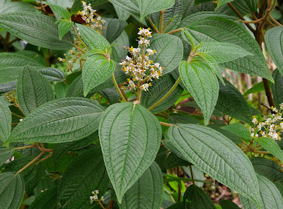
As currently recognised, Leandra includes over two hundred species with the highest diversity centred in southeastern Brazil. Leandra forms part of the tribe Miconieae, distinguished by flowers with more or less inferior ovaries and fleshy berry fruits. Genera within the Miconieae have historically been difficult to define; as early as 1891, the Belgian botanist Alfred Cogniaux declared that they were essentially arbitrary. Leandra was supposed to be defined by its acute petals and terminal inflorescences but it has not always been clear whether a given species can be said to possess these features or not. It should therefore come as no surprise that the genus Leandra proved to be polyphyletic with the advent of molecular analysis (Martin et al. 2008). Nevertheless, a large clade centered on southern Brazil has continued to be referred to as Leandra sensu stricto.

There appear to be few if any direct observations of pollination in Leandra but flower morphology and comparison with related genera suggests that they are buzz-pollinated with pollinators taking pollen as a reward (Reginato & Michelangeli 2016b; buzz-pollination referring to pollination by bees where the bee's buzzing induces the flower to release pollen). Apomixis, with seeds being produced directly from ovule tissue without pollination, is not uncommon and may even be the majority condition (Reginato & Michelangeli 2016a). Seeds are dispersed by birds feeding on the berries. Many Leandra species appear very localised in distribution and they are particularly diverse in a number of high altitude areas. Species vary in their preferred habitat from disturbed to undisturbed; those species found in undisturbed locations are rare components of the understory, but those found in disturbed habitats may be among the most abundant shrubs in the area.

REFERENCES

Martin, C. V., D. P. Little, R. Goldenberg & F. A. Michelangeli. 2008. A phylogenetic evaluation of Leandra (Miconieae, Melastomataceae): a polyphyletic genus where the seeds tell the story, not the petals. Cladistics 24: 315–327.

Reginato, M., & F. A. Michelangeli. 2016. Diversity and constraints in the floral morphological evolution of Leandra s.str. (Melastomataceae). Annals of Botany 118: 445–458.

Reginato, M., & F. A. Michelangeli. 2016. Untangling the phylogeny of Leandra s.str. (Melastomataceae, Miconieae). Molecular Phylogenetics and Evolution 96: 17–32.

The Sordariales: In the Soil and Under the Skin

Microfungi are a very important factor in our lives. They play a key role in assuring that we are not literally up to our armpits in shit. Their hungry little hyphae break down ordure, cleaning up the planet and unlocking nutrients that will then be made available to other organisms. And among the most significant lineages of these largely unseen decomposers are the members of the order Sordariales.

Lab culture of Sordaria fimicola, copyright BlueRidgeKitties.


Members of the Sordariales are, without exception, minute. Many species are coprophilous, growing on dung. Others may be found on rotting wood, or other decaying plant matter or soil. Fruiting bodies, when they appear, are flask-shaped perithecia protruding to a greater or lesser degree from the surface of their substrate. The walls of the perithecia are made up of large cells and have a membranous or coriaceous (leathery) texture. Within the fruiting body, the asci are single-walled and contain one- or two-celled ascospores that are often surrounded by a gelatinous sheath or bear various appendages. If the ascospores are two-celled, the cells are typically differentiated into an apical head and a basal tail (Kruys et al. 2015; Marin-Felix et al. 2020). Genera of Sordariales have historically been recognised on the basis of ascospore morphology but the advent of molecular data has indicated that such genera are highly polyphyletic. As a result, the Sordariales have seen (and are still seeing) a great deal of taxonomic reassessment. Miller & Huhndorf (2005) suggested that the structure of the fruiting body walls are more consistent with molecular phylogenies than ascospore morphology.

Cake of oncom-fermented beans, copyright Hariadhi.


Apart from their significant role as decomposers, most Sordariales have little direct impact on human economics. The mould Neurospora intermedia is used to make oncom, a fermented food similar to tempeh. A number of species of Sordariales such as Neurospora crassa and Sordaria fimicola have been widely used in genetic research, to the extent that they have been labelled the 'fruit flies of the fungal world'. Seriously, it's one of those expressions almost every publication seems obliged to crow-bar in somewhere. The analogy is made even more apropos by the fact that one of the most widely used species, Triangularia née Podospora anserina, has been made the subject of debate whether taxonomic considerations should be allowed to shake up the name of a popular model organism.

Molecular studies have also shown that the Sordariales encompass Madurella mycetomatis, a fungus causing subcutaneous inflammation in humans (van de Sande 2012). Seeing as sexual fruiting bodies are unknown in this species, and even asexual spore-producing structures are exceedingly rare, this organism would have previously been all but impossible to classify. Infection by M. mycetomatis is characterised by the production of granular swellings. It is most significant in central Africa but is also known from other tropical regions of the world. Madurella mycetomatis infects people via trauma such as animal bites and other wounds, and it has been isolated from soil and ant nests. In its normal state, M. mycetomatis is probably a quite innocent soil fungus. The trouble comes when it finds itself somewhere it shouldn't be.

REFERENCES

Kruys, Ã…., S. M. Huhndorf & A. N. Miller. 2015. Coprophilous contributions to the phylogeny of Lasiosphaeriaceae and allied taxa within Sordariales (Ascomycota, Fungi). Fungal Diversity 70: 101–113.

Marin-Felix, Y., A. N. Miller, J. F. Cano-Lira, J. Guarro, D. García, M. Stadler, S. M. Huhndorf & A. M. Stchigel. 2020. Re-evaluation of the order Sordariales: delimitation of Lasiosphaeriaceae s. str., and introduction of the new families Diplogelasinosporaceae, Naviculisporaceae, and Schizotheciaceae. Microorganisms 8: 1430.

Miller, A. N., & S. M. Huhndorf. 2005. Multi-gene phylogenies indicate ascomal wall morphology is a better predictor of phylogenetic relationships than ascospore morphology in the Sordariales (Ascomycota, Fungi). Molecular Phylogenetics and Evolution 35: 60–75.

van de Sande, W. W. J. 2012. Phylogenetic analysis of the complete mitochondrial genome of Madurella mycetomatis confirms its taxonomic position within the order Sordariales. PLoS One 7 (6): e38654.

The Age of the Perisphinctoid

During the Mesozoic era, the world's oceans were dominated by the ammonites. The coiled shells of these extinct cephalopods can be found preserved in rocks of this era around the planet, encompassing a bewildering array of species. During the latter half of the Jurassic, the most diverse ammonites were members of the superfamily Perisphinctoidea.

Likely Perisphinctes, copyright Spacebirdy.


Perisphinctoids first appear around the mid-point of the Jurassic, during what is known as the Bajocian epoch (Énay & Howarth 2019). As with other major ammonite groups, perisphinctoids are characterised by features of the folding around the edges of the septa that separate chambers of the shell. Perisphinctoids have basally five-lobed septa that differ from their ancestors in the Stephanoceratoidea in the loss of the UII lobe towards the outer edge of the whorl. The earliest perisphinctoids had more or less evolute shells (that is, later whorls did not significantly overlap the predecessors) with a rounded venter. Some later lineages would become more involute, with older whorls becoming partially hidden, and the venter might get sharper or flatter. Others would pretty much retain the original conformation to the end. The majority of perisphinctoids exhibited strong ribs on the outside of the shell, these ribs usually branching towards the outer rim of the whorl. Some forms developed further elaborations of the shells such as prominent nodules or spines.

Dimorphism was widespread in the perisphinctoids, if not universal. As with other dimorphic ammonites, populations included distinct microconches and macroconches (the majority interpretation is that macroconches were female and microconches male, but of course this is speculative). Macroconches usually had simple peristomes whereas microconches commonly had the mature shell aperture flanked by elongate lappets. The early Late Jurassic (Bathonian and Callovian) Tulitidae had a tendency in macroconches for the shell coiling to become eccentric in the outermost whorls, the peristome being distinctly skewed from the main plane of the shell.

Aspidoceras hirsutum, copyright Daderot.


Perisphinctoid faunas were often markedly provincial with many lineages being restricted to particular regions (such as the bipolar Perisphinctes or the western Eurasian Parkinsoniinae). They were mostly animals of shallower waters, perhaps foraging close to the bottom. This may go some way to explaining their high diversity but it can provide a challenge to their use in stratigraphy. Ammonites of the 'perisphinctoid' type would survive the end of the Jurassic but would fade from the fossil record not too long afterwards. Nevertheless, that would not be the end of their lineage: at the beginning of the Cretaceous, they would also spawn two derived descendants (Besnosov & Michailova 1991), the largely smooth-shelled Desmoceratoidea and the Ancyloceratoidea with four-lobed septa, that would continue to dominate the Mesozoic seas.

And while I'm on the subject of ammonites, I have another correction to make to an earlier post. However, while I was able to shift some of the blame for the correction in my last post onto my original source, in this case the blame is entirely mine. In a prior discussion of the live anatomy of ammonites, I discussed the evidence that the aptychus (a pair of calcified plates that probably functioned as an operculum) originated as a modification of the lower jaw. As such, I criticised reconstructions of ammonites that showed the aptychus articulating with the shell in the manner of a nautilus' hood. Unfortunately, I had overlooked a significant difference between ammonites and nautiluses. The coiled shell of a nautilus is exogastric—that is, when they evolved from their straight-shelled ancestors, the shell coiled upwards so the original lower edge corresponded to the outside of the whorl. However, the shell of ammonites was endogastric, with the shell coiled downwards so the original venter was on the inside (in the absence of preserved soft anatomy, we can infer this from the position of the siphuncle within the shell). This means that, even though the lower ammonite aptychus was anatomically on the opposite side of the animal from the upper nautilus hood, functionally they would have appeared in life to occupy much the same position. Entirely my mistake, and a reminder to me that describing orientation in coiled animals can be confusing.

REFERENCES

Besnosov, N. V., & I. A. Michailova. 1991. Higher taxa of Jurassic and Cretaceous Ammonitida. Paleontological Journal 25 (4): 1–19.

Énay, R., & M. K. Howarth. 2019. Part L, revised, volume 3B, chapter 7: Systematic descriptions of the Perisphinctoidea. Treatise Online 120: 1–184.

Ophiusini Corrections

Earlier this year, I presented a post on the noctuoid moth tribe Ophiusini. As it turns out, that post includes some notable errors. One of the main sources I used, Zahiri et al. (2012), stated that Ophiusini "have a strongly modified apex to the proboscis, with strong and enlarged spines and erectile, reversed hooks that are used in fruit-piercing or lachrymal-feeding behaviour". As reviewed by Zilli (2021), such hooks on the proboscis are unique to a separate subgroup of the family Erebidae, the Calpinae. Ophiusini have thin, nail-like spines on the proboscis but no erectile hooks. They are still fruit-piercers but no ophiusins have been observed to date engaging in lachrymal feeding.

Artena dotata, copyright Shipher Wu.


Zilli (2021) had further comments on the historically fraught concept of Ophiusini. As noted in my earlier post, 'Ophiusini' has historically been recognised as a cosmopolitan group of moths but molecular studies have lead to its restriction to the Old World, North American exemplars being transferred to the related tribe Poaphilini. However, though the two groups are each supported as monophyletic by molecular data, they are not well defined morphologically. Characters previously thought distinct to one or the other do not always hold true. Ophiusini have been described as having reduced coremata but some ophiusins have coremata larger than those of some poaphilins. Ophiusins have been supposed to lack the waxy bloom on the pupa found in other noctuoids but some species do indeed have such a bloom. Some have pointed to the use of Euphorbiaceae as host plants by Poaphilini but not Ophiusini, but not all poaphilins feed on Euphorbiaceae and their use of this plant family is generally correlated with species being more generalist feeders overall.

One character that may yet distinguish the two tribes is the location of the androteca, a groove along the top of one of the leg segments in the male that contains a long brush of dense hairs (I'm not sure just what the function of this structure is meant to be but I would suspect something to do with dispersing pheromones). In Ophiusini, this structure is found on the femur of the fore leg. In Poaphilini, it is on the tibia of the mid leg. Nevertheless, Zilli (2021) questions the reliability of this feature: both arrangments are found in other tribes and neither alone is diagnostic.

Conversely, molecular phylogenies support the two tribes as sister taxa, and they share a number of distinctive features of the terminalia. While he does not formalise the suggestion, Zilli (2021) seems to feel that we might be better served by a return to a broader Ophiusini uniting the two tribes as one. I commented in my previous post that noctuoid classification has been in a continuous flux for as long as it has been a thing. It would be presumptuous to believe that it has finally been settled.

REFERENCES

Zahiri, R., J. D. Holloway, I. J. Kitching, J. D. Lafontaine, M. Mutanen & N. Wahlberg. 2012. Molecular phylogenetics of Erebidae (Lepidoptera, Noctuoidea). Systematic Entomology 37: 102–124.

Zilli, A. 2021. Tabwecala robinsoni gen. nov., sp. nov., from Vanuatu and its systematic postion in the 'Ophiusini-Poaphilini' clade (Lepidoptera, Erebidae). Nota Lepidopterologica 44: 193–211.

Rasahus albomaculatus, the White-Spotted Corsair

Though the Hemiptera began their long evolutionary history as plant-feeders, many of their subgroups later switched to a predatory lifestyle, their suctorial mouthparts being just as suited for stabbing flesh as vegetation. Among the most successful of the predatory bugs where the assassin bugs of the family Reduviidae.

Image copyright Jacob Gorneau.


This is Rasahus albomaculatus, a widespread assassin of the Neotropical region, found from Mexico to Argentina (Coscaron 1983). Though not one of the largest members of its genus, R. albomaculatus is a decent-sized bug, growing close to an inch in length. Rasahus is a genus of the reduviid subfamily Peiratinae, commonly known as corsairs for their fearsome aspect. Features distinguishing Rasahus from other genera of corsairs include their large eyes, a deep grove across the head in front of the ocelli, long procoxae, and well-developed spongy pads on the fore- and mid-tibiae. Rasahus albomaculatus is distinguished from other species of the genus by its colour pattern. The body is mostly black with white patterning on the wings. Stripes along the top of the wing and across the mid-length form a crude H-shape when the wings are closed, with separate spots towards the base of the wing and towards the tip. Other noteworthy features include a lack of granulation on the pronotum, and a rounded apex to the scutellum (Swanson 2018).

Corsairs are mostly predators of other insects and not often dangerous to humans (though their bite is supposed to be very painful). Indeed, they may be beneficial to humans as among their prey are believed to be other reduviids of the subfamily Triatominae, the blood-sucking "kissing bugs" that spread Chagas disease (contrary to the Wikipedia page on the western corsair R. thoracicus, corsairs do not spread Chagas themselves). Rasahus albomaculatus may provide its vertebrate co-habitants with far more comfortable living conditions.

REFERENCES

Coscarón, M. del C. 1983. Revision del genero Rasahus (Insecta, Heteroptera, Reduviidae). Revista del Museo de La Plata (nueva serie) (Zoologia) 13: 75–138.

Swanson, D. R. 2018. Three new species of Rasahus, with clarification on the identities of three other Neotropical corsairs (Heteroptera: Reduviidae: Peiratinae). Zootaxa 4471 (3): 446–472.