Field of Science

Showing posts with label Foraminifera. Show all posts

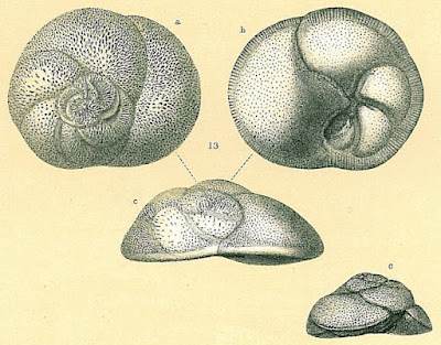
Lifestyles of the Rosalinidae

Among the modern foraminiferans, one of the most prominent radiations is among members of the Rotaliida, characterised by globose chambers and calcareous, hyaline test walls. Among the numerous families making up the Rotaliida are members of the Rosalinidae.

Benthic form of Rosalina globularis, from Brady (1884).


Rosalinids may be regarded as fairly typical-looking marine rotaliids with the test growing freely as a low trochospire (so a flattened cone or dish shape). The aperture of the test is a low slit on the interior margin along the umbilicus (Hansen & Revets 1992). Rosalinids have a complex life cycle involving both benthic and planktonic stages (Sliter 1965). The asexually reproducing diploid stage is benthic. Depending on conditions, diploid individuals may divide to produce other diploid individuals, resulting in several asexual generations. Eventually, however, the diploid generation will undergo meiosis to produce the haploid sexual generation (in the common species Rosalina globularis, this is induced by exposure to warmer water). In the sexual generation, a large globular chamber forms at maturity that covers the umbilical side of the test. This float chamber becomes filled with gas, allowing the foram to disperse planktonically before releasing gametes to produce the next diploid generation. Planktonic individuals are distinct enough in appearance from their benthic counterparts that they were long mistaken for distinct taxa before their identity was revealed by lab cultures.

Life cycle of Rosalina globularis, from Sliter (1965).


The majority of forams are particulate feeders. A network of filamentous pseudopodia radiating outwards from the cell body captures micro-organisms and other organic particles. However, one genus of rosalinids, Hyrrokkin, lives as parasites on sessile invertebrates (Cedhagen 1994). Species of this genus have variously been found on sponges, corals and bivalves. On sponges, they settle on the inhalent surface of the sponge and dissolve the underlying tissues. On bivalves, they form pits on the shell surface from which they bore holes through to the body cavity. Pseudopodia extended through this hole allow the foram to feed on host tissue. Infested hosts may bear multiple scars from the foram moving about on the outer surface. The forams may also feed on other animals such as polychaete worms or bryozoans attached to the surface of their primary host. In such cases, Hyrrokkin remains in its original pit but develops an irregularly shaped chamber with its aperture directed towards the alternate prey. Hyrrokkin species evidently do well from their rapacious lifestyle: whereas other rosalinids are only a fraction of a millimetre in diameter, Hyrrokkin sarcophaga is an absolute giant reaching around six millimetres across and with protoplasm containing thousands of nuclei. Proving once again that one may make a great deal of profit from the labour of others.

Cross-section of Hyrrokkin sarcophaga boring into shell of file clam Acesta excavata, from Schleinkofer et al. (2021).


REFERENCES

Cedhagen, T. 1994. Taxonomy and biology of Hyrrokkin sarcophaga gen. et sp. n., a parasitic foraminiferan (Rosalinidae). Sarsia 79: 65–82.

Hansen, H. J., & S. A. Revets. 1992. A revision and reclassification of the Discorbidae, Rosalinidae, and Rotaliidae. Journal of Foraminiferal Research 22 (2): 166–180.

Sliter, W. V. 1965. Laboratory experiments on the life cycle and ecologic controls of Rosalina globularis d'Orbigny. Journal of Protozoology 12 (2): 210–215.

The Bolivinitids

The Cretaceous was a period of significant innovation in the evolution of Foraminifera with a number of distinct new lineages making their appearance during this period. Among those, appearing in the latter part of the Cretaceous, were the first members of the modern family Bolivinitidae.

Bolivinita costifera, from the Smithsonian National Museum of Natural History.


The Bolivinitidae are free-living benthic forams with a calcareous, hyaline (glassy) test. The overall shape of the test is elongate with chambers arranged in biserial coils (that is, there are two chambers per loop). The terminal aperture is usually loop-shaped with a surrounding lip. Inside the chamber, a tooth plate (an inner protrusion of the test) runs from the aperture to the opening of the previous chamber and may protrude through the aperture (Revets 1996).

Representatives of the Bolivinitidae are found in a wide range of depths, from the shallow waters of the ocean to the bathyal zone. They may be among the most abundant forams in areas of low oxygen concentrations and are commonly associated with sustained organic matter input (Erdem & Schönfeld 2017). In other words, these are muck-lovers. Individuals growing in low oxygen conditions tend to show less pronounced surface sculpture on the test than those where the oxygen levels are higher. Conversely, individuals at deeper levels tend to be larger overall than those in shallower waters (Brun et al. 1984). As such, bolivinitids have received their fair share of attention as potential indicators of changes in environmental condition over time.

REFERENCES

Brun, L., M. A. Chierici & M. Meijer. 1984. Evolution and morphological variations of the principal species of Bolivinitidae in the Tertiary of the Gulf of Guinea. Géologie Méditerranéenne 11 (1): 13–57.

Erdem, Z., & J. Schönfeld. 2017. Pleistocene to Holocene benthic foraminiferal assemblages from the Peruvian continental margin. Palaeontologica Electronica 20.2.35A: 1–32.

Revets, S. A. 1996. The generic revision of the Bolivinitidae Cushman, 1927. Cushman Foundation for Foraminiferal Research Special Publication 34: 1–55.

The Glandulinid Position

In an earlier post, I described how the majority of modern multi-chambered foraminiferans can be divided between two lineages, the Tubothalamea and Globothalamea. The two groups generally differ in the shape of the first chamber following the proloculus (the central embryonic chamber of the test): in one, this chamber is tubular whereas in the other it is globular or crescent-shaped (guess which is which). But there is a third notable group of multi-chambered forams: the Nodosariata. In both tubothalameans and globothalameans, the chambers more or less coil around the proloculus to form a spiral. In the Nodosariata, the test is more or less linear with apical chamber apertures. The chambers may be successively stacked one after the other to form a uniserial test, or they may be arranged in a zig-zag or twirling arrangement to form biserial, triserial, etc. arrangments. In living Nodosariata, the wall of the test is made of a single layer of hyaline calcite though some earlier representatives (up to the end of the Jurassic) had differing wall make-ups (Rigaud et al. 2016). Among the numerous notable representatives of the Nodosariata in the modern fauna are representatives of the family Glandulinidae.

Series of Glandulina ovula, from Brady (1884).


Species have been assigned to the Glandulinidae going back to the Jurassic with the modern genus Glandulina recognisable in the Palaeocene (Loeblich & Tappan 1964). The test may be uniserial, biserial or polymorphine (more than two series); a common arrangement is for the test to start out biserial or polymorphine then become uniserial as the individual chambers become larger. In Glandulina, the microspheric generation starts biserial but the megalospheric form is uniserial throughout (Taylor et al. 1985). As the test grows, the internal walls between chambers may be resorbed. The terminal aperture of the test may be radial or slit-like. The most characteristic feature of the family is a tube running into the chamber from the inside of the aperture, referred to as the entosolenian tube. Some glandulinids have been described as lacking an entosolenian tube but such absences are likely artefacts of preservation: the delicate tube is easily dislodged during the fossilisation process (Taylor et al. 1985).

The overall relationships of the Nodosariata remain a question open to investigation. The classification of forams by Loeblich & Tappan (1964) included both multi-chambered and single-chambered (unilocular) forms within the Glandulinidae, with the unilocular forms placed in a subfamily Oolininae. Oolinines resemble glandulinids proper in a number of features including wall structure and the presence of an entosolenian tube. More recent authors, however, have rejected this relationship. Rigaud et al. (2016) entirely excluded unilocular forms from the Nodosariata as a whole, regarding it as improbable that single-chambered forms could have evolved from multi-chambered ancestors (as would seemingly be required by their relative appearances in the fossil record). Do the similarities between glandulinids and oolinines reflect a common ancestry, or are they the result of simple convergence? Unfortunately, with so few significant characters available to inform our understanding of foram higher relationships, the answer you prefer may come down to no more than your own personal feelings about which indicators are more reliable.

REFERENCES

Loeblich, A. R., Jr, & H. Tappan. 1964. Treatise on Invertebrate Paleontology pt C. Protista 2. Sarcodina: chiefly "thecamoebians" and Foraminiferida vol. 2. The Geological Society of America, and The University of Kansas Press.

Rigaud, S., D. Vachard, F. Schlagintweit & R. Martini. 2016. New lineage of Triassic aragonitic Foraminifera and reassessment of the class Nodosariata. Journal of Systematic Palaeontology 14 (11): 919–938.

Taylor, S. H., R. T. Patterson & H.-W. Choi. 1985. Occurrence and reliability of internal morphologic features in some Glandulinidae (Foraminiferida). Journal of Foraminiferal Research 15 (1): 18–23.

Pyrgoidae

I have referred in the past to there being something of a divide in approaches to the classification of the Foraminifera. This divide arises from disagreements such as the relative significance of various character complexes. One taxon that stands as an example of such disagreements is the subject of this post, the family Pyrgoidae as recognised by Mikhalevich (2005).

Pyrgo williamsoni, copyright Michael.


Pyrgoids are members of the group of forams generally recognised as the Miliolida, the porcelaneous forams. In this group, the wall of the test is composed of calcite but the calcite crystals are not regularly lined up with each other so the wall is not transparent. As a result, the wall of the test resembles porcelain in appearance. Most miliolidans have the chambers of the test coiling in a single plane. The Pyrgoidae were distinguished from other miliolidans by Mikhalevich (2005) by the overall structure of the test which is primarily biloculine (with the whorls of the test composed of two chambers). The family was divided into subfamilies by the nature of the test aperture: single with an inner tooth in Pyrgoinae, single with a flap in Biloculinellinae, and multiple (at least when mature) in Cribropyrgoinae and Idalininae. Idalininae also differed from other subfamilies in that the very last chamber was further enlarged to envelop the entire test. Members of the Pyrgoidae are known from the fossil record going back to the Jurassic period.

In the system of Loeblich & Tappan (1964), however, the pyrgoids were not recognised as a single group. Instead, they were dispersed among separate subfamilies of the family Miliolidae. Part of the reason was simply that Loeblich & Tappan did not divide the miliolidan families as finely as Mikhalevich later would but a bigger difference was one of priority. Loeblich & Tappan regarded the nature as an aperture as a more important feature taxonomically than the arrangement of chambers. Both classifications seem to have been constructed more from a diagnostic viewpoint than necessarily intended to reflect phylogenetic relationships.

Cribropyrgo aspergillum, from the National Museum of Natural History.


As with most other forams, pyrgoids exist in what are called megalosphaeric and microsphaeric forms. These forms represent alternate generations in the foram life cycle: microsphaeric forams are the sexually reproducing generation whereas megalosphaeric forams reproduce asexually. The names refer not to the overall size of the individuals but to the size of the proloculus, the very first embryonic chamber that sits at the center of the test. In megalosphaeric pyrgoids, the developing test is biloculine from the very start. In microsphaeric individuals, the earliest stages of the test are quinqueloculine (with five chambers per whorl) then become triloculine then finally biloculine (with a further progression for the idalinines, of course). The significance of the differences between the two forms has historically been the subject of discussion with some authors arguing that the microsphaeric forms represented a retention and overwriting of ancestral forms, or an expression of the trajectory the lineage might evolve along in the future (Loeblich & Tappan 1964). The most likely explanation, though, seems to me to be the simplest. The size of the proloculus correlates with the amount of cytoplasm in the young foram. Megalosphaeric pyrgoids start with fewer chambers per volution from the start for the simple reason that they don't have the space to pack in more.

REFERENCES

Loeblich, A. R., Jr, & H. Tappan. 1964. Treatise on Invertebrate Paleontology pt C. Protista 2. Sarcodina: chiefly "thecamoebians" and Foraminiferida vol. 1. The Geological Society of America, and The University of Kansas Press.

Mikhalevich, V. 2005. The new system of the superfamily Quinqueloculinoidea Cushman, 1917 (Foraminifera). Acta Palaeontologica Romaniae 5: 303–310.

Rugosofusulinids?

We return once again to the fusulinoids, large, complex Foraminifera of the late Palaeozoic. For this post, I'm taking a look at the Rugosofusulinidae, a group known from the last part of the Carboniferous and the earliest part of the Permian. Or to put it more technically, from the Gzhelian and Asselian epochs; their numbers collapsed at the end of the Asselian (Leven 2003).

Axial section of Rugosofusulina prisca, from Loeblich & Tappan (1964).


In an earlier post, I referred to a historical divide that has existed between American and Russian classifications of fusulinoids, with the Russian system recognising a more divided arrangement of taxa. The rugosofusulinids are one example of this: whereas Rauzer-Chernousova et al. (1996) recognise them as a distinct family in the order Schwagerinida, Loeblich & Tappan (1964) treated the entire group of 'schwagerinidans' as a subfamily Schwagerininae in the Fusulinidae (I believe more recent western authors might be inclined to at least treat Schwagerinidae as a separate family but would probably still not separate the rugosofusulinids). Whatever level you wish to place them at, the most distinctive feature of rugosofusulinids as a group is a distinct rugosity of the outer wall of the chambers. This may be due to undulations in the entire chamber wall or rugosity of the outer surface only. When first described, it was thought that this unevenness reflected ridges on the outer surface, but it was later observed that the rugosity looked much the same whatever angle the foram was cut at (remember, fusulinoids are most commonly studied in thin sections rather than as entire separated fossils) so probably represented more discrete ornaments. Skinner & Wilde (1966) suggested that "the outer surface [of Rugosofusulina] is scored by numerous sharp furrows which are directed both axially and sagittally, resulting in a surface which resembles a miniature cobblestone pavement".

The question of whether you wish to recognise rugosofusulinids as a distinct family is definitely not helped by a question hanging over recognition of the name Rugosofusulina. The problem is not really with Rugosofusulina itself but with another genus, Pseudofusulina, recognised in the Rauzer-Chernousova et al. (2007) system as type of another family of Schwagerinida, Pseudofusulinidae, and its type species P. huecoensis. Classically, this genus and family has been supposed to have a smooth rather than rugose outer tectum. However, the type specimen of P. huecoensis was re-examined by Skinner & Wilde (1966) who found that it did indeed have 'Rugosofusulina'-type external rugosities. They consequently synonymised the two genera with Pseudofusulina standing as the older name. The response of Russian authors to this challenge to their system, it seems, was generally to ignore it. Pseudofusulina and Rugosofusulina may still potentially be distinguishable as genera by degree of rugosity (Zhang et al. 2013) but this seems a weak basis for a full family distinction. Even if 'Rugosofusulina' is okay, 'Rugosofusulinidae' may not be.

REFERENcES

Leven, E. J. 2003. The Permian stratigraphy and fusulinids of the Tethys. Rivista Italiana di Paleontologia e Stratigrafia 109 (2): 267–280.

Loeblich, A. R., Jr, & H. Tappan. 1964. Treatise on Invertebrate Paleontology pt C. Protista 2. Sarcodina: chiefly "thecamoebians" and Foraminiferida vol. 1. The Geological Society of America, and The University of Kansas Press.

Rauzer-Chernousova, D. M., F. R. Bensh, M. V. Vdovenko, N. B. Gibshman, E. Y. Leven, O. A. Lipina, E. A. Reitlinger, M. N. Solovieva & I. O. Chedija. 1996. Spravočnik po Sistematike Foraminifer Paleozoâ (Èndotiroidy, Fuzulinoidy). Rossijskaâ Akademiâ Nauk, Geologičeskij Institut, Moskva "Nauka".

Skinner, J. W., & G. L. Wilde. 1966. Type species of Pseudofusulina Dunbar & Skinner. University of Kansas Paleontological Contributions 13: 1–7.

Zhang, Y.-C., Y. Wang, Y.-J. Zhang & D.-X. Yuan. 2013. Artinskian (Early Permian) fusuline fauna from the Rongma area in northern Tibet: palaeoclimatic and palaeobiogeographic implications. Alcheringa 37 (4): 529–546.

Chilostomellidae: Deep Forams

Holotype of Chilostomella serrata, from the Smithsonian National Museum of Natural History.


The specimen in the figure above is a fairly typical representative of the Chilostomellidae, a cosmopolitan family of forams known from the Jurassic to the present day. Members of this family have a translucent calcareous test with chambers arranged in a trochospiral (broad conical) or planispiral (flat spiral) pattern. The chambers of each spiral are expanded to cover over the prior spirals so only the outermost spiral is generally visible. The aperture of the test in the final chamber is a narrow slit along the margin with the underlying chamber (Loeblich & Tappan 1964).

Despite their long history and wide distribution, I get the general impression that chilostomellids are not usually abundant. They are generally restricted to deeper waters, more than 100 m below the surface (Cushman et al. 1954). Members of the genus Chilostomella, at least, have commonly been regarded as associated with low-oxygen environments. However, it has also been suggested that their favoured conditions are not so much a question of low oxygen as high organic flux (Jorissen 2002). Perhaps the best location to find chilostomellids would be around sites where dead animals and seaweeds have fallen to the deeper waters below.

REFERENCES

Cushman, J. A., R. Todd & R. J. Post. 1954. Recent Foraminifera of the Marshall Islands. Bikini and nearby atolls, part 2, oceanography (biologic). Geological Survey Professional Paper 260-H: 319–384, pls 82–93.

Jorissen, F. J. 2002. Benthic foraminiferal microhabitats below the sediment-water interface. In: Sen Gupta, B. K. (ed.) Modern Foraminifera pp. 161–179. Kluwer Academic Publishers: Dordrecht.

Loeblich, A. R., Jr & H. Tappan. 1964. Treatise on Invertebrate Paleontology pt C. Protista 2. Sarcodina: chiefly "thecamoebians" and Foraminiferida vol. 2. The Geological Society of America, and The University of Kansas Press.

Fusulinellidae, -inae, summat like that...

In an earlier post, I introduced you all to the fusulinids, a group of complex foraminiferans that were abundant during the later Palaeozoic. In that post, I alluded to the complex array of terminology that can be used when describing fusulinids but said that I would rather not cover it at that time. Well, this time I'm going to be dredging some of it up because I've drawn the Fusulinellidae as the topic for today's post.

Sectioned reconstruction of Fusulinella, from here. Labels: нк = primary chamber, са = septal folds, с = septa, сб = septal furrows, х = chomata, у = septal aperture, т = tunnel.


The Fusulinellidae as recognised by Vachard et al. (2013) are a family of fusulinids with fusiform or oblong tests known from the Middle to Late Pennsylvanian (during the later part of the Carboniferous). One genus, Pseudofusulinella, persists into the early Permian (Ross 1999). They are a part of the larger superfamily Fusulinoidea, a group of fusulinids characterised by what is known as a diaphanotheca. This is a thick, more or less translucent layer in the test wall. As noted in my earlier post, such a test structure may have functioned to allow light through to symbiotic microalgae (or possibly captured chloroplasts from algal prey) sheltered within. Fusulinellids are distinguished from other fusulinoids by the structure of the septa dividing chambers within the test, which are mostly flat except for some folding near the poles of the test (in the Fusulinidae, in contrast, the septal walls were folded throughout). As the test developed, sections of the septa were resorbed to form tunnels connecting adjacent chabers (and presumably allowing the transmission of materials between chambers in life). The course of the tunnels is commonly delimited within the chambers by chomata, discrete ridges of shell material. In other species, the chomata are absent but axial fillings of calcite were formed in the chambers instead.

How fusulinids are more commonly seen: sections of fusulinellid Dagmarella iowensis from Vachard et al. (2013). Image on left = subaxial section (scale bar = 0.1 mm); image on right, larger individual = tangential section (scale = 0.5 mm). The smaller individual on the right is a juvenile Profusulinella cf. fittsi, which depending on the author may or may not be considered a fusulinellid.


Being so widespread and abundant when they lived, fusulinellids are commonly used as index fossils for identifying when a deposit was formed. However, this process is complicated somewhat by ongoing debates about fusulinid systematics. Rauzer-Chernousova et al. (1996) proposed a classification of fusulinids that represented an extensive modification from previous systems. Part of this was simply a question of ranking, with Rauzer-Chernousova et al. recognising many groups at higher ranks than previously (so, for instance, recognising the separate family Fusulinellidae as opposed to its previous recognition as a subfamily of Fusulinidae). Nevertheless, some subsequent authors have felt that Rauzer-Chernousova et al. and their followers attribute too much significance to relatively minor variations. For instance, Kobayashi (2011) synonymised several genera under Profusulinella that Rauzer-Chernousova et al. regarded as belonging to distinct families (and Vachard et al. 2013 even placed in separate superfamilies). Some of the features regarded by Rauzer-Chernousova et al. as indicating separate genera were regarded by Kobayashi as representing variation within a single species. Indeed, there have even been arguments that some 'significant' features may represent post-mortem preservation artefacts (I've come across the term 'taphotaxa' used to refer to taxa based on such features). At present, my impression is that there is something of a geographical divide in preferred systems with eastern European authors following the lead of Rauzer-Chernousova et al. whereas authors from elsewhere may keep to a more conservative arrangement. The Berlin Wall may be down but the Fusulinid Cold War continues.

REFERENCES

Kobayashi, F. 2011. Two species of Profusulinella (P. aljutovica and P. ovata), early Moscovian (Pennsylvanian) fusulines from southern Turkey and subdivision of primitive groups of the family Fusulinidae. Rivista Italiana di Paleontologia e Stratigrafia 117 (1): 29–37.

Rauzer-Chernousova, D. M., F. R. Bensh, M. V. Vdovenko, N. B. Gibshman, E. Y. Leven, O. A. Lipina, E. A. Reitlinger, M. N. Solovieva & I. O. Chedija. 1996. Spravočnik po Sistematike Foraminifer Paleozoâ (Èndotiroidy, Fuzulinoidy). Rossijskaâ Akademiâ Nauk, Geologičeskij Institut, Moskva "Nauka".

Ross, C. A. 1999. Classification of the Upper Paleozoic superorders Endothyroida and Fusulinoida as part of the class Foraminifera. Journal of Foraminiferal Research 29 (3): 291–305.

Vachard, D., K. Krainer & S. G. Lucas. 2013. Pennsylvanian (Late Carboniferous) calcareous microfossils from Cedro Peak (New Mexico, USA). Part 2: smaller foraminifers and fusulinids. Annales de Paléontologie 99: 1–42.

Holding Forams Together

Nouria polymorphinoides, from Foraminifera.eu.


In past posts relating to the Foraminifera, I've made reference to the changes in classification undergone by this group over the years. Forams are unusual among unicellular organisms in producing a hard, often complex test that means they have both left an extensive fossil record and provided a number of characters on which to base a classification. However, there has been much disagreement over the relative attention due to particular features of the test. The classification used for forams in the Treatise on Invertebrate Paleontology by Loeblich & Tappan (1964), one of most influential sources in recent decades, made its primary divisions on the basis of the structure and chemistry of the test itself. Forams that produce a test by gluing together (agglutinating) sand particles and other foreign objects were treated as fundamentally distinct from those that secreted calcareous tests. Because the foram cell itself is amoeboid, there was an underlying assumption that the test architecture was too mutable to indicate anything more than low-level relationships.

However, there were some prominent inconsistencies with this assumption (Mikhalevich 2013). One is that the division between agglutinated and calcareous tests is not always perfect. Agglutinated forams might not secrete the bulk of the test themselves but they do secrete the cement used to hold the sand grains together, and there is a definite spectrum in the proportion of sand to cement used by a given foram. In some agglutinated forms, a distinct calcareous layer may underlie the agglutinated section of the test, and it is easy to envision how a progressive reduction in the proportion of agglutinated material could lead to the evolution of an entirely secreted test. This was not in itself fatal to the earlier system as it had generally been assumed that agglutinated forams were likely to represent a paraphyletic group. More problematic was the common appearance of foram species that were extremely similar in test architecture with the only really significant difference being that one was agglutinated and the other calcareous. This lead some authors to argue that whereas a small number of such cases might be accepted as the result of convergence, the abundance of such cases suggested that changes in test composition were more common than previously recognised. Molecular studies of forams are still in their infancy but have offered some support for the significance of test architecture, such as the division between globular and tubular forams (Pawlowski et al. 2013) that I referred to in an earlier post.

Liebusella goesi, from Foram Barcoding.


One effect of this change in focus is that the Mikhalevich (2013) classification divides the agglutinated forams between a number of groups that are not recognised in alternative systems. One such group is the Nouriida, known from the Cretaceous to the present day. Mikhalevich included the Nouriida in a larger group called the Hormosinana; at least one hormosinanan was placed by Pawlowski et al. (2013) at the base of the globular foram lineage. In contrast, Loeblich & Tappan (1964) included most of the nouriidans in the family Ataxophragmiidae, other members of which belong to the tubular forams. Nouriida and other Hormosinana are united by having the aperture of the test in a terminal position; in some nouriidans, it may be raised on a short neck. Nouriida differ from other hormosinanans in the arrangement of chambers in the test. In early stages they tend to be more or less trochospiral; with maturity, the number of chambers to a whorl decreases and the test may become biserial or uniserial. The two subfamilies recognised within the Nouriida by Mikhalevich differ in the internal structure of their chambers: Nourioidea have internally simple chambers but Liebuselloidea have the lumen of the chambers complexly subdivided.

I haven't found much about their ecological role; at least one modern species, Nouria polymorphinoides, seems to be not uncommon in shallower continental shelf waters worldwide. My general impression (just confirmed by asking a colleague who actually works on forams) is that agglutinated forams receive far less attention than calcareous ones. A big part of this is simply that they're harder to find: it takes a lot of practice to be able to pick out an actual agglutinated foram test from any other conglomeration of sand, and if they break apart during sample prep (which they often do) then there is little sign they were ever there to begin with.

REFERENCES

Loeblich, A. R., Jr, & H. Tappan. 1964. Treatise on Invertebrate Paleontology pt C. Protista 2. Sarcodina: chiefly "thecamoebians" and Foraminiferida vol. 1. The Geological Society of America, and The University of Kansas Press.

Mikhalevich, V. I. 2013. New insight into the systematics and evolution of the Foraminifera. Micropaleontology 59 (6): 493–527.

Pawlowski, J., M. Holzmann & J. Tyszka. 2013. New supraordinal classification of Foraminifera: molecules meet morphology. Marine Micropalaeontology 100: 1–10.

Meandering Forams

Specimen of Meandropsina vidali, showing the patterning on the external surface, from Loeblich & Tappan (1964).


There are some taxonomic names that just instantly bring up a mental image of the sort of organism to which they refer. For my part, I've always felt that Meandropsina is one of those names. The Meandropsinidae are another family of relatively large and complex foraminifera (growing up to a number of millimetres across) that are known only from the Upper Cretaceous. The several genera of the family are predominantly European, with only the genus Fallotia also known from the West Indies.

Cross-section of Meandropsina vidali, from Loeblich & Tappan (1964).


Meandropsinids are (as far as I know) more or less lenticular in shape with chambers enrolled in a flat spiral. The name of the type genus Meandropsina refers to the way that the outer margins of the chambers tend to meander irregularly around the test, giving it something of an ornate appearance. Both molecular and structural evidence indicate that multi-chambered forams arose from ancestors with undivided tests on more than one occasion, and the majority of multi-chambered forams can be assigned to two major lineages (Pawlowski et al. 2013). In one lineage, the Globothalamea (which includes, for instance, the rotaliids), the basic chamber shape is globular with successive chambers in the test being wider than long. In the other lineage, the Tubothalamea (including the miliolids and spirillinids), the basic chamber shape is tubular, and the test may grow through a number of spirals before it even starts to be divided into chambers (if at all). Members of the two lineages with calcareous tests may also be distinguished by their test structure: in calcareous globothalameans, the crystals making up the test are arranged regularly so the overall appearance of the test is hyaline (glass-like). In contrast, tubothalameans have the crystals of the test arranged irregularly so the appearance of the test is porcelaneous (like porcelain). Meandropsinids are unmistakeably tubothalameans in both regards.

Like other large forams of the Mesozoic, meandropsinids did not make it past the end of the Cretaceous. Early Palaeocene taxa that have been included in the families represent distinct lineages that evolved to take their place, occupying the ecological spaces opened up by the mass extinction ending the era.

REFERENCES

Loeblich, A. R., Jr, & H. Tappan. 1964. Treatise on Invertebrate Paleontology pt C. Protista 2. Sarcodina: chiefly "thecamoebians" and Foraminiferida vol. 1. The Geological Society of America, and The University of Kansas Press.

Pawlowski, J., M. Holzmann & J. Tyszka. 2013. New supraordinal classification of Foraminifera: molecules meet morphology. Marine Micropalaeontology 100: 1–10.

The Forams that Bind

Cross-section of Fabiania cassis, from BouDagher-Fadel (2008).


Here we see an example of Fabiania. Fabiania is a genus of foraminiferan known from the Eocene epoch that could reach a relatively large size as forams go, up to several millimetres across (nowhere near as large as some that I've covered on this site, maybe, but still respectable). It had a conical test with a rounded apex and a deeply excavated centre; depending on growing conditions, individual Fabiania might be a regular or a flattened cone. In its early stage, Fabiania had two globose thick-walled and perforate chambers; later chambers were cyclical and divided by horizontal and vertical partitions. The aperture of the test was a single row of pores opening into the large umbilicus. The wall of the test was thick and calcareous, and covered with coarse perforations on the upper side of the cone (BouDagher-Fadel 2008; Loeblich & Tappan 1964).

Fabiania lived in association with coral reefs, often preferring the undersides of corals and other sheltered locations. It was primarily found around the mid-depths, not too close to the water's surface but also not too deep (Bosellini & Papazzoni 2003). I've referred in an earlier post to another group of coral-encrusting forams, the acervulinids. Because reef forams tend to be cryptic (in more exposed parts of the reef they tend to get out-competed by coralline algae), and are often variable in morphology making them taxonomically difficult, they tend to be less studied than the reef's more prominent components. However, forams may play a not so insignificant role in developing the reef's structure, helping to bind the reef in place.

REFERENCES

Bosellini, F. R., & C. A. Papazzoni. 2003. Palaeoecological significance of coral-encrusting foraminiferan associations: a case-study from the Upper Eocene of northern Italy. Acta Palaeontologica Polonica 48 (2): 279–292.

BouDagher-Fadel, M. K. 2008. The Cenozoic larger benthic foraminifera: the Palaeogene. Developments in Palaeontology and Stratigraphy 21: 297–418.

Loeblich, A. R., Jr & H. Tappan. 1964. Treatise on Invertebrate Paleontology pt C. Protista 2. Sarcodina, chiefly "thecamoebians" and Foraminiferida vol. 1. The Geological Society of America and The University of Kansas Press.

Fusulinoids: Complex Forams of the Late Palaeozoic

Among the most characteristic fossils of the latter part of the Palaeozoic are the group of Foraminifera known as the fusulinoids. These forams, known from around the middle of the Carboniferous to the end of the Permian, can be extremely abundant. Indeed, I get the impression that some fossil deposits are pretty much made of fusulinoids. Fusulinoids did not merely thrive in their environment; they were the environment.

Limestone block dominated by fusulinids, copyright James St John. Field of view is about 3.9 cm across.


Fusulinoids are distinguished from other forams by their test composition, built from minute granules of calcite, and complex internal structure. Externally, fusulinoids (defined here to exclude their forerunners, the endothyroids) were fairly conservative, with a planispiral, usually involute test (that is, each successive whorl covers the last). The last whorl ended on a transverse wall without a defined aperture; instead, the only connection between the interior and exterior of the test was by a series of pores in said wall. Early forms were disc-shaped; later species could be more globular or fusiform. Some of the later fusulinoids also reached gigantic sizes by single-celled organism standards: whereas the earliest fusulinoids were only a fraction of a millimetre across, the late Permian Polydiexodina could be up to six centimetres along their longest axis (Loeblich & Tappan 1964). Internally, fusulinoids had an incredibly complicated and varied structure which I'm not going to go into too much detail about here, primarily because I barely understand a word of it myself. Any description of fusulinoid morphology quickly devolves into madly throwing about terms like chomata, parachomata, spirotheca, tectorium, and the like, and your humble narrator feeling the need to go look at something else.

Cutaway diagram of a fusulinid, showing an example of internal structure, from here.


I have to go into some detail, though, because some features of the fusulinoid wall structure may explain their success. The ancestral state for the fusulinoid test wall involved a thin layer of solid calcite, the tectum. In most species, the inside of the tectum was coated with a thicker, less dense layer. As the test wall becomes more derived, this inner layer becomes more or less translucent, or pierced by tubular alveoli to produce a honeycomb-like appearance. It has been suggested that these modifications may have been adaptations to accomodating symbiotic microalgae, striking a balance between maintaining the protective test and allowing optimal transmission of light. Microalgal associations with fusulinoids may be corroborated by the discovery of minute fossils of probable planktonic relationships such as Ovummuridae preserved within fusulinoid tests (Vachard et al. 2004).

Ecologically, fusulinoids were restricted to off-shore marine habitats, being mostly found preserved in limestones and calcareous shales. They are absent from deposits that would have been formed in brackish water, and while they may be found in sandstones it is debatable whether such occurrences represent life associations or post-mortem transport (Loeblich & Tappan 1964). Fusulinoids would therefore have been ecologically similar to the inhabitants of modern-day photic zone coral reefs, another reflection of their probable co-dependence with photosynthetic microalgae. However, as successful as the advanced fusulinoids were in their time, they did not make it past the massive extinction event at the end of the Permian. This was not the end of giant and complex forams entirely—indeed, some later forms such as the alveolinids would evolve morphologies very similar to those of fusulinoids—but it was the end of these particular giant forams.

REFERENCES

Loeblich, A. R., Jr & H. Tappan. 1964. Treatise on Invertebrate Paleontology pt C. Protista 2. Sarcodina, chiefly "thecamoebians" and Foraminiferida vol. 1. The Geological Society of America and The University of Kansas Press.

Vachard, D., A. Munnecke & T. Servais. 2004. New SEM observations of keriothecal walls: implications for the evolution of Fusulinida. Journal of Foraminiferal Research 34 (3): 232–242.

Forams with Teeth

Time for another foram post. The above image (copyright Robert P. Speijer, scale bar = 100 µm) shows Turrilina brevispira, a typical Eocene representative of the foram subfamily Turrilininae.

The Turrilininae are a group of calcareous forams that first appeared in Middle Jurassic (Loeblich & Tappan 1964). In most species, the test is what is called a 'high trochospiral' form: that is, it coils in a similar manner to, and overall looks rather like, a high-shelled snail. Each of these whorls is divided into at least three successive chambers, sometimes more. At the end of the test is a loop-shaped aperture. At least one species of turrilinine, Floresina amphiphaga, is a predator/parasite of other forams, drilling into their test to extract their protoplasm.

The turrilinines are most commonly classified in a broader foram superfamily known as the Buliminoidea or Bulimnacea. Other buliminoids commonly resemble turrilinines in their overall form. The group has commonly been defined, however, on the basis of what is called a 'tooth-plate'. This is an outgrowth of the internal wall of the test that runs between the apertures of each chamber. The exact appearance of the tooth-plate differs between taxa; in Turrilina, for instance, it is a trough-shaped pillar that is usually serrated along one end (Revets 1987). I have no idea what the function of the tooth-plate is, if indeed any is known, whether it provides an anchor for some cytoplasmic structure or anything else. However, in more recent decades a number of authors have questioned whether the tooth-plate is as significant a taxonomic feature as previously thought. For instance, Tosaia is a Recent genus of foram whose overall morphology and chamber arrangement is fairly typical for the Turrilininae but which lacks any sign of a tooth-plate (Nomura 1985). Excluding Tosaia from the buliminoids on this basis alone would imply a remarkably strong evolutionary convergence of every other feature of this genus.

REFERENCES

Loeblich, A. R., Jr & H. Tappan. 1964. Treatise on Invertebrate Paleontology pt C. Protista 2. Sarcodina, chiefly "thecamoebians" and Foraminiferida vol. 2. The Geological Society of America and the University of Kansas Press.

Nomura, R. 1985. On the genus Tosaia (Foraminiferida) and its suprageneric classification. Journal of Paleontology 59 (1): 222–225.

Revets, S. A. 1987. A revision of the genus Turrilina Andreae, 1884. Journal of Foraminiferal Research 17 (4): 321–332.

We've Got a Thing that's called Foram Love

Pileolina patelliformis, from Brady (1884).


It's been a while since we last had a foram post, so why don't we have one today? Ladies and gentlemen, I present to you: the Glabratellidae.

Glabratellids are a group of forams found in littoral habitats, first appearing in the fossil record in the Eocene (Loeblich & Tappan 1964). They secrete a calcareous test with a hyaline (glass-like) microstructure. By foram standards, glabratellids can be quite small: the smallest are well under 100 µm in diameter. They have a trochospiral body shape—that is, the body chambers are arranged in such a way that they spiral like a trochus or top shell—with a flat base. At the centre of the underside is an aperture or umbilicus. The spire may be fairly low, giving them what I always think of as a 'jelly mould' shape, or it may be high so their overall appearance is conical. In the genus Schackoinella, the test bears a spine on the outside of each of the body chambers.

The glory that is Schackoinella sarmatica, from the Geological Survey of Austria.


The most distinctive feature of glabratellids, perhaps, is their life cycle. We know the life cycles of relatively few foram species but as a rule they show a clear alternation of generations, with both well-developed haploid and diploid individuals. Haploid individuals (gamonts) produce gametes by nuclear mitosis that fuse to form zygotes that grow into mature diploid individuals (schizonts or agamonts); these latter produce haploid embryos via meiosis. The two generations may differ somewhat in appearance, and many foram species have had their gamonts and schizonts mistaken in the past for separate species. The most consistent difference between generations in all chambered forams is that the gamonts have a larger first chamber as a result of growing from larger embryos than the schizonts. In glabratellids, gamonts are also smaller and relatively higher-spired than schizonts, and the former are sinitrally coiled (to the left) while the latter are dextrally coiled (to the right).

The life cycle of Glabratellidae was described in detail by Loeblich and Tappan (1964) (the figure to the the left from therein shows the lifecycle of Pileolina patelliformis). Schizonts herald the production of offspring by wrapping themselves in a protective cover of dead diatoms and other rubbish. Young gamonts are formed by nuclei dividing in the test and each becoming surrounded by their own individual cell membranes. After they form, the embryonic offspring crawl around in the parent test feeding on any leftover cytoplasm and also on the test itself. By the time they grow to about two or three chambers in size, the gamonts dissolve the umbilical wall of the parent test and escape through the aperture.

As the gamonts themselves reach maturity, their thoughts no doubt turn to their own posterity. Whereas in some other forams the haploid generation simply releases their gametes into the water column to find their own way to fusion, sexual reproduction in glabratellids is a somewhat more intimate affair. Mature gamonts form into pairs, joined to each other via their umbilical surfaces from which they will resorb the test. Locked in their embrace, the pair become cemented to the substrate. Gametes, again, are formed by the production of plasma membranes around individual nuclei; these gametes move by means of three flagella instead of by pseudopodia. The two parents exchange gametes of which only about a tenth fuse to form zygotes; the remainder provide a food source for their developed siblings. Again, the young schizonts grow to about two or three chambers in size before being released by the dissolution of the cement holding the parent tests together.

This cosy mode of reproduction means that glabratellids may have the potential for greater population differentiation than other broadcast-spawning Foraminifera. Tsuchiya et al. (2003), in a study genetic diversity in representatives of the genus Planoglabratella collected around Japan, found evidence for cryptic speciation in P. opercularis. Some individuals of this 'species' were closer genetically to individuals of another species P. nakamurai than to other P. opercularis, and closer inspection revealed certain details of their morphology that were more nakamurai-like than opercularis-like. It may be that we have underestimated the diversity of glabratellids, and many more species of this group remain to be discovered.

REFERENCES

Loeblich, A. R., Jr & H. Tappan. 1964. Treatise on Invertebrate Paleontology pt C. Protista 2. Sarcodina: chiefly "thecamoebians" and Foraminiferida. The Geological Society of America, and The University of Kansas Press.

Tsuchiya, M., H. Kitazato & J. Pawlowski. 2003. Analysis of internal transcribed spacer of ribosomal DNA reveals cryptic speciation in Planoglabratella opercularis. Journal of Foraminiferal Research 33 (4): 285–293.

Hyperamminids: A Rough Retort

Hyperammina elongata, photographed by Onno Groß.


Agglutinated foram time again! In previous posts, I've described how these aquatic amoeboids construct coverings for themselves by cementing together particles from the surrounding environment. In the family of forams I'm presenting you with today, the Hyperamminidae, their choice of particle is generally sand grains, glued together with a relatively small amount of cement. As a result, hyperamminids often have a quite rough appearance to their walls. They live free, not cemented to their substrate. The test is not divided into chambers; instead, it starts as a globular chamber (the proloculus) that opens into an elongate tube. The overall appearance, therefore, is not dissimilar to one of those glass cylinders with a basal bulb (like an old thermometer). In species of Hyperammina, the tube is simple and tapers as it gets further from the proloculus. In contrast, the genus Saccorhiza has the tube more constant in diameter, and also has the tube branching dichotomously (Loeblich & Tappan 1964). Hyperamminids are abundant in the deep sea, and though certainly not among the largest forams, they can easily be a millimetre or more in size.

Agglomeration of Saccorhiza ramosa tubes, from here.


The classification of agglutinated forams presented by Kaminski (2004) lists six genera in the Hyperamminidae, with separate subfamilies for Hyperammina and Saccorhiza. These two genera are the only ones alive today; the remainder are all fossils. The record of hyperamminids stretches back some way: specimens have been assigned to Saccorhiza from the Lower Carboniferous, while Hyperammina is recorded from as far back as the Lower Ordovician. Yep, that's a single genus that goes back nearly 500 million years (really makes you wish that meant something). The genus Platysolenites, if correctly placed within the hyperamminids, is one of the oldest of all forams, known from the very early Cambrian. The other genera are also Palaeozoic; one of them, Sacchararena, had a test made with fine white sand, leading to its name: 'sugar sand' (Loeblich & Tappan 1984).

REFERENCES

Kaminski, M. A. 2004. The Year 2000 classification of the agglutinated Foraminifera. In: Bubík, M. & M. A. Kaminski (eds) Proceedings of the Sixth International Workshop on Agglutinated Foraminifera. Grzybowski Foundation Special Publication 8: 237-255.

Loeblich, A. R., Jr & H. Tappan. 1964. Treatise on Invertebrate Paleontology pt C. Protista 2. Sarcodina, chiefly "thecamoebians" and Foraminiferida, vol. 1. The Geological Society of America and The University of Kansas Press.

Loeblich, A. R., Jr & H. Tappan. 1984. Some new proteinaceous and agglutinated genera of Foraminiferida. Journal of Paleontology 58 (4): 1158-1163.

Porcelain Fans

Mature specimen of Rhapydionina deserta, from Loeblich & Tappan (1964).


Calcareous foraminiferans have been featured on this site before: planktic floaters, living stars, microscopic jelly moulds and gigantic reef-formers. All these forms have belonged to the group of calcareous forams known as the rotaliids. Today's subject is another group of forams, the Rhapydionininae, belonging to a different calcareous group, the Miliolida. Miliolids may have shell walls made of calcite like the rotaliids, but differ in the wall structure: while the walls of rotaliids are glass-like and porous, those of miliolids are structured like porcelain. Phylogenetic studies of forams have not placed the miliolids close to the rotaliids, and the two groups seem to have evolved their secreted shells independently (Sen Gupta 2002).

Rhipidionina liburnica, from Loeblich & Tappan (1964).


The Rhapydionininae were defined by Loeblich & Tappan (1964) as a group of miliolids with a conical test composed of broad chambers stacked one on top of another (the overall shape being kind of like a fan or an ice-cream cone), with each of these chambers subdivided by internal septa into multiple chamberlets (the difference between a 'chamber' and a 'chamberlet' being that the latter are not completely divided from each other by the walls). The opening of the test took the form of a sieve-like array of pores at the top end. However, subsequent researchers have discovered that Loeblich & Tappan's definition was inadequate. Rhapydioninines start life growing as a flat spiral, with growth becoming linearised at maturity. However, it turns out that not all Rhapydionininae become linear; some retain their juvenile coiling into maturity (Vicedo et al. 2011). At least some species are believed to have both a linear megalospheric form and a coiled microspheric form. To explain, forams can be divided between microspheric forms, in which the first chambers of a new test are much smaller, and megalospheric forms with larger initial chambers. In those relatively few forams whose life cycles have been studied in detail, these two forms correspond to an alternation of generations, with a mostly microspheric asexually-reproducing generation giving rise to the generally megalospheric sexually-reproducing phase. Loeblich & Tappan's (1964) concept of rhapydionines, therefore, would have potentially placed members of a single species into separate families.

Diagram of internal structure of two adult chambers of Cuvillierinella, from Vicedo et al. (2011). Key to abbreviations: ap f = apertural face, c chl = cortical chamberlets, flo = floor, m chl = medullar chamberlet, prp = preseptal space, rpi = residual pillars, s = septum, sl = septulum.


Rhapydionines are best known as fossils, with a definite range from the Upper Cretaceous to the mid-Eocene (Loeblich & Tappan 1984). Believe it or not, whether there are still rhapydioninines in the world is something of an open question. Loeblich & Tappan (1964) listed two Recent genera in the Rhapydionininae, each represented by only a single known specimen. Ripacubana conica was originally described from sand deposits in Cuba; however, Loeblich & Tappan (1964) suggested that Ripacubana may actually represent what has been referred to as a 'zombie taxon'. Some of you may be familiar with the palaeontological concept of a 'Lazarus taxon', where a species disappears from the fossil record only to reappear at a later date. What has actually happened in these cases is that the species had only become locally extinct, but survived in some other locality that has not been preserved, subsequently recolonising its old range. A 'zombie taxon', however, is one that has genuinely become extinct at the earlier date, but its fossilised remains have since been transported into a younger sediment deposit, giving the impression that it survived later than it did*. In the case of Ripacubana, it is difficult to know just how long a foram shell buried in sand has been lying there.

*Identifications of Lazarus taxa also have to be on the look-out for 'Elvis taxa': where the more recent population does not in fact represent the same species, but a different species that has convergently evolved similar features.

Craterites rectus, from Loeblich & Tappan (1964).


Loeblich & Tappan (1964) did not express the same reservations about Craterites rectus, described from a beach on Lord Howe Island east of Australia. Craterites was later separated as its own subfamily by Loeblich & Tappan (1984) on the basis of its being attached to the substrate, and so differing from other free-living Rhapydionininae. Nevertheless, they kept the two subfamilies together as the family Rhapydioninidae, so Craterites may still be the only known survivor of the rhapydioninine lineage. However, with only one known specimen, the details of the internal structure of Craterites remain unknown.

REFERENCES

Loeblich, A. R., Jr & H. Tappan. 1964. Treatise on Invertebrate Paleontology pt C. Protista 2. Sarcodina, chiefly "thecamoebians" and Foraminiferida, vol. 1. The Geological Society of America and The University of Kansas Press.

Loeblich, A. R., Jr & H. Tappan. 1984. Suprageneric classification of the Foraminiferida (Protozoa). Micropaleontology 30 (1): 1-70.

Sen Gupta, B. K. 2002. Modern Foraminifera. Springer.

Vicedo, V., G. Frijia, M. Parente & E. Caus. 2011. The Late Cretaceous genera Cuvillierinella, Cyclopseudedomia, and Rhapydionina (Rhapydioninidae, Foraminiferida) in shallow-water carbonates of Pylos (Peloponnese, Greece). Journal of Foraminiferal Research 41 (2): 167-181.

Acervulinids: Reef Forams

Regular readers of this site will know that, contrary to common belief, not all representatives of the vaguely defined category of organisms known as 'protozoa' are too small to be seen with the naked eye. Some are very visible indeed, and many of the more visible forms can be found among the shell-constructing amoeboids known as Foraminifera. One group of giant forams, the xenophyophores, has become fairly famous in internet circles as one of the contenders for the title of 'largest single cell', though, as noted in the post linked to, the question is kind of a pointless one with regard to forams. Besides, as described later in this post, xenophyophores may not have even have always been the largest forams.

Encrusting nodules of Acervulina inhaerens from Rhodes, from M. Hesemann.


The Acervulinidae are a family of reef-inhabiting forams belonging among the rotaliids. Juvenile chambers of newly growing acervulinids are arranged in a flat spiral, but chambers of mature specimens may be arranged in one to several layers. The chambers do not have regular apertures, and instead their walls are only pierced by coarse pores (Perrin 1994). Genera and species of acervulinids are distinguished by the presence, arrangement and shapes of layers and chambers, but defining distinctions appropriately is challenging. Acervulinids do not have a determinate 'adult morphology'; instead, the final adult appearance can be affected by factors such as substrate relief and water movement. Properly identifying acervulinids therefore requires identification of features independent of these external factors.

Living crust of Gypsina plana, photographed by Hal Ray Tichenor.

Acervulinids can be abundant on tropical coral reefs, and may play a not insiginificant role in reef formation as binding organisms. They tend to be particularly prominent in deeper parts of the reef, as they can tolerate lower light levels than other organisms such as coralline algae; in shallower parts of the reef, they are found in more cryptic locations among the coral. Acervulinids may be free-living, or they may be directly attached to their substrate. Like the star-shaped calcarinids, their primary food source is benthic diatoms (that they may or may not live with symbiotically), and the abrupt disappearance of the modern Acervulina inhaerens below depths of 130 m probably corresponds to the lower limit of that food source (Bosellini & Papazzoni 2003).

Fossilised nodules from a Solenomeris reef, photographed by Stefano Dominici. Note that Stefano identifies these as Acervulina; due to the complications in distinguishing acervulinid taxa, it remains contentious whether Solenomeris and Acervulina can be reliably separated.


Attached acervulinids may form either nodules or spreading crusts, depending on species and/or growth conditions (Perrin 1994). Such nodules or crusts may have diameters in the millimetre range, but some living species may be within the decimetre range. The most dramatic expression of acervulinid potential, however, was known from the Tethyan region during the Eocene period (the Tethys, for those unfamiliar with it, was the sea that connected the Atlantic and Indian Oceans north of Africa, before the northward movement of that continent closed off the Mediterranean at the eastern end). Here was found Solenomeris ogormani, initially interpreted as a red alga but since reidentified as an acervulinid. Solenomeris was primarily an encrusting form, but large growths would also produce tightly packed branches one or two centimetres in diameter. Over time, Solenomeris formed massive metre-sized domes, and these domes together would form entire reefs stretching over multiple kilometres: reefs formed not of coral, or of algae, but purely of forams!

REFERENCES

Bosellini, F. R., & C. A. Papazzoni. 2003. Palaeoecological significance of coral−encrusting foraminiferan associations: a case−study from the Upper Eocene of northern Italy. Acta Palaeontologica Polonica 48 (2): 279-292.

Perrin, C. 1994. Morphology of encrusting and free living acervulinid Foraminifera: Acervulina, Gypsina and Solenomeris. Palaeontology 37 (2): 425-458.

The Osangulariidae: Deep-Water Trochospires

Dorsal (spiral side), lateral and ventral (umbilical side) views of an Osangularia specimen, from here.


For today's post, I'm presenting for your consideration the Osangulariidae, a family within the rotaliid Foraminifera (see here for my post introducing the Rotaliida). These are benthic forams, mostly found in intermediate waters within the top few centimetres of sea floor sediment (Kaiho 1998). The Osangulariidae were first established as a distinct family of Foraminifera by Loeblich & Tappan (1964) to include trochospiral forams with bilamellar walls, with an important distinguishing feature separating osangulariids from related families being their granular rather than radial test wall structure. However, Loeblich & Tappan were criticised by Kaiho (1998) for their utilisation of this character. In developing a more lineage-based classification of the osangulariids and related taxa, Kaiho concluded that "radial-granular texture has no taxonomic significance in the suprageneric classification of calcareous trochospiral benthic foraminifera". Instead, Kaiho defined the Osangulariidae as trochospiral forams with an aperture on the umbilical side of the test, an angular periphery and strongly oblique sutures on the spiral side.

The Coniacian (Late Cretaceous) Globorotalites multisepta, from Loeblich & Tappan (1964).


The Osangulariidae first appeared in the Early Cretaceous, during the Aptian epoch. Kaiho (1998) recognised two subfamilies within the Osangulariidae, the Osangulariinae and the Globorotalitinae (not to be confused with the Globorotaliinae), regarding the slightly earlier-appearing Globorotalitinae as probably ancestral to the Osangulariinae. The Globorotalitinae possessed a test with a strongly inflated umbilical side, and can basically be described as looking like a jelly mould. Osangulariids of the globorotalitine type became extinct during the Palaeocene.

Nuttallides rugosus, from Todd 1965.


The Osangulariinae, on the other hand, have survived to the present day. Their most obvious distinction from the Globorotalitinae is the reduction of the umbilical side of the test, so that osangulariines tend to be more discus-shaped than jelly-mould-shaped. The earliest osangulariine genus, Protosangularia, appeared in the Aptian and survived until the Cenomanian in the early Late Cretaceous. At the end of the Cenomanian, a major anoxic event took place in the ocean followed by a reduction in world ocean temperatures. After this, Protosangularia was replaced by a number of other osangulariine genera appearing from the Turonian to the early Campanian (Kaiho 1998). Two of these genera, Osangularia and Nuttallides, are the family's modern representatives.

REFERENCES

Kaiho, K. 1998. Phylogeny of deep-sea calcareous trochospiral benthic Foraminifera: evolution and diversification. Micropaleontology 44 (3): 291-311.

Loeblich, A. R., Jr & H. Tappan. 1964. Treatise on Invertebrate Paleontology pt C. Protista 2. Sarcodina. Chiefly "thecamoebians" and Foraminiferida vol. 2. The Geological Society of America and The University of Kansas Press.

The Rotaliida: Building a Wall

Tests of Elphidium crispum, photographed by Spike Walker.


The foraminifers have been featured on this site a number of times before, when various members of this diverse group of unicellular organisms have been introduced. Today, I thought I'd take a look at the broader classification of forams through the lens of one of their major subgroups, the rotaliidans.

The earliest classifications of forams divided them on the basis of the number and arrangement of chambers within the test, but over time the composition of the test walls came to be also recognised as an important feature (Haynes 1990). This reached an apotheosis of sorts in the Treatise on Invertebrate Paleontology classification of Loeblich and Tappan (1964), in which the forams were divided between five suborders primarily on the basis of test composition. These were the Allogromiina (with membranous or chitinous tests), Textulariina (with agglutinated tests) and three suborders with calcareous tests but differing wall structures: the microgranular Fusulinina, the porcelaneous, imperforate Miliolina and the hyaline, perforate Rotaliina (other authors would treat these groups as orders, with the suffix -ida instead of -ina). However, later authors (including Loeblich and Tappan themselves) regarded this classification as somewhat oversimplified, and divided groups such as the planktonic rather than benthic Globigerinida, the aragonitic rather than calcitic Robertinida, the Lagenida with monolamellar rather than bilamellar walls, and the high-spired or serial rather than planispiral Buliminida from the Rotaliida proper (Haynes 1990). However, many of the subdivisions within forams remained somewhat artificial, and potentially did not reflect true evolutionary relationships.

Bulimina marginata, photographed by Fabrizio Frontalini.


Molecular phylogenetics of forams got off to a fairly rocky start. For various reasons, extraction of reliably genetic samples from forams is a difficult process (for instance, their tendency to live in symbiotic associations makes contamination a continuing issue). However, studies have progressed to the point where a broad outline is beginning to emerge. One significant agreement between studies has been the monophyly of forams with a perforate calcareous test (Flakowski et al. 2005; Schweizer et al. 2008). The 'Globigerinida' and 'Buliminida' have both been shown to fall within this clade, and should probably not be distinguished from the Rotaliida. No representatives of the Lagenida or Robertinida appear to have been analysed molecularly; the lagenidans may be an independent lineage, while the robertinidans may be closely related to the Rotaliida (Sen Gupta 2002).

Suggested relationships of major foraminiferan groups from Sen Gupta (2002). This arrangement, which is somewhat concordant with available molecular data, proposes two separate lineages of multi-chambered forams, with calcareous members in each.


Within the Rotaliida, the most extensive molecular analysis has been that of Schweizer et al. (2008), whose results support a division between three main clades. Though reasonably well supported molecularly, these clades do not correspond to morphological divisions: the 'Buliminida', for instance, are divided between at least two clades. The planktonic forms may also be polyphyletic within the Rotaliida, though analyses have been inconsistent on their exact position in the clade (and Schweizer et al. do not include any globigerinidans in their analysis). Not all molecular results clash with the morphology, though: the Nummulitidae, a group of giant discus-shaped forams that get up to five centimetres in diameter, are one of a number of families that remain supported by either data source.

Internal chambers of Nummulites gizehensis, from the Natural History Museum. This species is most famous for being the major component of the limestone used in the construction of the pyramids of Giza.


REFERENCES

Flakowski, J., I. Bolivar, J. Fahrni & J. Pawlowski. 2005. Actin phylogeny of Foraminifera. Journal of Foraminiferal Research 35 (2): 93-102.

Haynes, J. R. 1990. The classification of the Foraminifera—a review of historical and philosophical perspectives. Palaeontology 33 (3): 503-528.

Loeblich, A. R., Jr & H. Tappan. 1964. Treatise on Invertebrate Paleontology pt C. Protista 2. Sarcodina, chiefly "thecamoebians" and Foraminiferida, 2 vols. The Geological Society of America and The University of Kansas Press.

Schweizer, M., J. Pawlowski, T. J. Kouwenhoven, J. Guiard & B. van der Zwaan. 2008. Molecular phylogeny of Rotaliida (Foraminifera) based on complete small subunit rDNA sequences. Marine Micropaleontology 66: 233-246.

Sen Gupta, B. K. 2002. Modern Foraminifera. Springer.