Field of Science

Showing posts with label Brachiozoa. Show all posts

The Huenellidae

Researchers who deal with the modern marine fauna are used to thinking of brachiopods as a marginal group, their diversity greatly overshadowed on a global scale by the superficially similar bivalves. However, modern brachiopods are but a shadow of their former selves; for much of the Palaeozoic era, their relationship with the bivalves was the inverse of today. Many are the brachiopod lineages that came and went over this time.

External views of ventral (left) and dorsal valves of Huenella triplicata, from Walcott (1924).


The Huenellidae were an assemblage of brachiopods that lived during the late Cambrian and early Ordovician (Amsden & Biernat 1965). They represent early representatives of the Pentamerida, a Palaeozoic order of fairly generalised-looking brachiopods. Within the Pentamerida, they fall within the suborder Syntrophiidina. Syntrophiidinans as a whole are rarely found in the fossil record and as a result remain poorly known. Members of the suborder share a distinctive shape with biconvex valves marked by a dorsal fold and ventral sulcus. That is, the midline of the shell is raised above either side with the ventral valve forming a 'valley' to match the raised 'hill' of the dorsal valve. What, if anything, was the purpose of this arrangement I wouldn't know but modern brachiopods often inhabit locations with a lot of organic silt and/or fine sediment. Perhaps the uneven level of the syntrophiidinan shell helped protect it from burial by a shifting substrate.

Interior view of ventral valve of Radkeina taylori, from Laurie (1997), with scoop-shaped spondylium at upper midline.


Families of Syntrophiidina may be distinguished based on the development of the spondylium, an internal projection at the base of the ventral valve that provided an attachment site for the shell muscles. Members of the Huenellidae possessed either a sessile spondylium or a pseudospondylium, a spondylium-type structure rising from the internal surface of the valve itself rather than from the hinge. Amsden & Biernat (1965) recognised a division of the huenellids between two subfamilies based on the development of the brachiophore plates, projections on the inside of the dorsal valve that would have supported the lophophore. Members of the Huenellinae possessed more developed brachiophores than members of the Mesonomiinae. Outer ornament of the huenellid shell varied from more or less smooth with weak concentric ridges to costate with distinct radiating ridges.

Phylogenetic relationships within the Syntrophiidina do not seem to have been established in detail but the early appearance in the fossil record of huenellids at least raises the question of whether they included the ancestors of later families. As well as other families of the Syntrophiidina, candidates for descent would include members of the suborder Pentameridina as well as of the related order Rhynchonellida. This latter order includes species which survive to the present day so the possibility exists that while the huenellids themselves may be long gone, their legacy may yet live on.

REFERENCE

Amsden, T. W., & G. Biernat. 1965. Pentamerida. In: Moore, R. C. (ed.) Treatise on Invertebrate Paleontology pt H. Brachiopoda vol. 2 pp. H523–H552. The Geological Society of America, Inc.: Boulder (Colorado), and The University of Kansas Press: Lawrence (Kansas).

The Oligorhynchiidae

Dorsal view of Oligorhynchia subplana gibbosa, from Cooper (1935).


From Oligochiton, we move onto Oligorhynchia. The Oligorhynchiidae are a family of very small brachiopods known from the Middle and Late Ordovician. They were among the earliest representatives of the Rhynchonellida, a major group of brachiopods that survives to the present day. Rhynchonellidan shells are usually characterised by a strong beak associated in life with a well-developed pedicel. In oligorhynchiids, this beak is suberect and the shell as a whole is an elongate subtriangular shape. The valves of the shell are folded into coarse plicae (ridges). At least towards the base of the shells, the major folds are in what is called an inverted arrangement, with a ridge in the dorsal valve matched by a valley in the ventral valve (Schmidt & McLaren 1965). Other structural features defining the group include small plates projecting into the pedicel opening, distinct vertical dental plates and divided hinge plates in the valve articulation, and the usual absence of a median septum or cardinal process inside the shell (Savage 1996).

The oligorhynchiids first arose in the east of what was then the continent of Laurentia (corresponding to modern North America). They subsequently spread across the Iapetus Ocean to the continents of Baltica and Kazakhstan (Jin 1996). The end of the Ordovician saw their replacement by other rhynchonellid families. Nevertheless, their genetic lineage would continue for some time yet as they have been identified as ancestors of later families: the Trigonirhynchiidae and Camarotoechiidae (Jin 1989). The brief oligorhynchiid spark would blossom into later rhynchonellid success.

REFERENCES


Jin, J. 1989. Late Ordovician–Early Silurian rhynchonellid brachiopods from Anticosti Island, Quebec. Biostratigraphie du Paléozoïque 10: 1–127, 130 pls.

Jin, J. 1996. Ordovician (Llanvirn–Ashgill) rhynchonellid brachiopod biogeography. In: Copper, P., & J. Jin (eds) Brachiopods pp. 123–132. CRC Press.

Savage, N. M. 1996. Classification of Paleozoic rhynchonellid brachiopods. In: P. Copper, & J. Jin (eds) Brachiopods pp. 249–260. CRC Press.

Ice-cream Cones of the Early Palaeozoic

It's time for something I haven't done in a very long time... (credit to Niel from Microecos):


I briefly described tentaculitoids on this site way back in September 2007. These narrowly conical shells of uncertain affinities were prominent members of the marine fauna during the Silurian and the Devonian, only to then disappear without a trace. No direct evidence is available for the soft-body appearance of the animals that produced them nor are we overly certain on their lifestyle. But at least one of the major subgroups of the tentaculitoids, the Dacryoconarida, are held to be of palaeontological significance due to their ubiquity and cosmopolitan distribution at the species level making them of use in biostratigraphy.

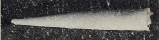
Reconstruction of Nowakia elegans, from Berkyová et al. (2007).


Dacryoconarids have generally been presumed to be planktonic in some way, owing to the aforementioned tendency of individual species to be found more or less worldwide, together with their small size (generally about the centimetre range). Dacryoconarids are distinguished from other tentaculitoids by the apical portion of their shell ending in a small globular bulb, presumed to represent the embryonic or larval shell of the original animal (Farsan 2005). A more or less distinct constriction or 'neck' separates this embryonic bulb from the remainder of the shell. In those forms with more heavily ornamented shells such as the genus Nowakia, a distinct juvenile section of the shell is visible immediately following the embryonic bulb in which the adult ornament is absent or weakly developed; said adult ornament, when it appears, takes the form of rounded transverse ridges and troughs, often associated with longitudinal and/or transverse striae. In other forms, such as the genus Styliolina, the outside of the shell is flat and ridgeless, with at most the only ornamentation present being striae. The inside of the shell may be rippled to follow the exterior ornamentation or it may be perfectly smooth (Fisher 1962).

Dacryoconarids are first recorded from the Late Ordovician but they remained at relatively low diversity until the Devonian which saw a notable radiation (Wittmer & Miller 2011). Nevertheless, they declined rapidly towards the end of the Devonian. It has been suggested that their extinction by the end of that period may be related to the appearance of more actively swimming predatory fish before which the tentaculitoids may have been relatively defenceless. Other early Palaeozoic planktic groups such as the graptoloids experienced a similar collapse at about this time, though the disappearance of the dacryoconarids may have lagged behind that of the graptoloids.

Styliolina clavulus, from Fisher (1962).


Over the years, a wide range of suggestions have been made about the affinities of the tentaculitoids, ranging from jellyfish to annelids. Perhaps the most persistent association has been made with molluscs but there really is little to support such a premise than the possession of a calcareous shell, a feature that is hardly unique to molluscs even among living animals. The structure of the tentaculitoid shell is most similar to that of some brachiopods (Fisher 1962) and some sort of brachiozoan affinity is perhaps the currently most favoured concept. As noted above, we know nothing about the tentaculitoid anatomy other than what we can infer from the nature of the shells themselves. In some larger tentaculitoids (though not among the dacryoconarids so far as we know) the apical parts of the shell may become walled off by solid septa so the living animal presumably didn't occupy the entire shell. Fisher (1962) described the tentaculitoids as "presumably tentacle-bearing" but I have no idea on what basis he made that statement (as I've noted before, the name 'tentaculitoid' itself comes not from a belief that they possess tentacles but from the mistaken interpretation of the first specimens named as being themselves the tentacles of larger animal). Tentacles would be a not unreasonable method of capturing the smaller micro-plankton on which the dacryoconarids presumably fed but it is not impossible that some other structure served this purpose.

REFERENCES

Farsan, N. M. 2005. Description of the early ontogenetic part of the tentaculitids, with implications for classification. Lethaia 38: 255–270.

Fisher, D. W. 1962. Small conoidal shells of uncertain affinities. In: Moore, R. C. (ed.) Treatise on Invertebrate Paleontology pt W. Miscellanea: Conodonts, Conoidal Shells of Uncertain Affinities, Worms, Trace Fossils and Problematica pp. W98–W143. Geological Society of America, and University of Kansas Press.

Wittmer, J. M., & A. I. Miller. 2011. Dissecting the global diversity trajectory of an enigmatic group: the paleogeographic history of tentaculitoids. Palaeogeography, Palaeoclimatology, Palaeoecology 312: 54–65.

Dalmanellidae

The photo above (copyright Dave), may or may not show Dalmanella, a brachiopod originally described from the later Ordovician of Sweden. Dalmanella belongs to the Orthida, one of the earliest groups of articulate brachiopods to appear in the fossil record ('articulate' meaning that the two valves of the shell are hinged together, not that they are particularly well spoken). The Dalmanellidae, the family to which Dalmanella belongs, are known from the lower Ordovician to the lower Carboniferous (Williams & Wright 1965).

Over the years, numerous fossil brachiopods from Europe and North America have been assigned to Dalmanella, leading Jin & Bergström (2010) to describe it as "perhaps one of the most commonly reported orthide brachiopods". However, if truth be told, the main reason Dalmanella is so widely recognised is because of how perfectly unremarkable it is. It is small and unspecialised, and the genera within Dalmanellidae have mostly been separated by somewhat vague characters such as shell shape and ribbing pattern. Some studies of variation in dalmanellid populations have questioned whether characters used to separate genera can even be used to separate species or whether they may vary within a single population.

This uncertainty lead Jin & Bergström (2010) to restudy the original type species of Dalmanella, D. testudinaria. Their conclusion was that D. testudinaria was morphologically distinct from North American species attributed to the genus: for instance, the midline of the dorsal valve bore an interspace (the furrow between two costae) in D. testudinaria but a raised costa in the American species. The myophore, a process associated with the hinge to which the muscles responsible for opening the shell would have attached in life, is much narrower in D. testudinaria than in the American species. Not only were the morphologically distinct, they were ecologically distinct as well: D. testudinaria being found in cooler, deeper waters while the American species basked in tropical shallows. Not for the first time, it appears that an external sameyness masks an internal divergence.

REFERENCES

Jin, J., & J. Bergström. 2010. True Dalmanella and taxonomic implications for some Late Ordovician dalmanellid brachiopods from North America. GFF 132 (1): 13–24.

Williams, A., & A. D. Wright. 1965. Orthida. In: Moore, R. C. (ed.) Treatise on Invertebrate Paleontology pt H. Brachiopoda vol. 1 pp. H299–H359. The Geological Society of America, Inc.: Boulder (Colorado), and The University of Kansas Press: Lawrence (Kansas).

The Acrotretids: Micro-brachiopods from the Dawn of... Brachiopods

Ventral valve of Acrotreta sp., copyright Ivo Paalits / TÜ geoloogiamuuseum.


When brachiopods have been featured on this site before, they have generally been representatives of the group known as the articulates. Today's subjects, the Acrotretidae, are instead members of the inarticulate brachiopods. Whereas the shells of articulate brachiopods have a hinge connecting the two valves, the shells of inarticulates do not. Instead, the valves of inarticulates are held together purely by the muscle and tissue around them. Fewer of the living brachiopods are inarticulates than articulates, and the inarticulates have been less diverse over most of brachiopod history.

The Acrotretidae are one of the earliest known families of brachiopods in the fossil record, first appearing in the early Cambrian. They were most diverse in the later Cambrian and early Ordovician, becoming less so in the later Ordovician. Only a single genus is known to have survived into the Silurian (Holmer & Popov 2000). This may be something of a pseudo-extinction: the 'Acrotretidae' as currently defined is probably ancestral to other families of the order Acrotretida that post-dated it. Nevertheless, the acrotretid lineage as a whole became extinct during the Devonian. At one time it was thought that some living brachiopod families (the craniids and discinids) might be descendants of the acrotretids; they are now believed to not be closely related.

Reconstruction of the anatomy of the acrotretid Linnarssonia constans (with a boring parasite at lower left) from Bassett et al. (2004).


The first feature that springs to attention about the acrotretids is that they were tiny. In general, their shells were only one or two millimetres across. The two valves of the shell were generally quite distinct for each other. The dorsal valve was generally low and convex, whereas the ventral valve was more or less a deep lop-sided cone. A rounded or oval opening was present in the ventral valve, usually just behind the point of the cone. In life, this would have been the opening through which extended the pedicel, the fleshy stalk that would have attached the stalk to its substrate. In brachiopods as small as acrotretids, the lophophore would have been fairly simple. Living forms with such simple lophophores open the shell wide when feeding and hold the lophophore filaments in a bell-shape; water containing food particles is drawn into the centre of the 'bell' and pushed out laterally through the filaments (Rudwick 1965).

An alternate model of the acrotretid anatomy was proposed by Chuang in the early 1970s. He compared acrotretids to the living inarticulate brachiopod Lingula, in which the pedicel does not pass through an opening in the ventral valve but instead is positioned in the centre rear of the animal, passing between the two valves. Chuang suggested that the acrotretid pedicel did likewise, and that the opening in the conical valve (which he interpreted as dorsal rather than ventral) was used to expel water after it was drawn over the lophophore. In support of this model, he conducted an experiment in which he drilled holes in a comparable position in the dorsal valve of living craniid brachiopods (demonstrating once again the concept that one can get away with anything so long as one is experimenting on 'lower lifeforms'), through which the brachiopods did indeed expel water. However, Chuang's model was dismissed by Rowell (1977) who identified a number of features confirmed that the perforate valve of acrotretids was indeed ventral. Lingula, despite being the best-known inarticulate in the modern brachiopod fauna, is a poor model for acrotretids due to its adaptations to an infaunal lifestyle buried in mud, including the modification of the pedicel into a supersized structure for digging and anchoring itself. As for Chuang's experimental observations, Rowell argued that the only thing they demonstrated was that "a system under pressure leaks when perforated", noting that "This relationship... applies equally to bicycle tires and brachiopods".

So how did acrotretids make their living? The impression I've gotten while researching this post is that they are common in deposits that would have been part of the outer continental shelf. In particular, they are often found in black shales, a rock type that was originally formed from anoxic mud. Obviously, few animals are actually able to make a living in an environment lacking oxygen. Some do, such as the "rat-tailed maggot" larvae of hoverflies that possess a long breathing tube with which to obtain air, but it is difficult to imagine acrotretids functioning in this way. The other animals found fossilised in black shales alongside acrotretids are planktonic and nektonic forms, such as graptolites or cephalopods. It is possible that many acrotretids were pseudoplankton, living attached to other organisms or objects floating in the water, such as floating seaweeds (not floating wood, though, because wood didn't exist yet). When the acrotretid died, or its host substrate disintegrated, then it would begin the long descent towards eventual fossilisation in the black muds deep below.

REFERENCES

Bassett, M. G., L. E. Popov & L. E. Holmer. 2004. The oldest-known metazoan parasite? Journal of Paleontology 78 (6): 1214–1216.

Holmer, L., & L. Popov. 2000. Lingulata. In: Kaesler, R. L. (ed.) Treatise on Invertebrate Paleontology pt H. Brachiopoda, Revised vol. 2. Linguliformea, Craniiformea and Rhynchonelliformea (part) pp. 30–146. Geological Society of America: Boulder, and University of Kansas: Lawrence.

Rowell, A. J. 1977. Valve orientation and functional morphology of the foramen of some siphonotretacean and acrotretacean brachiopods. Lethaia 10: 43-50.

Rudwick, M. J. S. 1965. Ecology and paleoecology. In: Moore, R. C. (ed.) Treatise on Invertebrate Paleontology pt H. Brachiopoda vol. 1 pp. H199–H214. The Geological Society of America, Inc., and The University of Kansas Press.

The Cancellothyridids: A Modern Success Story

Northern lamp shels Terebratulina septentrionalis, from Oceana.


As has been noted on this site more than once before, brachiopods are a group of animals probably more familiar to the student of palaeontology than of zoology. From the brief gloss that tends to be their only coverage in textbooks, one might be forgiven for thinking them all but inconsequential in the modern fauna. But where conditions suit them (usually sheltered locations where low levels of light and water flow favour their slow metabolisms over the higher energy requirements of bivalves), brachiopods can still be abundant, and even dominant.

One of the most diverse families of brachiopods in the modern fauna is the Cancellothyrididae. Cancellothyridids first make their appearance in the Jurassic, becoming widespread in the Cretaceous (Cooper 1973). Members of this family have shells with a large foramen (the opening at the rear of the shell through which passes the pedicel or stalk by which the brachiopod is attached to its substrate), usually with the deltidial plates surrounding the foramen greatly reduced. The main defining feature of the Cancellothyrididae is the structure of the brachidium, the skeletal structure that provides the support for the base of the lophophore, the tentacle-like feeding structures. In cancellothyridids, the two sides of the brachidium coalesce in the middle to form a tube.

Dorsal valve of the Cretaceous cancellothyridid Cricosia filosa in (A) lateral, (B) ventral and (C) posterior views, from Cooper (1973), showing the tubular brachidium.


The brachidium does not extend into the arms of the lophophore, which are instead strengthened by unattached spicules. The tubular shape of the brachidium distinguishes the Cancellothyrididae from the closely related family Chlidonophoridae, whose members share the large posterior foramen but have the two sides of the brachidium open in back. Cooper (1973) recognised two subfamilies of cancellothyridids, the living Cancellothyridinae and the Cretaceous Cricosiinae; the cricosiines have the tubular section of the brachidium longer and narrower than the cancellothyridines.

Modern cancellothyridids are found in the Indo-Pacific and the North Atlantic, but seem to be absent from the South Atlantic. The majority of living species are included in the widespread genus Terebratulina, with the other living genera all having restricted distributions in the Indo-Pacific. However, a molecular phylogenetic analysis of species of Terebratulina and the Australian genus Cancellothyris by Lüter & Cohen (2002) indicated that both Atlantic Terebratulina and Cancellothyris were nested within Pacific Terebratulina. Paraphyly of the widespread genus would also correlate with its palaeontological distribution: while the other genera are known only from the Recent fauna, Terebratulina has a fossil record dating right back to the origins of the cancellothyridids in the Jurassic (Muir-Wood 1965). Lüter & Cohen (2002) tentatively suggested the possibility of a North Pacific origin for Terebratulina (and, by implication, for Cancellothyrididae as a whole), with dispersal to the North Atlantic occurring through the gap between North and South America before formation of Central America. Their preference for this option rather than the alternative of dispersal through the Tethys (the seaway that once separated Africa from Eurasia) was based on their estimate via molecular clock of a separation of about 100 million years between the Atlantic and Pacific species, supposedly too early for the Tethys option. However, it must be stressed that their sampling of even modern cancellothyridid diversity was not comprehensive. A trans-Tethys dispersal of cancellothyridids may also be indicated by the presence of the fossil genus Rhynchonellopsis in the lower Oligocene of northern Europe (Muir-Wood 1965). Of course, there is no inherent reason why cancellothyridids could not have travelled in both directions!

REFERENCES

Cooper, G. A. 1973. Fossil and recent Cancellothyridacea (Brachiopoda). Tohoku Univ., Sci. Rep., 2nd Ser. (Geol.), Special Volume 6: 371–390.

Lüter, C., & B. L. Cohen. 2002. DNA sequence evidence for speciation, paraphyly and a Mesozoic dispersal of cancellothyridid articulate brachiopods. Marine Biology 141: 65–74.

Muir-Wood, H. M. 1965. Mesozoic and Cenozoic Terebratulidina. In: Moore, R. C. (ed.) Treatise on Invertebrate Paleontology pt H. Brachiopoda vol. 2 pp. H762–H816.

The Rhipidothyrididae: Brachiopods of the Devonian

Specimen of Rhenorensselaeria, copyright Miguasha National Park.


In the modern world, the brachiopods are an unfamiliar group to most people. To most, they would probably not be readily distinguished from the much more abundant bivalves that they superficially resemble (a resemblance that is literally only skin deep: brachiopods and bivalves are in no way close relatives, and their internal anatomy is fundamentally different). However, this was not always the case. If one was to travel back to some point in the Palaeozoic era, one would find the situation reversed. At this time, it was the brachiopods that dominated the world's seas, while the bivalves were relegated to a minor supporting role. Their respective fortunes changed around the beginning of the Mesozoic, though whether that was because changing conditions favoured the bivalves, or whether the bivalves simply got a head start in recovering from the Rocks Fall, Everyone Dies clusterf*** that was the end-Permian extinction event, I couldn't tell you.

The fossil shown at the top of this post is one of these Palaeozoic brachiopods, a member of the family Rhipidothyrididae. Rhipidothyridids were among the earliest families of the order Terebratulida, which includes the majority of surviving brachiopods but in the Palaeozoic was just one group among many. Half a dozen genera from the Devonian period have been assigned the Rhipidothyrididae (Lee 2006). They often occur in mass assemblages, with a low diversity of other fossils (Boucot & Wilson 2004). That these assemblages represent their habits in life is indicated by the fact that the individual brachiopods in them are usually articulated; because the shells lacked a toothed hinge, the valves would soon become disassociated if transported after death.

The relationships of the rhipidothyridids are somewhat uncertain. A significant feature used in terebratulid classification is the morphology of the loop, a calcified ring at the base of the shell that provides part of the support for the lophophore in life. In some terebratulids, the loop is long and provides most of the lophophore support; in others, the loop is much shorter and lophophore support is partially taken over by free spicules embedded in the lophophore itself. However, because the loop is a quite delicate structure, its study in fossil taxa requires careful sectioning of specimens, with due consideration of the possibility of post-mortem damage. To date, this has not yet been done for the rhipidothyridids, so their loop morphology remains unknown.

REFERENCES

Boucot, A. J. & R. A. Wilson. 1994. Origin and early radiation of terebratuloid brachiopods: thoughts provoked by Prorensselaeria and Nanothyris. Journal of Paleontology 68 (5): 1002–1025.

Lee, D. E. 2006. Stringocephaloidea. In: Kaesler, R. L. (ed.) Treatise on Invertebrate Paleontology pt H. Brachiopoda (Revised) vol. 5. Rhynchonelliformea (part) pp. 1994–2018.

The Athyrididae: Spiralia and Lamellae

A specimen of the Devonian Athyris fultonensis photographed by Kentuckiana Mike. This specimen has part of the shell broken away to expose the calcified spiralium underneath.


The Athyrididae were a family of brachiopods that lived from the Silurian to the Permian, or until the end of the Triassic if the Diplospirellidae and Retzioidea are derived from the athyridids (Alvarez et al. 1998). Many athyridids possessed concentric lamellae on the outside of the shell; in the Upper Devonian to Permian Cleiothyridina, these lamellae were developed into a dense forest of flat spines. Specimens of the Devonian species Athyris vittata with preserved colour patterns indicate that the presence of radial stripes (Blodgett et al. 1988).

The Athyrididae are members of the order Athyridida, one of a number of brachiopod groups to possess a calcified spiral support (called, funnily enough, a spiralium) for the lophophore (see the link above for an explanation of the brachiopod lophophore). In the 1965 Treatise on Invertebrate Paleontology volume for brachiopods (Moore 1965), all the spiralia-possessing brachiopods were combined as the Spiriferida; however, other features of the shell are not consistent with a single origin for the spiralium, and they are now divided between the Spiriferida, Atrypida and Athyridida.

The Lower Carboniferous lamellate athyridid Cleiothyridina sublamellosa, photographed by Dwergenpaartje. The fine spines projecting from the lamellae in this genus have been mostly worn off in this specimen.


Many spiralium-bearing species appear to have lived on soft sediments, and it is possible that the spiralium was developed primarily as an adaptation for such habitats (Alvarez & Brunton 1990). For filter-feeders living in such habitats, the greatest challenge for feeding is not so much taking in food particles, but keeping the filter (in this case, the lophophore) from becoming clogged by indigestible particles such as sand. Reversing the direction of beat of the lophophore cilia moves such particles back towards the shell opening, where rapidly closing the valves will give the final impetus to 'spit out' the offending particles. In those species with lamellae, the slow-down of water currents as they hit the lamellae before the water enters between the valves may have also reduced the amount of particulate matter getting in. The lamellae may have also helped to support the shell opening above the sediment surface, though more significant in this regard would have been the pedicle, the fleshy stalk emerging from an opening in the back of the shell that anchored the athyridid in the sediment.

REFERENCES

Alvarez, F., & C. H. C. Brunton. 1990. The shell-structure, growth and functional morphology of some Lower Devonian athyrids from northwest Spain. Lethaia 23: 117-131.

Alvarez, F., Rong J.-Y. & A. J. Boucot. 1998. The classification of athyridid brachiopods. Journal of Paleontology 72 (5): 827-855.

Blodgett, R. B., A. J. Boucot & W. F. Koch II. 1988. New occurrences of color patterns in Devonian articulate brachiopods. Journal of Paleontology 62 (1): 46-51.

Moore, R. C. (ed.) 1965. Treatise on Invertebrate Paleontology pt H. Brachiopoda, vol. 2. The Geological Society of America, Inc., and the University of Kansas Press.

More Crunchy Scleritome Goodness

Yep, it's time for another installment on my favourite assemblage of polyphyletic problematica. Two significant new additions have been made to the repertoire of articulated scleritomes:


An assortment of articulated Lepidocoleus (each about an inch long). Take especial note of figure d! From Högström et al. (in press).


Firstly, does anyone remember machaeridians? The animals for which I labelled the discovery of a specimen preserving soft tissue as "the greatest announcement of 2008"? (And now that 2008 has been and gone, I wholeheartedly support that designation.) Well, there's more. Högström et al. (in press) have described a collection of articulated machaeridians from the Devonian Hunsrück Slate in Germany, and among them is a second specimen with soft-tissue remains!

There are a few reasons why this is a very satisfying discovery. Firstly, the specimen supports the annelid affinities proposed for machaeridians by Vinther et al. (2008) when the first soft-tissue specimen was described from the Ordovician Fezouata Formation of Morocco. Secondly, the Hunsrück specimens represent a different family (Lepidocoleidae) from the Fezouata specimen (Plumulitidae), which confirms that the machaeridians do represent a monophyletic grouping, and are not unrelated taxa that have convergently developed similar sclerite morphologies (always a possibility with animals only known from disarticulated sclerites). [I should point out that articulated lepidocoleid scleritomes have been found before, but not preserving any soft tissue.] Whereas plumulitids appear to have had rather loosely articulated sclerites, giving them an ornamental spined appearance, lepidocoleids had a much more tightly-woven, armour-plated scleritome (see the earlier post for a comparative picture).

And thirdly, just as a kind of cherry on the top, one of the other Hunsrück specimens, while it may not have soft-tissue remains, has something else to commend it that some palaeontologists would probably find even more exciting. It's sitting neatly positioned at the end of a well-preserved trail. Trace fossils are often the best evidence you can get for working out the behaviour of extinct animals, but it can be a frustrating exercise because often the best conditions for preserving traces are not very good for preserving the animals that made them, and vice versa. When I was on a palaeontology field trip as an undergrad, I was told that one of the lecturers had a standing offer of a crate of beer for anyone who found a body fossil in association with a trace fossil. Finding a machaeridian in association with a trace fossil, I feel, would have warranted at least two.


Tommotiids. On the left, the articulated Eccentrotheca. On the right, sclerites of Micrina placed to show their suggested life positions. Photo from here.


The second big announcement comes from the Cambrian Arrowie Basin of South Australia - another articulated tommotiid! Last year, I reviewed a new reconstruction of the tommotiid Micrina presented by Holmer et al. (2008). This reconstruction, with two valves on either side of an attached stalk (though do note, it was a reconstruction rather than a description of an articulated specimen, so it's not immune to revision), was intriguing in its resemblance to a basal brachiopod (to which group of Recent animals tommotiids are almost certainly related, sharing a very similar shell microstructure). However, it was in fairly stark contrast to the previously described articulated tommotiid Eccentrotheca, which has its sclerites stacked one above another to form a tubular structure (Skovsted et al., 2008). The new articulated tommotiids described by Skovsted et al. (in press) may just go some way towards bridging the divide.


Paterimicra. On the left, apical (above) and lateral (below) views of the large sclerite S1. On the right, S1 in suggested life position with an S2 sclerite within the triangular notch. Scale bars for this and the next figure = 200 μm. Figures from Skovsted et al. (in press).


Skovsted et al. (in press) have described articulated scleritomes of the tommotiid Paterimitra. Like Eccentrotheca, Paterimitra had more sclerites in its scleritome than the two suggested for Micrina. However, unlike the tubular Eccentrotheca, Paterimitra had the scleritome dominated by a single basal S1 sclerite, shaped a bit like a wonky four-sided pyramid with one side extended out further than the other. On each of these two opposing sides was a deep notch or sinus, with the notch on the steeper side much deeper and with an outwards-pointing flange at the bottom. Inside this deeper notch would sit the smaller, triangular S2 sclerite, which also had an outwards-pointing flange at its bottom end that lined up with the flange of the S1 to form a loose protective tube. There were also a number of smaller, twisted-plate-shaped L sclerites. I have to confess, I'm still trying to work those out to some extent, but as far as I can tell they stacked on one side on the top of the S1 to form some degree of protective covering for the opening of the pyramid.


Lateral view of a partially-articulated Paterimicra specimen with L sclerites fused to the top of the S1 sclerite. It is noteworthy that the available articulated Paterimicra specimens with fused sclerites (this one, which is the only one to retain the L sclerites in place, particularly) show signs of injury or pathology at some point in development. This suggests that sclerite fusion was a pathological response in these individuals, not a normal part of scleritome development, which may partially explain why articulated specimens are so rare. Figure from Skovsted et al. (in press).


Skovsted et al. (in press) suggest a sessile life position for Paterimitra with the S1+S2 pyramid standing point-downwards, attached to the substrate by an organic stalk (like the pedicle of modern sessile brachiopods) passing between the flanges of the sclerites. They suggest Paterimitra may have been derived from an Eccentrotheca-like ancestor by the enlargement of the basal sclerites. Another Eccentrotheca-type lineage may have lost the sclerites entirely to give rise to the modern worm-like phoronids (though note that a few recent authors have suggested, based on soft-body characters shared with linguloids, that phoronids may be derived from within brachiopods). Micrina (in its suggested form) could be derived from a Paterimitra-type animal essentially by the loss of the L-sclerites. After that, it's simply a matter of extending the two remaining sclerites so that the shell is able to fully close (both Micrina and Paterimitra would have been permanently open to some degree, though Holmer et al., 2008, suggested a protective guard of long setae for Micrina), and what you've got is a quite passable basal brachiopod!

REFERENCES

Högström, A. E. S., D. E. G. Briggs & C. Bartels (in press, 2009) A pyritized lepidocoleid machaeridian (Annelida) from the Lower Devonian Hunsrück Slate, Germany. Proceedings of the Royal Society of London Series B - Biological Sciences.

Holmer, L. E., C. B. Skovsted, G. A. Brock, J. L. Valentine & J. R. Paterson. 2008. The Early Cambrian tommotiid Micrina, a sessile bivalved stem group brachiopod. Biology Letters 4 (6): 724-728.

Skovsted, C. B., G. A. Brock, J. R. Paterson, L. E. Holmer & G. E. Budd. 2008. The scleritome of Eccentrotheca from the Lower Cambrian of South Australia: lophophorate affinities and implications for tommotiid phylogeny. Geology 36 (2): 171-174.

Skovsted, C. B., L. E. Holmer, C. M. Larsson, A. E. S. Högström, G. A. Brock, T. P. Topper, U. Balthasar, S. Petterson Stolk & J. R. Paterson. (in press, 2009). The scleritome of Paterimitra: an Early Cambrian stem group brachiopod from South Australia. Proceedings of the Royal Society of London Series B - Biological Sciences.

Vinther, J., P. Van Roy & D. E. G. Briggs. 2008. Machaeridians are Palaeozoic armoured annelids. Nature 451 (7175): 185-188.

More Giant Larvae

Just a brief post - the air-conditioning in our office hasn't been working these past two days, I'm currently sitting in thirty degree-plus heat, and consequently I simply haven't the mental strength to compose anything more. Weather in Perth is evil.

I just thought that I'd show you something that I alluded to briefly nearly a year and a half ago in my post on Planctosphaera. This is the giant phoronid larva described by Temereva et al. (2006 - phoronids are small filter-feeding worms related to brachiopods), as illustrated in a figure from that paper:


For comparison, the animals to its right are more normal phoronid larvae (Actinotrocha is a form genus for such larvae, as it is not generally possible to identify a particular larva with its mature adult form). Phoronids are not the only marine animals for which such giant larvae have been found. If you've read the other post, you may recall that Planctosphaera was such an example. There's also the famed giant leptocephalus larvae, similar to the ten-centimetre (at most) leptocephalus larvae of eels or tarpons but reaching lengths of over six feet. Findings of giant larvae have lead to speculations about the existence of truly gigantic adults (particularly, it hardly needs saying, in the case of the leptocephali), but these adults remain as yet undiscovered. Many researchers suspect that giant larvae are not spawned from giant adults, but instead are pathological larvae of more normal-sized species that have failed to mature in the proper manner.

Even if the majority of giant larvae are merely abortive freaks, they are not without interesting implications for our understanding of evolution. Temereva et al.'s giant phoronid larva differed from other phoronid larvae in more than mere size. It also possessed a more fully developed circulatory system, as well as rudimentary gonads (which normally don't appear in phoronids until maturity). It takes little imagination to see the next step leading to a phoronid larva attaining full maturity while maintaining its larval form. It would not be the first known case - in 1928, Heath described Graffizoon lobatum, an animal very similar to the larva of a polyclad flatworm except for its possession of fully-developed gonads (as a reminder of our lack of familiarity with marine life, Graffizoon does not seem to have been recorded since).

For comparison, this is what adult phoronids look like (Phoronopsis viridis, from UCMP):


How difficult would it be to recognise the relationship between animals potentially only separated by a single generation?

REFERENCES

Heath, H. 1928. A sexually mature turbellarian resembling Müller's larva. Journal of Morphology and Physiology 45 (1): 187-207.

Temereva, E. N., V. V. Malakhov & A. N. Chernyshev. 2006. Giant actinotroch, a larva of Phoronida from the South China Sea: the giant larva phenomenon. Doklady Akademii Nauk 410 (5): 712-715 (transl. Doklady Biological Sciences 410: 410-413).

Back to the Scleritome - Tommotiids Revealed!


Disarticulated mitral sclerites of Micrina xiaotanensis. Image from GeoScience World.


Back in January, I brought you Scleritome Week where I looked at a range of fossil organisms that were originally described from bits of disassociated external skeleton. Some of these were still of unknown live appearance, some had turned out once soft-body fossils were discovered to look very different from what anyone had imagined. One such group that I didn't cover (though I did refer to them in passing) was tommotiids. Tommotiids are part of the Cambrian assemblage of scleritome animals that may or may not be related to each other, and may or may not include basal lophotrochozoans (the clade that includes brachiopods, annelids and molluscs). Tommotiids in particular have been suggested to be related to brachiopods, with which they share a similar shell ultrastructure (Holmer et al., 2002). Until recently, no articulated tommotiid specimens had been found, but comparisons of tommotiid sclerites with those of Halkieria and Wiwaxia had led to suggested reconstructions of tommotiids as bilateral armoured slug-like animals. A new paper by Holmer et al. (in press, 2008) suggests a quite different image.

An articulated tommotiid scleritome was recently described by Skovsted et al. (2008), though frustratingly I don't have access to the paper. Far from the imagined bilateral slug, sclerites of the tommotiid Eccentrotheca were joined into an expanding tube-shaped structure. Skovsted et al. inferred that Eccentrotheca was a sessile, vermiform (worm-shaped) filter-feeder. Such an interpretation, they argued, fit well with the potential brachiozoan (brachiopod + phoronid) affinities of tommotiids, though Eccentrotheca may have been more similar in appearance to the worm-like phoronids rather than the brachiopods.

The reconstruction of Holmer et al. (2008) focuses on another tommotiid, Micrina, which is the most brachiopod-like of the tommotiids. Micrina possessed two types of sclerite, the smaller and flatter sellate and the larger, cap-shaped mitral. By comparison with Halkieria, Williams & Holmer (2002) suggested that the two sclerites could have been situated at either end of a slug-shaped animal. However, the revelation from Eccentrotheca that at least some tommotiids might be sessile suggested that this reconstruction should be re-examined.


The new reconstruction of Micrina from Holmer et al. (2008) is shown above in a figure from that paper. Rather than being a slug-like animal, Micrina is reconstructed as a sessile filter-feeder like Eccentrotheca. However, Micrina differs from Eccentrotheca in being cup-shaped rather than vermiform. What it does bear a distinct resemblance to is a basal brachiopod similar to the diagram I used in an earlier post, which are also sessile filter feeders attached to a substrate by a short pedicle. In contrast to brachiopods, the two valves of Micrina would not have been able to form a sealed chamber, but the shell ultrastructure of Micrina does suggest the presence of a fringe of setae that Holmer et al. suggest could have served a protective function. One potential issue with the reconstruction is that mitral valves are generally preserved in much greater numbers than sellate valves when the reconstruction suggests they should be equally abundant, but this may be a preservation artefact resulting from the smaller and lighter construction of the sellate valve.

A sessile reconstruction for tommotiids has interesting implications for the interpretation of other Cambrian scleritome animals. Halkieria was suggested as a stem-brachiopod by Conway Morris & Peel (1995), but this was debated by Vinther & Nielsen (2005) who interpreted Halkieria as closer to molluscs. While the sessile reconstruction of tommotiids does not entirely rule out a halkieriid ancestry for brachiozoans (one could still potentially argue that halkieriids were ancestral to a tommotiid + brachiozoan clade), it does make it significantly less likely. Conway Morris & Peel (1995) suggested that the two large subterminal sclerites at each end of Halkieria could have been brought into apposition to form the two valves of the brachiopod shell, but the sessile tommotiids suggest that the equal-sized valves of brachiopods could have been derived from an unequally-valved ancestor.

They are also interesting by way of analogy with the chancelloriids, those incredibly confusing Cambrian animals whose sclerite structure demands they be lophotrochozoans, but whose sessile habit and radial organisation screams non-bilaterian. While there is no reason to suggest an actual phylogenetic connection between tommotiids and chancelloriids, the presence of a sessile habit in the former, which are almost undeniably lophotrochozoans, suggests that the radial nature of the latter may not be so difficult to resolve with a lophotrochozoan ancestry after all.

REFERENCES

Conway Morris, S., & J. S. Peel. 1995. Articulated halkieriids from the lower Cambrian of North Greenland and their role in early protostome evolution. Philosophical Transactions of the Royal Society of London Series B – Biological Sciences 447: 305-358.

Holmer, L. E., C. B. Skovsted, G. A. Brock, J. L. Valentine & J. R. Paterson (in press, 2008) The Early Cambrian tommotiid Micrina, a sessile bivalved stem group brachiopod. Biology Letters.

Holmer, L. E., C. B. Skovsted & A. Williams. 2002. A stem group brachiopod from the Lower Cambrian: Support for a Micrina (halkieriid) ancestry. Palaeontology 45 (5): 875-882.

Skovsted, C. B., G. A. Brock, J. R. Paterson, L. E. Holmer & G. E. Budd. 2008. The scleritome of Eccentrotheca from the Lower Cambrian of South Australia: lophophorate affinities and implications for tommotiid phylogeny. Geology 36 (2): 171-174.

Vinther, J., & C. Nielsen. 2005. The Early Cambrian Halkieria is a mollusc. Zoologica Scripta 34: 81-89.

Williams, A., & L. E. Holmer. 2002. Shell structure and inferred growth, functions and affinities of the sclerites of the problematic Micrina. Palaeontology 45 (5): 845-873.

Taxon of the Week: The Lamp (Shell) Post


This week's highlight taxon (sorry it's a little late, my partner's been sick) is the Rhynchonellata. Rhynchonellata are one of the major clades of the brachiopods, marine animals that look rather like bivalves (clams, mussels, etc.) on the outside, but completely different on the inside. Textbooks will usually tell you that brachiopods are commonly known as "lamp shells" (supposedly because the shell of some species bears a vague resemblance to an archaic lamp), but I can't say as I know if anyone actually uses that name. Rather, as with bryozoans supposedly being "moss animals", this seems to be a name promulgated in textbooks only. Anybody actually interested enough to discover the existence of brachiopods is usually interested enough to not be too scared by technical terminology to refer to brachiopods as anything other than "brachiopods". Living brachiopods are few and far between (Prothero, 1998, gives the figure of less than 120 living genera - the photo at the top of this post from Treasures of the Sea shows a cluster of one example, Liothyrella neozelanica) but were one of the dominant life-forms in the Palaeozoic. I must confess ignorance (but a fair amount of curiosity) as to exactly why brachiopods failed to hold onto their position of dominance. Successive mass extinctions (particularly the major die-off at the end of the Permian) seem to have decimated the brachiopods. Perhaps the main clue lies not in the failure of the brachiopods, but in the success of the bivalves - during the Palaeozoic, brachiopods excluded bivalves from all but the more marginal habitats, but bivalves sailed through the end-Permian event relatively unaffected, seemingly to claim the niches the brachiopods had left vacant before the brachiopods were able to reclaim their territory.


The diagram above (from Palaeos) shows the internal anatomy of a brachiopod, and just how different it is from a bivalve. Notably, while the two shells of a bivalve are actually on the left and right side of the animal, the shells of a brachiopod are actually dorsal and ventral. Many brachiopods are attached to the substrate by a pedicle, a fleshy stalk emerging from the back end of the shell through an opening called the delthyrium. The majority of the brachiopod's organs take up very little space in the shell, with most of the space being taken up by the lophophores, pinnate tentacle-like structures that are used to filter food particles out of the water. Because of the lophophore, brachiopods were previously regarded as forming a clade with bryozoans and phoronids (also possessing lophophore-type structures) called Lophophorata. Though the Lophophorata concept was accepted for a great many years, recent analyses have failed to support it. The lophophore in bryozoans is derived embryonically in a decidedly different manner from that of brachiopods and phoronids (Nielsen, 2002 - brachiopods and phoronids still form a clade, the Brachiozoa). Notably, the pterobranchs, a group of small sessile organisms undoubtedly belonging to the deuterostomes, actually have a lophophore-type structure much more like the brachiozoan structure than the bryozoan structure is. Molecular and possibly palaeontological* data place brachiopods somewhere close to the molluscs and annelids, but exact relationships between the three are still unclear.

*Depending on whether the suggested relationship between the brachiopods and the Cambrian halkieriids (e.g. Holmer et al., 2002) holds up.

Rhynchonellata are the major (and the only living) clade of the articulate brachiopods, distinguished from the paraphyletic "inarticulate" brachiopods by the development of proper hinge with sockets and teeth holding together the valves (inarticulate brachiopods use muscles to hold the valves together). Rhynchonellates are distinguished from the Strophomenata, the other (extinct) clade of articulate brachiopods by the absence of a pseudodeltidium (an internal shell plate partially covering the delthyrium) and the presence of a pedicle emerging posteriorly through the delthyrium (Sutton et al., 2005 - ummm, wouldn't these be plesiomorphic characters? Palaeos lists characters of the articulation supporting Rhynchonellata, but unfortunately the source sites linked to appear to be no longer available).


The majority of living rhynchonellates belong to the Terebratulida, including Liothyrella. The Palaeozoic rhynchonellates, of course, showed a much greater diversity, with some eight or so orders (one small living order, the Thecideida, didn't actually appear until during the Mesozoic). One extinct order, the Spiriferida, actually survived the end-Permian event and even made something of a go of it in the Triassic, not fizzing out until sometime in the Jurassic. Spiriferids are probably my favourite group in the brachiopods, going in for a decidedly baroque turn in presentation. As shown in the picture of Mucrospirifer above (from Wikipedia), some spiriferids developed greatly elongated hinge lines, giving them a distinct winged appearance. It is possible that these "wings" supported the brachiopod on soft sediment, allowing the spiriferid to live floating on mud.

REFERENCES

Holmer, L. E., C. B. Skovsted & A. Williams. 2002. A stem group brachiopod from the Lower Cambrian: Support for a Micrina (halkieriid) ancestry. Palaeontology 45 (5): 875-882.

Nielsen, C. 2002. The phylogenetic position of Entoprocta, Ectoprocta, Phoronida, and Brachiopoda. Integrative and Comparative Biology 42 (3): 685-691.

Prothero, D. R. 1998. Bringing Fossils to Life: An introduction to paleobiology. WCB McGraw-Hill: Boston.

Sutton, M. D., D. E. G. Briggs, D. J. Siveter & D. J. Siveter. 2005. Silurian brachiopods with soft-tissue preservation. Nature 436 (7053): 1013-1015.