Field of Science

Showing posts with label Euarthropoda. Show all posts

Paramesochra acutata

Copepod taxonomy, it seems, is largely about counting setae. In his review of relationships within the interstitial harpacticoid family Paramesochridae, Huys (1987) recognised four species groups within the genus Paramesochra (which previously got a look-in at this site here). One of these groups, labelled the P. acutata-group, was characterised by reductions in numbers of setae, having lost the inner setae on the first segments of the endopods on the third and fourth legs.

Paramesochra taeana, a close relative of P. acutata, from Back & Lee (2010).


The group takes its name from the species Paramesochra acutata, described by Klie in 1935 from samples taken from coastal groundwater near the town of Schilksee on the northeastern coast of Germany, in the state of Schleswig-Holstein. Other notable features of P. acutata include the presence of four setae on the antennary exopod, well-developed narrow, triangular endopodal lobes on the modified fifth legs of the females, and conical caudal rami produced into spinose processes (Back & Lee 2013). I haven't been able to find whether P. acutata has been collected much beyond its initial locality but other members of its species group have been found around the world. One of these, P. hawaiensis, from (nach) Hawaii, is similar enough that it was until recently treated as a subspecies of P. acutata.

Appendages of female Paramesochra acutata, from Klie (1935).


So what, if anything, does all this mean? That, I'm afraid, is getting a bit beyond me. The fifth legs are used in spermatophore transfer and differences between species might presumably function in recognising suitable mates. Regarding the details of setation and ramus appearance, one wonders if there could be any relation to preferred micro-habitat. Are harpacticoids with fewer setae and more robust rami adapted for crawling among coarser sand grains? Honestly, I have no idea. Anyone care to find out?

REFERENCES

Back, J., & W. Lee. 2010. A new species of the genus Paramesochra (Copepoda: Harpacticoida) from Korean waters. Proceedings of the Biological Society of Washington 123 (1): 47–61.

Back, J., & W. Lee. 2013. Three new species of the genus Paramesochra T. Scott, 1892 (Copepoda: Harpacticoida: Paramesochridae) from Yellow Sea, Korea with a redescription of Paramesochra similis Kunz, 1936. Journal of Natural History 47 (5–12): 769–803.

Huys, R. 1987. Paramesochra T. Scott, 1892 (Copepoda, Harpacticoida): a revised key, including a new species from the SW Dutch coast and some remarks on the phylogeny of the Paramesochridae. Hydrobiologia 144: 193–210.

Klie, W. 1935. Die Harpacticoiden des Küstengrundwassers bei Schilksee (Kieler Förde). Schriften des Naturwissenschaftlichen Vereins für Schleswig-Holstein 20 (2): 409–421.

Kirkby's Small Ostracods (or Small Kirkby's Ostracods)

I do not envy those who find themselves working with ostracods. These minute crustaceans, typically less than a millimetre in length, seem altogether too fiddly to handle. Nevertheless, the long history of ostracods, together with their diversity and the high fossilisation potential of their calcified carapace valves, have made them a common focus for studying biostratigraphy and historical environments. The classification of modern ostracods is commonly informed by features of the legs and other appendages but such characters are not commonly preserved in fossil representatives. As a result, there are many groups of ostracods known from the Palaeozoic whose relationships remain uncertain.

Left valve of Kirkbyella delicata, from Hoare & Merrill (2004).


One such group is classified by Liebau (2005) as the superfamily Kirkbyelloidea. Members of this group are small ostracods with reticulate valves. The dorsal and ventral margins of the valves tend to be more or less straight. They are commonly impressed with a single dorsal sulcus, extending downwards from the dorsal margin about halfway along the valve's length. Below this sulcus is a protruding horizontal lobe ending in members of the family Kirkbyellidae in a small spine. Evidence of sexual dimorphism, a not-uncommon feature of Palaeozoic ostracods, is not known from kirkbyelloids.

Definite kirkbyelloids are known from the Devonian to the Permian. If the earlier family Ordovizonidae is included, their record extends all the way back to the Ordovician. As noted above, it is unclear where kirkbyelloids sit in the ostracod family tree. Becker (1994) suggested a relationship via Ordovizona to the Ordovician Monotiopleuridae which resemble kirkbyelloids in the outline of the carapace valves and features of the adductor muscle scars. Though long-lived, kirkbyelloids don't seem to have ever been massively diverse, and they can probably be counted among the many lineages of organisms that never made it past the end of the Palaeozoic.

REFERENCES

Becker, G. 1994. A remarkable Ordovician ostracod fauna from Orphan Knoll, Labrador Sea. Scripta Geologica 107: 1–25.

Hoare, R. D., & G. K. Merrill. 2004. A Pennsylvanian (Morrowan) ostracode fauna from Texas. Journal of Paleontology 78 (1): 185–204.

Liebau, A. 2005. A revised classification of the higher taxa of the Ostracoda (Crustacea). Hydrobiologia 538: 115–137.

Platybunus: the Wide-Eyed Harvestmen of Europe

The western Palaearctic region (that is, Europe and the immediately adjacent parts of Asia and northern Africa) is home to a diverse and distinctive fauna of harvestmen. Among the various genera unique to this part of the world are the forest- and mountain-dwellers of the genus Platybunus.

Platybunus pinetorum, copyright Donald Hobern.


Platybunus species are moderate-sized long-legged harvestmen of the family Phalangiidae, the central body in larger individuals being about eight millimetres long (Martens 1978). Their most characteristic feature is a relatively large eye-mound, distinctly wider than long and occupying a large section of the anterior carapace. As with other European phalangiids, they eye-mound is ornamented with a row of denticles each side though the body lacks denticles over the remainder of the dorsum. The body is often comparatively slender, tapering towards the rear (particularly in males), and is marked on the dorsum by a darker median band. The pedipalps have a pair of well-developed setose apophyses on the inner distal ends of the patella and tibia, and a series of long spine-like tubercles on the underside of the femur. These tubercles presumably function in the capture of prey, forming a basket that can be closed around the harvestman's victims. External sexual dimorphism in Platybunus is fairly minimal though females are overall larger and fatter. The penis is notably long and slender with a relatively small glans, offset from the shaft by a more or less marked constriction.

Platybunus bucephalus, copyright Adrian Tync.


Martens (1978) recognises four species of Platybunus found in higher altitude regions of central Europe with the species P. bucephalus and P. pinetorum occupying much of the genus' range. Platybunus bucephalus may be distinguished from P. pinetorum by, among other features, its relatively shorter legs. Platybunus pallidus is endemic to the Carpathians, and the tiny P. alpinorelictus inhabits the Garda Mountains of northern Italy. Another species, P. anatolicus, was described from Turkey by Roewer (1956)*. In general, Platybunus species inhabit alpine and subalpine forests, being found among the herbaceous undergrowth, under bark or on rock faces. Where their ranges overlap, P. bucephalus is more accustomed to extending beyond the forest margins than P. pinetorum and may be found above the tree-line. In recent years, the range of P. pinetorum has extended northwards, being first recorded from the UK in 2010 and Sweden in 2015 (Fritzén et al. 2015). At least some populations of P. pinetorum are capable of reproducing parthenogenetically and this may have played a part in its spread.

*Platybunus mirus was described by Loman (1892) on the basis of two male specimens that supposedly came from Sumatra. Though the identity of this species has never been resolved (Loman's illustration of the penis is at least suggestive of a true Platybunus), the claimed locality seems almost certain to be an error of some kind.

The internal classification of the Phalangiidae remains in need of further investigation. Platybunus has been recognised by some authors as forming a subfamily Platybuninae with a cluster of other western Palaearctic genera bearing similar ventrally spined pedipalps (Zhang & Zhang 2012). However, other authors have not separated this group from the subfamily Phalangiinae. The platybunines may represent a phylogenetically coherent grouping, or their shared features may reflect adaptations to a similar life style. The genital morphology of Platybunus is recognisably distinct from that of other platybunines which may argue against any relationship (Martens 1978). On the other hand, platybunines might possibly be distinguished from phalangiines by the chemical composition of their repugnatorial gland secretions (Raspotnig et al. 2015). A formal analysis of the family's evolution would be a welcome advance.

REFERENCES

Fritzén, N. R., V. Rinne, M. Sunhede, A. Uddström, S. Van de Poel & P. De Smedt. 2015. Platybunus pinetorum (Arachnida, Opiliones) new to Sweden. Memoranda Soc. Fauna Flora Fennica 91: 37–40.

Loman, J. C. C. 1892. Opilioniden von Sumatra, Java und Flores. In: M. Weber (ed.) Zoologische Ergebnisse einer Reise in Niederländisch Ost-Indien vol. 3 pp. 1–26, pl. 1. E. J. Brill: Leiden.

Martens, J. 1978. Spinnentiere, Arachnida: Weberknechte, Opiliones. Gustav Fischer Verlag: Jena.

Raspotnig, G., M. Schaider, P. Föttinger, V. Leutgeb & C. Komposch. 2015. Benzoquinones from scent glands of phalangiid harvestmen (Arachnida, Opiliones, Eupnoi): a lesson from Rilaena triangularis. Chemoecology 25: 63–72.

Roewer, C. F. 1956. Über Phalangiinae (Phalangiidae, Opiliones Palpatores). (Weitere Weberknechte XIX). Senckenbergiana Biologica 37 (3–4): 247–318.

Zhang, C., & F. Zhang. 2012. On the subfamilial assignment of Platybunoides (Opiliones: Eupnoi: Phalangiidae), with the description of a new species from China. Zootaxa 3190: 47–55.

In Honour of Amblyseius

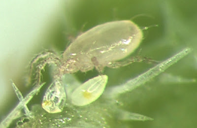
At this point in time, the Phytoseiidae are one of the most intensely studied families of mites. They are the only group of mesostigmatan mites to have significantly diversified among the foliar environment (on and around plant leaves) where they are mostly predators on other small invertebrates. The taxonomic history of phytoseiids is storied and complex but one taxon that has been consistently recognised as a major part of the family is the genus Amblyseius.

Swirski mite Amblyseius swirskii, from here.


When reviewed by Chant & McMurtry in 2004, Amblyseius was a sizeable assemblage of close to 350 known species (I quite expect that number to have expanded by now). Species of Amblyseius are lightly sclerotised, mostly pale in colour, and usually have a smooth shield covering most of the dorsum. The genus is characterised by the presence of eighteen or nineteen pairs of setae on the dorsum of the idiosoma (the central body) with three sublateral pairs being particularly long: one about the level of the third pair of legs (referred to as the s4 pair) and the other two towards the rear of the body. Except for a few pairs forward of the s4 setae, the remaining dorsal setae are all minute.

The primary focus of human interest in phytoseiids has been their role as predators of crop pests. I described some of the ways in which phytoseiids have been commercially utilised in an earlier post. Species used in this way include several Amblyseius though matters are complicated slightly by changes in taxonomy (for instance, one species which has been widely traded as Amblyseius cucumeris is now placed in the genus Neoseiulus). One of the most widely used of the commercial phytoseiids in recent years has been Amblyseius swirskii, commonly known as the Swirski mite (E. Swirski being an acarologist after whom the species was named). This species was first described in 1962 from almond trees in Israel and subsequently identified from a wide range of plant and crop species. Its history in pest control has been described in detail by Calvo et al. (2015).

The Swirski mite feeds on a range of prey, including mite, thrips and whitefly species, as well as on pollen and micro-fungi. It was first promoted as a commercial control for silverleaf whitefly Bemisia tabaci in the early 2000s. However, it did not get taken up in a big way until media publicity about pesticide residues on capsicum crops in Spain led to a crash in demand. Farmers in that country were forced to look for alternative means of pest control and found great success with A. swirskii (previous attempts to use the cooler-clime preferring Neoseiulus cucumeris in Spain had not been promising). Since then, the Swirski mite has been adopted in numerous countries for use on a range of crops to control various pests such as western flower thrips Frankliniella occidentalis. Because of its ability to grow and thrive on non-insect foods, including artificial diets, this mite is easily cultured commercially. It may also be released on crops before pest infestations develop, building up numbers on a diet of pollen until suitable prey presents itself. For the same reason, Swirski mite populations do not crash before pest control is complete. Overall, a remarkable success and a prime example of the value of Amblyseius species to mankind.

REFERENCES

Calvo, F. J., M. Knapp, Y. M. van Houten, H. Hoogerbrugge & J. E. Belda. 2015. Amblyseius swirskii: what made this predatory mite such a successful biocontrol agent? Experimental and Applied Acarology 65: 419–433.

Chant, D. A., & J. A. McMurtry. 2004. A review of the subfamily Amblyseiinae Muma (Acari: Phytoseiidae): part III. The tribe Amblyseiini Wainstein, subtribe Amblyseiina n. subtribe. International Journal of Acarology 30 (3): 171–228.

The Dermacentor Ticks

Pacific Coast tick Dermacentor occidentalis, copyright Jerry Kirkhart.


Among the ticks of most concern to humans are species of the genus Dermacentor. This genus of about forty known species is widely distributed in Africa, Eurasia and the Americas. Examples include the meadow tick D. reticulatus in Europe, and the wood tick D. variabilis and Rocky Mountain wood tick D. andersoni in North America. They are parasites of mammals, including both generalist and more host-specific species; records of Dermacentor individuals from reptiles and even carpenter bees (Goddard & Bircham 2010) presumably represent incidental and/or accidental associations. Species of Dermacentor are responsible for the spread of bacteria causing diseases such as Rocky Mountain spotted fever (which, despite sounding like a 1950s dance craze, is presumably not much fun), Q fever and tularemia. The ticks can also be more directly hazardous, as their bites inject a toxin that can cause tick paralysis.

Distinguishing features of Dermacentor species relative to other ticks include a rectangular base to the capitulum, relatively short, broad palps, well-developed eyes and the presence of festoons (impressed divisions of the posterior margin of the body) (Keirans 2009). Most are ornate—that is, marked on the dorsum with contrasting pale patterns—with the notable exception of the tropical horse tick D. nitens of the Americas (until recently, often treated as forming its own genus Anocentor). The function of such markings is unknown though suggestions include environmental protection, warning predators of distastefulness, or sexual signalling.

Meadow tick Dermacentor reticulatus, copyright Ferran Turmo Gort.


The majority of Dermacentor species have a three-host life cycle, dropping off the host between each of the life stages of larva, nymph and adult, and seeking out a new host after moulting. However, at least two New World species, the aforementioned D. nitens and the winter tick D. albipictus (a parasite of deer), are one-host ticks that remain on their original host between instars. In general, Dermacentor species are more resilient to dry climates than many other tick species. Individual species can differ in their climate tolerance, however. In North America, the geographical divide between D. variabilis in the east of the continent and D. andersoni in the west seems to be driven by the need for the latter of drier conditions (Yoder et al. 2007). Older instars also tend to be hardier than younger. Females of the ornate sheep tick D. marginatus, a European species, leave their host after gorging at the beginning of winter and then wait for more amoenable spring conditions before laying their delicate eggs (Dörr & Gothe 2001).

Higher relationships within the genus do not appear to have been extensively studied. A preliminary molecular phylogeny of hard ticks has suggested the possibility of a basal division between Afrotropical, Eurasian and New World lineages (Barker & Murrell 2004). Comparison with related tick genera raises the possibility of an Afrotropical origin for Dermacentor, though the genus has only a relictual presence in that continent now. However, with only a handful of species subjected to broad phylogenetic analysis to date, further testing is demanded. Does the continental divide hold true? Do the one-host species form a single clade within the genus? Inquiring minds wish to know.

REFERENCES

Barker, S. C., & A. Murrell. 2004. Systematics and evolution of ticks with a list of valid genus and species names. Parasitology 129: S15–S36.

Dörr, B., & R. Gothe. 2001. Cold-hardiness of Dermacentor marginatus (Acari: Ixodidae). Experimental and Applied Acarology 25: 151–169.

Goddard, J., & L. Bircham. 2010. Parasitism of the carpenter bee, Xylocopa virginica (L.) (Hymenoptera: Apidae), by larval Dermacentor variabilis (Say) (Acari: Ixodidae). Systematic and Applied Acarology 15: 195–196.

Keirans, J. E. 2009. Order Ixodida. In: Krantz, G. W., & D. E. Walter (eds) A Manual of Acarology 3rd ed. pp. 111–123. Texas Tech University Press.

Yoder, J. A., D. R. Buchan, N. F. Ferrari & J. L. Tank. 2007. Dehydration tolerance of the Rocky Mountain wood tick, Dermacentor andersoni Stiles (Acari: Ixodidae), matches preference for a dry environment. International Journal of Acarology 33 (2): 173–180.

Pied Harvestmen of the Antilles

Harvestmen of the Neotropical family Cosmetidae have been featured on this site a couple of times before. Each time, I've commented on the dire taxonomic state of this diverse family, with many genera being poorly or inaccurately defined. Thanks to extensive (and continuing) studies in recent years by Braxilian researchers and their associates, this situation has been progressively improving, but we still have a lot to learn.

Cynortoides sp., copyright Damion Laren Whyte.


Cynortoides is a genus currently holding ten species of cosmetid. Most of these are found on the islands of the Greater Antilles—Cuba, Jamaica and Hispaniola—though the genus has also been recorded from adjoining regions of Mexico and Venezuela (Kury 2003). As with other cosmetids, Cynortoides has historically been defined largely be features of the external spination, including a lack of spines on the legs, two pairs of spines in the rear part of the dorsal scutum, and no spines on the free abdominal segments (Mello-Leitão 1933). Also as with other cosmetid genera, Cynortoides species are colourfully patterned. The name of one species, C. v-album, refers to its characteristic bright white V marking on the back (though personally, I would describe the pattern as more of a Y).

Though this genus does not yet appear to have been revised in detail, some of its species were included in a recent broader study of cosmetid phylogeny by Medrano et al. (2021). They found strong support for an association between the Cuban C. cubanus and the Hispaniolan C. v-album, together with two other Cuban species previously included in the related genus Cynorta. These last two species were consequently transferred to Cynortoides though Medrano et al. did not comment on whether this affected the genus' established diagnosis. The authors speculated that further studies might prove Cynortoides to be a strictly Greater Antillean genus with mainland records being misplaced. Cynortoides would not be unique in this regard: the islands of the Caribbean are home to a number of lineages found nowhere else, reflecting a long history independent of the adjoining continents.

REFERENCES

Kury, A. B. 2003. Annotated catalogue of the Laniatores of the New World (Arachida, Opiliones). Revista Ibérica de Aracnología, special monographic volume 1: 1–337.

Medrano, M., A. B. Kury & A. C. Mendes. In press 2021. Morphology-based cladistics splinters the century-old dichotomy of the pied harvestmen (Arachnida: Gonyleptoidea: Cosmetidae). Zoological Journal of the Linnean Society.

Mello-Leitão, C. F. de. 1933. Notas sobre os opiliões do Brasil. Descritos na obra postuma de Sörensen: "Descriptiones Laniatorum". Boletim do Museu Nacional 9 (1): 99–114.

The Microzetid Enigma

The armoured mites of the Oribatida include their fair share of ornately ornamented species but perhaps the most grotesque of all are to be found under members of the family Microzetidae. These typically fairly small oribatids (the average size is about a third of a millimetre) are primarily found in soil and litter deposits around the world. They include a handful of species found in the far north but are primarily found in warmer regions with the greatest known diversity in the Neotropics (Woas 2002).

Dorsal, ventral and lateral views of Acaroceras galapagoensis, from Heinrich Schatz & Jose Palacios-Vargas.


The microzetids are primarily distinguished by elaborate outgrowths of the cuticle around the front of the body. In many oribatids, a pair of thin lamellae run down either side of the prodorsum (the part of a mite that might at first glance be taken for the 'head'). In microzetids, these lamellae have become massively enlarged and detached from the prodorsum over much of their length. As a result, they form a kind of hood over the front of the body. They are flanked on either side by similar lateral extensions called tutoria. The prodorsum as a whole is often remarkably large compared to the rear part of the dorsum, the notogaster. Indeed, the notogaster is often as wide as or wider than it is long. A pair of wing-like extensions, pteromorphs, extend on either side of the front of the notogaster; in microzetids, the pteromorphs are typically sharply pointed. To top all these excrescences off, the insertions of the first pair of legs are also shielded by well-developed flanges called pedotecta.

What, if anything, is the purpose of all these anatomical extravagances is a question I am unable to answer: whether they are related in some way to defense or water retention, for instance. They also make it difficult to understand the position of microzetids relative to other oribatids. The presence of pteromorphs has commonly been thought characteristic of a group of oribatids that have been referred to as the Poronoticae. However, microzetids lack any sign of another distinctive feature of poronotic oribatids: the array of glandular openings on the cuticle known as the octotaxic system. Some oribatids are known to have reduced octotaxic systems, and microzetids do bear a certain resemblance to a definitely poronotic family in the Oribatellidae, so it is possible they represent poronotic mites in which the octotaxic system has been lost. However, other features of microzetids further support affinities outside the Poronoticae. In particular, nymphs of microzetids carry scalps. As they moult from one instar to the next, the shed cuticle of the notogaster is retained in place like a cap. Over successive instars, this cap becomes a stack of scalps that potentially assist in defence (a would-be predator attempting to grab onto the notogaster finds itself holding only an empty scalp). This is generally thought to be a primitive bahaviour that was lost in the ancestor of the poronotics. So are the microzetids primitive relatives of the poronotics, descended from ancestors that had acquired pteromorphs but not yet lost the scalp-carrying habit? Are they derived poronotics that eschewed the octotaxic system and taken up their scalps once more? Further research into oribatid phylogeny is needed to know.

REFERENCE

Woas, S. 2002. Acari: Oribatida. In: Adis, J. (ed.) Amazonian Arachnida and Myriapoda: Identification keys to all classes, orders, families, some genera, and lists of known terrestrial species pp. 21–291. Pensoft: Sofia.

The Diosaccinae: Worldwide Sediment Dwellers

The harpacticoid copepods have been featured on this site a reasonable number of times now. These tiny crustaceans are among the most numerous animals in the world, both in terms of numbers of individuals and (in certain habitats) numbers of species. And among the most widespread representatives of the harpacticoids are members of the subfamily Diosaccinae.

Diosaccus tenuicornis, from Sars (1906).


The Diosaccinae are currently recognised as members of the family Miraciidae; earlier sources will usually refer to a family Diosaccidae but the recognition of the pelagic Miraciinae as derived members of this group (Willen 2000) requires use of the older name. Distinctive features of the Miraciidae compared to other harpacticoids include the presence of a relatively large, mobile rostrum and a number of distinctive arrangements of setae, including the inner seta on the basal endopodal segment of the first peraeopod (trunk leg) arising distally (Nicholls 1941, Willen 2000). Miraciids are also unusual in that females carry paired egg-sacs laterally; most other harpacticoid families carry only a single median egg-sac. Miraciids are divided between three subfamilies of which the Diosaccinae are the most diverse. Diosaccines are most readily distinguished by their retention of a number of plesiomorphic features such as crawling legs and relatively short caudal rami (Nicholls 1941; this author divided the current diosaccines between two subfamilies, the Diosaccinae sensu stricto and Amphiascinae, based on the presence or absence, respectively, of a clear distinction in breadth between the metasome and urosome, or 'trunk' and 'abdomen', but this division does not appear to have been recognised at this level by any subsequent authors). The great majority of diosaccines are marine, free-living and benthic. A handful of species have been described as associates of lobsters, whether commensals or semi-parasites. A small radiation of species of the genus Schizopera is known from Lake Tanganyika, and Karanovic & Reddy (2004) described a species Neomiscegenus indicus from subterranean fresh water in India. Marine diosaccines are found at all depths from the intertidal zone to the deep abyss. I don't know for sure but, though they are sediment dwellers, I don't get the impression (I could be wrong) that they are strictly meiofaunal. As noted earlier, many do not have the vermiform body shape characteristic of interstitial copepods. Many species also are around the half-millimetre size range, which I think may be relatively large for meiofauna?

Four species of Schizopera collected from Korea, from Karanovic & Cho (2016). Left to right: S. yeonghaensis, S. daejinensis, S. gangneungensis, S. sindoensis.


The other two subfamilies of Miraciidae are the aforementioned Miraciinae and the Stenheliinae, which have the endopod of the first peraeopod adapted for swimming rather than grasping and longer caudal rami. Though potential synapomorphies of the Diosaccinae were identified by Willen (2000), they're a bit weaksauce. There is a distinct possibility that further studies may identify the diosaccines as paraphyletic to the other two subfamilies. In particular, some diosaccines say a very unusual form of nauplius larva with the Stenheliinae, in which the body is strongly foreshortened and crab-like (Dahms et al. 2005). These nauplii also move sideways in a crab-like fashion and do not swim in the water column like the nauplii of other species. Practical considerations have lead most investigators of crustacean phylogeny to emphasis adult over larval morphology but the larval morphology of diosaccines raises some interesting questions.

REFERENCES

Karanovic, T., & Y. R. Reddy. 2004. A new genus and species of the family Diosaccidae (Copepoda: Harpacticoida) from the groundwaters of India. Journal of Crustacean Biology 24 (2): 246–260.

Nicholls, A. G. 1941. A revision of the families Diosaccidae Sars, 1906 and Laophontidae T. Scott, 1905 (Copepoda, Harpacticoida). Records of the South Australian Museum 7 (1): 65–110.

Willen, E. 2000. Phylogeny of the Thalestridimorpha Lang, 1944 (Crustacea, Copepoda). Cuvillier Verlag: Göttingen.

Where There's a Whip, There's a Scorpion

As our understanding of the higher relationships between organisms has improved vastly in recent decades, the arachnids have remained an intransigent bunch. Proposed connections between the various historically recognised orders have remained poorly supported and, even now, there are few that do not continue to jump about with gleeful abandon with each successive analysis. One small bastion of reliable support, however, has been been the tropical clade known as the Pedipalpi.

Whip spider Phrynus exsul, copyright Michel Candel.


Members of the Pedipalpi have traditionally been divided between two or three distinct orders: the whip spiders or tailless whip scorpions of the Amblypygi, the whip scorpions of the Uropygi, and the micro-whip scorpions of the Schizomida (alternative classifications have combined the last two in a single order Uropygi or Thelyphonida). All have a broad distribution in tropical and subtropical regions of the world. Representatives of the Pedipalpi are active hunters, united by the possession of large, raptorial pedipalps used in the capture of prey. All three groups also have the first pair of legs modified to become elongate and whip-like (Shultz 2007). These legs are not used in walking but are held forwards to function like antennae. The Uropygi and Schizomida are further united by the possession of a terminal appendage on the body, the 'whip' of a 'whip scorpion'. There is also a general agreement in recent years that the Pedipalpi are in turn the sister lineage to the spiders. Some researchers have argued for a closer relationship of the Amblypygi to the spiders rather than the whip scorpions, reflecting their (among other things) similar habitus, but this remains a minority view.

Syntype (one of the original described specimens) of Paracaron caecus, from Garwood et al. (2017). Scale bar = 5 mm.


Globally, the Pedipalpi are not a hugely diverse lineage, with a bit more than 600 known species overall. About 190 species belong to the Amblypygi, the whip spiders. As noted above, these arachnids are quite spider-like in appearance owing the lack of a terminal flagellum and the presence of a well-defined waist between cephalothorax and abdomen, but they lack the poison fangs and spinnerets of a spider. Most whip spiders have a distinctly flattened habitus, allowing them to enter narrow spaces under bark or between rocks. They also have the most remarkably elongate first legs among the Pedipalpi. Living whip spiders can be divided between two lineages, referred to as the Paleoamblypygi and Euamblypygi (Garwood et al. 2017). The Paleoamblypygi are represented in the modern fauna by only a single known (but little known) species, Paracharon caecus, a blind inhabitant of termite nests in western Africa. Paracharon caecus differs from other living whip spiders in retaining a vertical plane of motion of the pedipalps, like those of whip scorpions. In the Euamblypygi, the orientation of the pedipalps has shifted so they move in a horizontal plane only. In some whip spiders, the pedipalps have become remarkably long, perfect for clasping prey in a fatal hug.

Giant whip scorpion Mastigoproctus giganteus, copyright David Bygott.


The whip scorpions of the Uropygi are the least diverse of the three lineages of Pedipalpi, with about 110 known species. They are large, robust arachnids characterised by their long, filamentous terminal flagellum. Glands at the base of the flagellum produce noxious chemicals used in defense, giving some species the alternative name of 'vinegaroons'. The Schizomida are the most diverse subgroup of the Pedipalpi, including about half the known species. Some species have become widespread as a result of human transportation in association with greenhouses whereas others have even been collected among ice and snow in California (Harvey 2003). Schizomids are smaller and softer-bodied than the Uropygi and the terminal flagellum is shorter (as in Uropygi, the flagellum is flanked by repugnatorial glands). In male schizomids, the flagellum is often distinct in shape from that of the females, becoming bulbous. Schizomids also differ from most other arachnids in the presence of visible dorsal divisions between the segments of the cephalothorax.

Female schizomid Hubbardia briggsi, copyright Marshal Hedin.


In all subgroups of the Pedipalpi, reproduction involves mating displays in which the male deposits a spermatophore on the ground and then guides the female over it (Harvey 2003). The exact manner in which the male guides the female differs between subgroups. In schizomids, the female grasps onto the male's flagellum and he leads her. In Uropygi, the male grasps the female's fore legs with his pedipalps before turning to face the same direction as her with himself in front, and pulls her over the spermatophore. He then turns, embraces her abdomen with his pedipalps, and manually inserts the spermatophore into her genital operculum. Amblypygi have perhaps the most graceful option of the three: the male stands facing the female then gently beckons her forward, allowing her to approach and collect the spermatophore of her own volition.

REFERENCES

Garwood, R. J., J. A. Dunlop, B. J. Knecht & T. A. Hegna. 2017. The phylogeny of fossil whip spiders. BMC Evolutionary Biology 17: 105.

Harvey, M. S. 2003. Catalogue of the Smaller Arachnid Orders of the World: Amblypygi, Uropygi, Schizomida, Palpigradi, Ricinulei and Solifugae. CSIRO Publishing.

Shultz, J. W. 2007. A phylogenetic analysis of the arachnid orders based on morphological characters. Zoological Journal of the Linnean Society 150 (2): 221–265.

Sea Spiders

With arthropods being such a massively diverse sector of the global biota (and even that feels like an understatement; describing arthropods as 'very diverse' seems a bit like describing the Andromeda Galaxy as 'very far away'), it is only to be expected that it contains some very weird corners. And definitely among the weirder of those corners are the Pycnogonida, commonly known as the 'sea spiders'.

Anoplodactylus evansi, copyright Mick Harris & Claudia Arango.


Pycnogonids are a group of marine arthropods found around the world (not actual spiders, of course, though honest-to-goodness marine spiders are a thing that does exist). Their relationships to other arthropods have long been in dispute but the majority view is that they are distant relatives of the terrestrial arachnids. Pycnogonids are not uncommon in both coastal and deep-sea habitats but tend to go unnoticed: they feed on rock-encrusting colonial animals such as hydrozoans and are often coloured to disguise themselves against their prey. If one ever does see a sea spider, the first thing to stand out about them is how they are made of legs. The central body is often remarkably small compared to its limbs, to the extent that the dubbing of pycnogonids as 'no-bodies' by an early 20th Century author has become something of a cliché. Certain major organs, such as the gonads and parts of the digestive system, have even been diverted into the legs to make up for the lack of space in the body. Most pycnogonids possess four pairs of walking legs though there are species with more. At the front of the body on the underside of the head is a large proboscis that is used for sucking the juices out of prey, flanked by pairs of pincer-bearing chelifores and/or palps used for tearing it open. Near the first pair of walking legs there is often a pair of slender leg-like appendages known as the ovigers, used for carrying bundles of eggs until they hatch. The greater part of the body behind the head is taken up by the leg-bearing thorax; the legless abdomen is reduced to the merest nub like the docked tail of a dog.

Close-up on preserved male Anoplodactylus lentus, from Florida Museum of Natural History.


One of the largest recognised genera of pycnogonids is Anoplodactylus, with over 130 species worldwide and many continuing to be described (Lucena et al. 2015). This genus can be distinguished by the possession of chelifores with functional chelae (pincers) but palps are absent or reduced to buds. Both the chelifores and the proboscis are relatively short (Child 1998). Ovigers are five- or six-segmented and present in males only (male care of eggs is the standard pattern among pycnogonids). Species vary from 0.6 to 6 millimetres in body length. The majority of species of Anoplodactylus are found in shallow waters in temperate and tropical regions with a smaller number of species found in polar and deep waters. Alvarez & Ojeda (2018) record finding a single specimen of the species A. batangensis among vegetation on the surface of an anchialine pool in the Yucatan Peninsula of Mexico. Though the surface of these pools is more or less fresh water, deeper sections are saline owing to subterranean connections to the sea. The collection of a pycnogonid near the surface of this pool suggests an ability to adjust to very low salinity though one questions whether it would be able to survive indefinitely.

Larvae of Anoplodactylus are very small compared to those of other pycnogonids and have what has been termed an 'encysting' development (Burris 2011). As bizarre as the appearance of adult pycnogonids is, their larvae are arguably even weirder, being essentially nothing more than a head bearing chelifores, proboscis, and two pairs of undifferentiated appendages. The remaining segments of the body are added over the course of development. In Anoplodactylus, the larvae develop as parasites, forming a cyst in the gastrocoel (the stomach cavity) of cnidarians (having presumably been placed there somehow by their fathers, though I haven't found if we know how). They become free-living upon reaching the first juvenile stage, emerging from their host to pursue their predatory lives.

REFERENCES

Alvarez, F., & M. Ojeda. 2018. First record of a sea spider (Pycnogonida) from an anchialine habitat. Latin American Journal of Aquatic Research 46 (1): 219–224.

Burris, Z. P. 2011. Larval morphologies and potential developmental modes of eight sea spider species (Arthropoda: Pycnogonida) from the southern Oregon coast. Journal of the Marine Biological Association of the United Kingdom 91 (4): 845–855.

Child, C. A. 1998. The Marine Fauna of New Zealand: Pycnogonida (Sea Spiders). National Institute of Water and Atmospheric Research (NIWA).

Lucena, R. A., J. F. de Araújo & M. L. Christoffersen. 2015. A new species of Anoplodactylus (Pycnogonida: Phoxichilidiidae) from Brazil, with a case of gynandromorphism in Anoplodactylus eroticus Stock, 1968. Zootaxa 4000 (4): 428–444.

Mites of Southern Sediment

Water mites of the clade Hydrachnidiae are one of the few groups of arachnids that have not only adopted an aquatic lifestyle but have thrived and diversified there. Over fifty families are currently recognised within this clade, some of which can be found in almost every body of fresh water worldwide. Others, however, are notable for their restricted ranges. One of these latter examples is the Omartacaridae.

Ventral view of female Omartacarus elongatus, from Cook (1963).


Omartacaridae is a small family currently recognised as including only two genera, Omartacarus and Maharashtracarus. They have a somewhat elongated body with a soft integument, contrasting with the more globular form of many other water mites. They are also distinguished by the arrangement of the coxae (the basal segments of the legs on the underside of the body) which are clustered together with the medial edges of the anterior pairs much longer than those of the posterior pairs (Walter et al. 2009) so the third pair of coxae are triangular in shape. As far as is known, omartacarids are restricted to interstitial habitats or the hyporheic zone of sediment beneath and alongside stream beds. I am unaware of any direct observations of omartacarid behaviour but they are presumably predators like other water mites. Most of the (rather limited) attention that has been given to omartacarids has focused on discussions of their distribution. Species of Omartacarus are found in South and southern North America, as well as in Australia. Maharashtracarus species are known from India and Costa Rica. It has been presumed that this reflects an ancestral Gondwanan distribution, spreading into North America from South America as the continents joined.

The larval stage of omartacarids is, to date, unknown. Larvae of other water mites live as parasites of water-associated insects such as midges and omartacarid larvae are presumably also parasitic. But in what capacity? Do mature omartacarids emerge from their subterranean habitats at some particular time of year in search of a host for their eggs? Do they somehow manage to find a host while remaining safely sequestered underground? The secret remains to be uncovered.

REFERENCE

Walter, D. E., E. E. Lindquist, I. M. Smith, D. R. Cook & G. W. Krantz. 2009. Order Trombidiformes. In: Krantz, G. W., & D. E. Walter (eds) A Manual of Acarology 3rd ed. pp. 233-420. Texas Tech University Press.

Atropacarus

The little guy pictured above (photo copyright Scott Justis) is a representative of the box mite genus Atropacarus, members of which can be found in most parts of the world. Atropacarus is a genus of the Phthiracaroidea, a group of box mites characterised by the plates on the underside of body being relatively wide, in contrast to the narrow ventral plates of its sister group, the Euphthiracaroidea (members of which have featured on this site before: here and here). The difference in configuration of these plates reflects a difference in the way that the body is contracted to allow legs and prosoma to be withdrawn beneath the protective cover of the notogaster. In euphthiracaroids, the sides of the notogaster are contracted inwards; in phthiracaroids, the ventral plates of the body are lifted upwards (Schmelzle et al. 2015).

The classification of phthiracaroids is subject to conflict with two main systems in the recent literature. In one, championed by the Polish acarologist Wojciech Niedbała, the phthiracaroids are divided between two families with Atropacarus in the Steganacaridae. Species of Atropacarus have the surface of the notogaster extensively covered with dimples. The dorsal seta on the tibia of the fourth leg is short and closely associated with a solenidion (a type of specialised sensory hair). The setae of the genital plate are arranged in a more or less straight row along the inner margin of the plate with the fifth and sixth setae further apart than the fourth and fifth (Niedbała 1986). Niedbała divides Atropacarus between two subgenera. In Atropacarus sensu stricto, there are sixteen or more pairs of setae on the notogaster and the second adanal seta is moved inwards on the ano-adanal plate to form a more or less straight line with the anal setae. In Hoplophorella, there are fifteen pairs of setae on the notogaster and the second adanal seta is distinctly laterally placed relative to the anal setae.

The super-hairy Atropacarus niedbalai, from Liu & Zhang (2013). Scale bar = 100 µm.


In the competing system, used for instance by Subías (2019), Atropacarus and Hoplophorella are treated as distinct genera and each is in turn divided into subgenera by the number of setae on the ano-adanal plate. To a certain extent, of course, the question of whether to treat Atropacarus and Hoplophorella as genera or subgenera is arbitrary. Nevertheless, this arguably cosmetic distinction does relate to an underlying difference in theory. The classification of phthiracaroids used by Subías (2019) is a largely diagnostic one, inspired by a desire to facilitate specimen identifications. Niedbała's classification, in contrast, is intended to reflect phylogenetic relationships. Simple setal counts may be convenient when composing keys but one might question its overall phylogenetic significance. Neotrichy (increases in setal count by multiplication of the original setae) is not uncommon in phthiracaroids, particularly on the notogaster. Setal counts may vary between individuals of a single species and overall neotrichy reaches an extreme in the New Zealand species Atropacarus niedbalai. In this species, the basic count of fifteen or sixteen pairs of notogastral setae has been increased to 109 or 115 pairs, with further neotrichy on the prodorsum and ventral plates (Liu & Zhang 2013). Subías (2019) defends his choice of classification by arguing that Niedbała's key features are often difficult to discern. I sympathise with the difficulty but, as a wise man once said, species are under no obligation to evolve with regard to the convenience of taxonomists.

REFERENCES

Liu, D., & Z.-Q. Zhang. 2013. Atropacarus (Atropacarus) niedbalai sp. nov., an extreme case of neotrichy in oribatid mites (Acari: Oribatida: Phthiracaridae). International Journal of Acarology 39 (6): 507–512.

NiedbaÅ‚a, W. 1986. Système des Phthiracaroidea (Oribatida, Euptyctima). Acarologia 27 (1): 61–84.

Schmelzle, S., R. A. Norton & M. Heethoff. 2015. Mechanics of the ptychoid defense mechanism in Ptyctima (Acari, Oribatida): one problem, two solutions. Zoologischer Anzeiger 2015: 27–40.

Subías, L. S. 2019. Nuevas adiciones al listado mundial de ácaros oribátidos (Acari, Oribatida) (14a actualización). Revista Ibérica de Aracnología 34: 76–80.

Austrotritia: Jack-in-the-Box Mites

We just keep coming back to the oribatids, don't we?

In an earlier post, I introduced you to Oribotritia, one of the genera of box mites. These, you may recall, are the armoured mites that have evolved the ability to curl the front of the body under themselves and tuck back their legs to form a solid case (in the Oribotritiidae, that mechanical defense is supplemented by the production of a defensive chemical, chrysomelidial, from glands in the cuticle—Shimizu et al. 2012). In the earlier post, I also gave you a quick overview of the families of what are known as the 'true' box mites. Today's post is for another component of the family Oribotritiidae, the genus Austrotritia.

Austrotritia lebronneci, copyright R. Penttinen.


Austrotritia accounts for nearly twenty species of box mite, the great majority of which are found in Australasia and southern and eastern Asia (Liu et al. 2009). Outliers are A. engelbrechti in South Africa, A. herenessica in the Canary Islands and, most unexpected of all, A. finlandica in Finland. Austrotritia differs from all other oribotritiids except the small Bornean genus Terratritia in lacking any division between the genital and aggenital plates on the underside of the body. The distinction between Austrotritia and Terratritia perhaps requires reassessment: Niedbała (2000) distinguished them by the presence of five-segmented palps and a single pair of exobothridial setae in Austrotritia versus three-segmented palps and two pairs of exobothridial setae in Terratritia (the bothridia are the structures bearing large sensory setae on the prodorsum of the mite; exobothridial setae are thus setae sitting alongside the bothridia). However, Liu & Zhang (2014) redescribed the widespread species Austrotritia lebronneci as having three-segmented palps but only a single pair of exobothridial setae. Note that classification of oribatids has mostly been conducted from a diagnostic rather than a phylogenetic perspective; it would not surprise me if Terratritia turned out to be a derived subgroup of Austrotritia.

Schematic of jump performance by Indotritia cf. heterotrichia from Wauthy et al. (1998); the solid line represents observed jumps, the dashed lines modelled jumps. Line drawings represent (a) body posture when beginning jump, (b) rotation during jump, and (c) enclosed posture after jumping.


As well as the aforementioned defenses standard for box mites, Austrotritia and the related genus Indotritia stand out from other oribotritiid genera in that at least some species have the ability to jump. The mechanics of jumping were described for a species of Indotritia by Wauthy et al. (1998) who recorded the mites jumping nearly a centimetre in height over a distance of just under an inch (for perspective, the mite itself is about half a millimetre in length). Jumping was preceded by compressing the notogaster while raising the ventral plates under the opisthosoma, together with lowering the prosoma and bringing the legs together under the body. Small hooks at the end of femur of the first pair of legs were used to catch ridges on the side of the prodorsum in order to hold the body compression. The force for the jump was presumably supplied by the release of the hydraulic compression of the body fluids when the legs disengaged from the prodorsum, propelling the mite backwards while the body rolled forwards: essentially, the mite would star-jump away. The mite would curl up after jumping to lie in an enclosed state.

Whether all Austrotritia species are jumpers is not entirely certain. The femoral hooks that seem to play a significant role in jumping have not been described in all species. However, it is not clear if this lack of observation represents an actual absence or whether this minute feature has simply been overlooked. I also wonder whether the aforementioned fusion of the ventral plates in Austrotritia is related to their jumping abilities (Indotritia species also have the genital and aggenital plates fused anteriorly though they retain a degree of separation at the rear of the plates; non-jumping Oribotritia have the plates entirely separated). As always, there's still a lot we could potentially find out.

REFERENCES

Liu, D., J. Chen & G. Qiao. 2009. Review of Austrotritia (Acari: Oribatida: Oribotritiidae), with descriptions of two new species from China. Zootaxa 2144: 54–64.

Liu, D., & Z.-Q. Zhang. 2014. Redescription of Austrotritia lebronneci (Oribotritiidae) and descriptions of two new species of Euphthiracaridae (Acari, Oribatida) from Australian region. International Journal of Acarology 40 (1): 43–51.

Niedbała, W. 2000. The ptyctimous mites fauna of the Oriental and Australian regions and their centre of origin (Acari: Oribatida). Polskie Towarzystwo Taksonomiczne: Wrocław (Poland).

Shimizu, N., R. Yakumaru, T. Sakata, S. Shimano & Y. Kuwahara. 2012. The absolute configuration of chrysomelidial: a widely distributed defensive component among oribotritiid mites (Acari: Oribatida). Journal of Chemical Ecology 38: 29–35.

Mites of Marine Sands

Mites may be the most ecologically diverse group of animals on the planet. It is something of a challenge to think of a habitat supporting complex life in which mites may not be found. Nevertheless, it can fairly be said that the marine environment has provided them with a challenge. Though a wide variety of mites can be found in habitats along the shoreline, few lineages have learnt to make a life for themselves beyond the littoral fringe. The most diverse group of truly marine mites is the Halacaridae, of which the genus Simognathus is a representative.

Simognathus sp., from Banks (1915).


Halacarids are notably armoured mites, their bodies protected by an array of reticulate plates. They are found in a wide range of marine habitats and pursue the gamut of lifestyles: representatives of halacarids include algal grazers, micropredators, and parasites. Despite their aquatic lifestyle, they are not swimmers. Instead, they cling to their substrate and crawl slowly on legs bearing large claws. The diversity of halacarid morphologies is reflected in their classification with over a dozen subfamilies currently recognised.

Simognathus is a genus of halacarids found around the world though the greater diversity of species are known from the Southern Hemisphere. They are found at depths ranging from near the low tide mark to around 500 m, and from the full range of tropical, warm-temperate and cold-temperate waters. Bartsch (2005) speculated that the only reason they are not known from even colder waters may be a question of sampling effort rather than true absence. Most Simognathus species are known to live among coarse sand, or in other interstitial microhabitats such as among coral rubble, among colonies of sessile animals such as barnacles or tubeworms, or within algal holdfasts. I haven't come across any specific comments on their diet but their robust chelicerae and grasping fore legs leads me to suspect that Simognathus species are probably micropredators.

Simognathus and the closely related genus Acaromantis form the subfamily Simognathinae. Simognathines differ from other halacarids in their spindle-shaped body with short rostrum, reflecting their interstitial habitat. The first leg ends in a pincer arrangement formed from the terminal claw and a spine on the underside of the tibia. Acaromantis species have a two-segmented palp, no lateral claws at the end of the first leg, and a spinose seta on the genu (the segment between the femur and tibia) of the first leg. Simognathus species have a three-segmented palp, a pair of slender lateral claws on the first leg as well as the terminal claw, and no spinose seta on the first genu. The defining features of Simognathus are all likely to be primitive relative to those of Acaromantis and it has been suggested for some time that Acaromantis may be a derived subgroup of Simognathus. This suggestion is bolstered by a recent molecular analysis of halacarids by Pepato et al. (2018) which found the two Simognathus representatives included to be paraphyletic to the included species of Acaromantis.

REFERENCES

Bartsch, I. 2005. Lohmannella and Simognathus (Halacaridae: Acari) from Western Australia: description of two new species and reflections on the distribution of these genera. Records of the Western Australian Museum 22: 293–307.

Pepato, A. R., T. H. D. A. Vidigal & P. B. Klimov. 2018. Molecular phylogeny of marine mites (Acariformes: Halacaridae), the oldest radiation of extant secondarily marine animals. Molecular Phylogenetics and Evolution 129: 182–188.

Pseudogagrella: A Harvestman Torn

The Sclerosomatidae are one of the most diverse of the currently recognised harvestmen families, and one of the most problematic when it comes to classification. In various earlier posts, I have noted the challenges that bedevil sclerosomatid systematics, many reflecting a historical focus on superficial external features of questionable evolutionary significance. Perhaps no taxon more neatly exemplifies the problems with higher sclerosomatid classification than the eastern Asian genus Pseudogagrella.

Pseudogagrella sakishimensis, copyright Tomoya Suzuki.


Historically, the greater number of sclerosomatids have been divided between two major subfamilies, the Leiobuninae and Gagrellinae. The Leiobuninae have mostly been recognised as living in the northern temperate regions whereas the Gagrellinae were mostly tropical. The division between the two subfamilies has long been regarded as more than a little fuzzy, and has usually hinged on a single feature: the presence (Gagrellinae) or absence (Leiobuninae) of rings of flexible integument (noduli) in the femora of the legs. Pseudogagrella is a genus of sclerosomatid harvestmen recognised from Japan, Taiwan, China and Sumatra (Chen & Shih 2017). Members of this genus lack leg noduli so have historically been included in the Leiobuninae. The problem is that their overall appearance, with a tendency to bold coloration, a tall median spine rising from the hardened scute covering most of the abdomen, and legs that are not merely long but ludicrously so (even by harvestman standards), is extremely similar to species of Gagrellinae. So much so, in fact, that some species currently placed in Pseudogagrella were long included in the archetypical gagrelline genus, Gagrella (Suzuki 1977).

With the distinction between the two subfamilies being so vague, I don't think it really came as that much surprise to anyone when molecular phylogenetics underlined the need for a thorough re-working of sclerosomatid systematics. Though the analysis conducted by Hedin et al. (2012) did not support the prior distinction between 'leiobunines' and 'gagrellines', it did suggest the existence of distinct lineages occupying distinct geographical regions. One species of Pseudogagrella included in the analysis (the southern Japanese P. amamiana) was placed in a cluster of eastern Asian species including other Asian 'gagrellines', but also the 'leiobunine' 'Leiobunum' japonicum. So it seems likely that, should subfamilies of Sclerosomatidae continue to be recognised, Pseudogagrella will indeed be a member of Gagrellinae, but Gagrellinae itself shall not quite be what people think of it as being.

Pseudogagrella dorsomaculata, copyright Tyus Ma.


There is also, of course, the question of whether Pseudogagrella itself is a coherent unit. Hedin et al. (2012) included only the one Pseudogagrella species in their analysis and the need for an extensive revision of the Asian sclerosomatid genera still remains. A study of Chinese species assigned to the genus Melanopa (Zhang & Zhang 2013), which is primarily distinguished from Gagrella by having relatively shorter legs, suggested the possibility of this 'genus' being divided between groups of Palaearctic and Indo-Malayan species, and I've wondered if this division might carry further (unfortunately, I'm not aware of any Indo-Malayan 'gagrellines' being included in molecular phylogenies; I think all the Asian species covered by Hedin et al. were Palaearctic). The majority of Pseudogagrella species, found in Japan and Taiwan, can be comfortably compared to other sclerosomatids from that region, but the Sumatran P. multimaculata, and possibly the southern Chinese species, might turn out to be closer to their own geographical peers. As always, a great deal of research remains to be done.

REFERENCES

Chen, S.-L., & H.-T. Shih. 2017. Descriptions of three new species of the harvestmen genus Pseudogagrella (Opiliones: Sclerosomatidae: Gagrellinae) from Taiwan, supported by morphological and molecular evidence. Zootaxa 4268 (1): 34–52.

Hedin, M., N. Tsurusaki, R. Macías-Ordóñez & J. W. Shultz. 2012. Molecular systematics of sclerosomatid harvestmen (Opiliones, Phalangioidea, Sclerosomatidae): geography is better than taxonomy in predicting phylogeny. Molecular Phylogenetics and Evolution 62 (1): 224–236.

Suzuki, S. 1977. Opiliones from Taiwan (Arachnida). Journal of Science of the Hiroshima University, Series B, Division 1 (Zoology) 27 (1): 121–157.

Zhang, C., & F. Zhang. 2013. Notes on some species of the genus Melanopa (Opiliones: Sclerosomatidae: Gagrellinae) from China, with description of a new species. Journal of Arachnology 41: 306–318.

The Splanchnotrophidae: Comfy inside a Sea Slug

In previous posts, I've referred to the great significance of the minute crustaceans known as copepods to aquatic ecosystems. At the time, I was referring to free-living members of this group but the copepods also include a wide range of parasitic forms. Some of these parasitic copepods have evolved into forms so derived and bizarre that they are barely recognisable as crustaceans. One example of this is the family Splanchnotrophidae.

Sea slug Janolus fuscus with protruding egg sacs of a splanchnotrophid copepod, probably Ismaila belciki, copyright Michael D. Miller.


Splanchnotrophids are a group of copepods endoparasitic on two orders of shell-less marine gastropods (sea slugs), the Nudibranchia and Sacoglossa. They are characterised by reduced mouthparts and appendages though they retain a distinct pair of claw-like antennae. These antennae seem to be used to hold the copepod in place in their preferred location within the body cavity of their host. Though the exact means of feeding by splanchnotrophids is not certain, their rudimentary mouthparts, combined with a rarity of observations of actual tissue damage in parasitised hosts, indicate that they probably suck nutriment from their host's haemolymph. Females and males live in association within the host, the minute (and slightly more recognisably copepod-y) males holding close to their comparatively gigantic mates. As well as their size, female splanchnotrophids differ from males in the possession of elongate, tubular dorsal outgrowths of the thorax. These are most commonly presumed to function to provide more space for the female's enlarged ovaries, though some have suggested additional functions such as maintaining position within the host, respiration or absorbing nutrients (Anton & Schrödl 2013). The female's tubular egg-sacs extend through an opening in the host's body wall to release eggs into the water column. Usually, these egg-sacs will emerge close to some outgrowth of the host's own body, such as gills or papillae, and may be coiled if relatively long; these measures presumably help protect the egg-sacs from external damage. How the released larvae find and colonise new hosts remains unknown but it is possible the antennules (the smaller second pair of antennae possessed by most crustaceans) are used to locate hosts chemically, with their reduced condition in adults the result of a halt to development once their purpose has been fulfilled.

Female (left) and male Ismaila aliena dissected out from host, from Anton & Schrödl (2013).


Relatively few splanchnotrophids have been recognised to date, maybe about a dozen species divided between five genera. A few other species that had earlier been included in the family on little more grounds than that they were endoparasites of gastropods were excluded by Huys (2001)*. A sixth genus and species Chondrocarpus reticulosus is of uncertain relationships. If correctly associated with the splanchnotrophids, it is of interest in parasitising a different group of sea slugs (the pleurobranchids) and in its massive size (growing to twelve millimetres vs only a few millimetres for females of the other genera), but the only available description is inadequate for its proper characterisation. In some localities, splanchnotrophids have proven to be surprisingly abundant. A once-off survey of potential host species in Oregon found no less than 62% of individuals of one species to be infected (25 other potential host species were completely free of parasites), whereas a longer-term survey off the coast of Chile found an overall infection rate of 13% with some particular host species approaching 100% infection (Schrödl 2002). Host specificity seems to vary within the family: a study by Anton et al. (2018) found that species of the genus Ismaila tended to restrict themselves to a single host species, whereas species of Splanchnotrophus are more catholic and undiscriminating. Nevertheless, a lack of correlation between relationships of splanchnotrophid species and those of their host species suggests that, even in the more discriminating Ismaila, host changes may not have been uncommon.

*As a concise indication of just how sloppy some of the earlier work on 'splanchnotrophids' had been, one misattributed species was re-identified by Huys (2001) as having been based on the detached head of a pelagic amphipod.

The broader relationships of splanchnotrophids within copepods also remain poorly understood. A phylogenetic study by Anton & Schrödl (2013) suggested that Splanchnotrophidae may form a clade with another genus of copepods endoparasitic in gastropods, Briarella, with this clade being in turn derived from ectoparasitic ancestors. However, by the authors' own admission, this study was heavily biased in both taxon and character coverage to the Splanchnotrophidae, and may have been affected by insufficient scrutiny of non-splanchnotrophid taxa. Though derivation of the endoparasitic splanchnotrophids from ectoparasitic ancestors has a definite intuitive appeal, further study is required before we can feel confident about it.

REFERENCES

Anton, R. F., D. Schories, N. G. Wilson, M. Wolf, M. Abad & M. Schrödl. 2018. Host specificity versus plasticity: testing the morphology-based taxonomy of the endoparasitic copepod family Splanchnotrophidae with COI barcoding. Journal of the Marine Biological Association of the United Kingdom 98 (2): 231–243.

Anton, R. F., & M. Schrödl. 2013. The gastropod-crustacean connection: towards the phylogeny and evolution of the parasitic copepod family Splanchnotrophidae. Zoological Journal of the Linnean Society 167: 501–530.

Huys, R. 2001. Splanchnotropid systematics: a case of polyphyly and taxonomic myopia. Journal of Crustacean Biology 21 (1): 106–156.

Schrödl, M. 2002. Heavy infestation by endoparasitic copepod crustaceans (Poecilostomatoida: Splanchnotrophidae) in Chilean opisthobranch gastropods, with aspects of splanchnotrophid evolution. Organisms, Diversity & Evolution 2: 19–26.

The Camisiids: Cryptic Inhabitants of Soil and Wood

Various views of Camisia biverrucata, copyright Pierre Bornand.


The animal in the above pictures is a typical representative of the Camisiidae, a widely distributed family of oribatid mites. Members of this family can be found in soil, on the trunks of trees, or hidden among mosses and lichens. They are slow-moving animals and are often concealed from potential predators by an encrusting layer of dirt and organic debris. Carrying this encrusting layer may be related to a reduction in the offensive chemical-producing glands that are used by many other oribatids for defense (Raspotnig et al. 2008). In members of the genus Camisia, the openings of these glands are completely covered by dirt, but in the genera Platynothrus and Heminothrus the openings still protrude above the encrustation. The recently described Paracamisia osornensis, which does not carry an encrusting layer, retains a large offensive gland (Olszanowski & Norton 2002).

Close to 100 species have been assigned to this family; though found in most parts of the world, camisiids are most diverse in the Northern Hemisphere. One species in particular, Platynothrus peltifer, is almost global in distribution and the range of habitats in which it has been found includes soil, litter, peat and even aquatic habitats (Norton & Behan-Pelletier 2009) When one is as small and metabolically undemanding as these animals are, there may be surprisingly little difference between being out in the air or immersed in water, and even primarily terrestrial oribatids may survive submersion almost indefinitely. Genetic studies of P. peltifer have identified a high level of within-species divergence and it has been calculated on this basis that this species may have survived almost unchanged in external appearance for some 100 million years (Heethoff et al. 2007).

The ubiquitous Platynothrus peltifer, copyright Centre for Biodiversity Genomics.


The Camisiidae are closely related to another oribatid family, the Crotoniidae, that is found in South America and Australasia. One of the more significant differences between the two families is that whereas the camisiids appear to be entirely parthenogenetic, crotoniids reproduce sexually. Recent analyses, both molecular and morphological, indicate that the 'camisiids' are paraphyletic with regard to the crotoniids, leading Colloff & Cameron (2009) to treat the latter as a subfamily, Crotoniinae, of the former. This re-classification has been accepted by other authors though the law of priority requires that the combined family should be known as the Crotoniidae, not Camisiidae. The nested position of the sexual crotoniines within the asexual 'camisiids', with other related oribatid families also being asexual, has led to the suggestion that the crotoniines have somehow re-evolved sexuality. This would be fascinating if true, seemingly violating the usual principle that complex features can't be re-evolved once lost. Personally, I tend to be sceptical of claims like this (see this old post, for instance). I would like to see evidence beyond simple phylogenetic position to indicate if this is a true re-evolution rather than an historical bias towards loss of sexuality giving a misleading image.

REFERENCES

Colloff, M. J., & S. L. Cameron. 2009. Revision of the oribatid mite genus Austronothrus Hammer (Acari: Oribatida): sexual dimorphism and a re-evaluation of the phylogenetic relationships of the family Crotoniidae. Invertebrate Systematics 23: 87–110.

Heethoff, M., K. Domes, M. Laumann, M. Maraun, R. A. Norton & S. Scheu. 2007. High genetic divergences indicate ancient separation of parthenogenetic lineages of the oribatid mite Platynothrus peltifer (Acari, Oribatida). Journal of Evolutionary Biology 20: 392–402.

Norton, R. A., & V. M. Behan-Pelletier. 2009. Suborder Oribatida. In: Krantz, G. W., & D. E. Walter (eds) A Manual of Acarology 3rd ed. pp. 430–564. Texas Tech University Press.

Olszanowski, Z., & R. A. Norton. 2002. Paracamisia osornensis gen. n., sp. n. (Acari: oribatida) from Valdivian forest soil in Chile. Zootaxa 25: 1–15.

Raspotnig, G., E. Stabentheiner, P. Föttinger, M. Schaider, G. Krisper, G. Rechberger & H. J. Leis. 2008. Opisthonotal glands in the Camisiidae (Acari, Oribatida): evidence for a regressive evolutionary trend. Journal of Zoological Systematics and Evolutionary Research 47 (1): 77–87.

Metavononoides: Retreating from the Coast

I've commented before on the taxonomic issues bedevilling the study of South American harvestmen, particularly members of the diverse family Cosmetidae. Recent years have seen researchers make gradual but steady progress towards untangling these multifarious snarls by more firmly establishing the identities of this family's many genera.

Metavononoides guttulosus photographed by P. H. Martins, from Kury & Medrano (2018).


The genus Metavononoides was established by Roewer in 1928 for two species from south-eastern Brazil. As with other Roewerian genera, its definition was not exactly robust, being based on a combination of tarsal segment count together with the presence of a pair of large spines on the dorsal scutum. The genus was later re-defined by Kury (2003) who used it for a group of species found in the Brazilian Atlantic Forest region around Rio de Janeiro. Members of this group shared a number of distinctive features including the presence of a distinctive U-shaped marking (later dubbed a 'lyre mask' or 'lyra')on the scutum. A number of species previously placed in other genera were transferred to Metavononoides, and the next few years saw the description of a couple more species in the genus. And then Paecilaema happened.

The genus Paecilaema was first established by C. L. Koch in 1839 but a poor description of its type species P. u-flavum lead to confusion about its identity. Over time, Paecilaema became associated with a large number of species over a range stretching from Mexico to Brazil (as an aside, it doesn't help matters that Paecilaema has been one of those names that taxonomists have found themselves chronically uncertain how to spell). When Kury & Medrano (2018) recently set out to determine the exact identity of Paecilaema by determining that of its type, they fixed P. u-flavum as a species that was common around Rio de Janeiro and that corresponded to one of the species included by Kury (2003) in Metavononoides. As a result, many of the species shifted by Kury (2003) into Metavononoides were shifted once again into Paecilaema. Many of the species assigned to Paecilaema from outside the Atlantic Forest Region remain unrevised but will almost certainly prove to require re-classification.

Metavononoides barbacenensis photographed by P. H. Martins, from Kury & Medrano (2018).


Metavononoides was not outright synonymised with Paecilaema, though. Among the group of species possessing the aforementioned lyra on the scutum, Kury & Medrano (2018) identified two distinct subgroups. In one, corresponding to Paecilaema, the lyra is made up of two components. Part of the lyra is composed of light coloration on the plane of the scutum itself while another part is raised granules. In some species, these granules are particularly concentrated along the margins of the lyra (you can see an example on this on Flickr, photographed by Mario Jorge Martins; though labelled Metavononoides, this individual is now identifiable as Paecilaema u-flavum). In the second subgroup, corresponding to Metavononoides, the differentiated coloration on the plane of the scutum is absent and the lyra is composed solely of raised granules. Not only are the two genera morphologically distinct, they are also more or less geographically distinct. Whereas Paecilaema is found in the moist broadleaf forests closer to the coast, Metavononoides is now restricted to species largely found in the grasslands and shrublands further inland, corresponding to the Cerrado region. Though more depauperate of species than it was before, the identity of Metavononoides is certainly firmer.

REFERENCES

Kury, A. B. 2003. Annotated catalogue of the Laniatores of the New World (Arachida, Opiliones). Revista Ibérica de Aracnología, special monographic volume 1: 1–337.

Kury, A. B., & M. Medrano. 2018. A whiter shade of pale: anchoring the name Paecilaema C. L. Koch, 1839 onto a neotype (Opiliones, Cosmetidae). Zootaxa 4521 (2): 191–219.

Roewer, C. F. 1928. Weitere Weberknechte II. II. Ergänzung der: "Weberknechte der Erde", 1923. Abhandlungen der Naturwissenschaftlichen Verein zu Bremen 26 (3): 527–632, 1 pl.