Field of Science

Mites in Red Velvet

Adult Platytrombidium fasciatum, copyright Walter Pfliegler.


Mites in red velvet,
decorated with stripes.
Completing their diet,
hunting down eggs from flies*.

*With apologies to Justin Hayward.

Among the mites most likely to be seen by the casual observer are the various species of active predators known as red velvet mites. They grow to relatively large sizes for a mite (the species in the photo above can get up to 2.5 mm long), they are brightly coloured and they can often be seen moving about in search of food. As well as the colour, the name 'red velvet mite' refers to their dense covering of setae giving them almost a teddy-bearish appearance. There aren't many mites that could be described as cuddly, but these are arguably among them (at least as adults, as explained below).

Red velvet mites form a number of families in the mite clade Parasitengonina. Earlier posts on this site (here and here) have already described the somewhat complicated life cycles of parasitengonines, but to recap briefly: parasitengonines start their lives as parasitic larvae, followed by a dormant 'pupa-like' stage, followed by an active predatory nymph, then another dormant 'pupa', and finally the active predatory adult. Whereas differences between the active nymphs and adults are slight (kind of raising the question as to why the intervening dormant phase), differences between adults and larvae are significant. From their appearance alone, there is no way of telling whether a given larval form corresponds to a given adult, and connecting the two requires challenging indirect methods such as brood-rearing. Nevertheless, both forms are commonly encountered: not only are adults significant micro-predators, the larvae are often found attachned to insects and other arthropods. Some larval species, commonly known as chiggers, attack vertebrates such as humans and so are even more well-studied. Because of the resulting need to classify both adults and larvae without an easy way to connect the two, a kind of double taxonomy has developed with many parasitengonines. Adults and larvae are treated as if they were separate 'genera' and 'species', with separate names for each. Sometimes a larval 'species' may be successfully connected to an adult 'species' and the two can be synonymised, but many taxa remain that are known only from one or the other.

The genus Platytrombidium, belonging to the velvet mite family Microtrombidiidae, was established in 1936 on the basis of adults, but its larval form was not described until 2005. A number of species have been assigned to this genus from various parts of the world but, as a result of obtaining better descriptions of both adult and larva, Gabryś et al. (2005) restricted it to three species known from the Palaearctic region (Europe and northern Asia). Adult Platytrombidium are characterised by an even covering of stout, uniform setae covered with delicate setules; when alive, they are even more readily recognised from their transverse white stripes across the body. As adults and active nymphs, Platytrombidium fasciatum (the best-known species in the genus and the only one with known larvae) feed on fly eggs. Their larvae are also parasites on drosophilids and similar small flies, most often found attached to the dorsal surface of the abdomen (Gabryś et al. recorded one larva found attached to its host's eye).

Most of the confusion about the taxonomy of Platytrombidium has revolved around its relationship to the very similar genus Atractothrombium. For a long time, the only recognised difference between the two was whether the setae on the body were pointed (Platytrombidium) or blunt (Atractothrombium). Needless to say, this was not a very clear character, and might appear to vary even over the surface of a single individual. Nevertheless, Gabryś et al. (2005) found that they were able to distinguish the type species of the two genera by features of the adult palps and larval claws (Atractothrombium sylvaticum is also evenly dark red, lacking the white stripes of Platytrombidium fasciatum). They also differ in habits: both are predators and parasites of flies but whereas P. fasciatum is found in drier habitats such as gardens and parks, A. sylvaticum prefers damp habitats that flood regularly, such as reed beds and salt marshes.

REFERENCES

Gabryś, G., A. Wohltmann & J. Mąkol. 2005. A redescription of Platytrombidium fasciatum (C. L. Koch, 1836) and Atractothrombium sylvaticum (C. L. Koch, 1835) (Acari: Parasitengona: Microtrombidiidae) with notes on synonymy, biology and life cycle. Annales Zoologici 55 (3): 477–496.

Stars and Blessings

Yellow starthistle Centaurea solstitialis, copyright Franco Folini.


The first thing that struck me when I was looking up material on Centaurea was how evocative some of the vernacular names associated with this genus are: starthistle, blessed thistle, dusty miller, sweet sultan. Centaurea, the starthistles and knapweeds, is a genus of composite-flowered plants native to Eurasia and northern Africa, with the highest diversity of species in the Mediterranean region. A handful of species have been spread to other parts of the world in association with humans; a handful of these are significant pasture pests such as spotted knapweed C. maculosa and yellow starthistle C. solstitialis, whereas others such as dusty miller C. cineraria are grown as garden plants. Centaurea is a large genus: depending on how you count them, it may contain anywhere between 300 and 700 species. The greater number of species are perennial herbs, but the genus varies from small spiny shrubs to low spreading annuals (Wagenitz 1986). Some arise from a single central tap-root; others grow from spreading rhizomes. Some species have spiny leaves and conform to our general idea of a 'thistle'; others do not. The leaves are often deeply divided at the base of the plant, becoming entire towards the top. Flowerheads may be borne singly or in a corymbiform arrangement (a flat-topped cluster); the phyllaries (the bracts surrounding the flowerhead) often extend outwards around the head, and may be themselves tipped with spines.

Squarrose knapweed Centaurea triumfettii, copyright Kristian Peters.


With a genus of this size, it should be hardly surprising that taxonomic complications are involved. Long recognised as morphologically diverse, it has been confirmed as polyphyletic by more recent molecular analyses (Garcia-Jacas et al. 2001). The majority of Centaurea species fall within a single derived clade within the composite subtribe Centaureinae, united both by molecular data and by a number of morphological synapomorphies including adaptations for myrmecochory, dispersal of the seeds by ants (the seeds carry an attached oily body called an elaiosome; ants carry the seeds back to their nest where they may eat the elaiosome but leave the seed to sprout). A handful of species, though, lack these synapomorphies and lie in scattered segregate clades among the remainder of the Centaureinae. Some of these segregate clades, such as the former section Psephellus, have been straightforwardly promoted to the status of separate genera. One small segregate clade, however, is a little more problematic because it happens to include the north African Centaurea centaurium, the original type species of the genus Centaurea. Under normal circumstances, then (other than lumping the entirety of centaureines in a single genus), the name Centaurea would apply only to this small clade (including only about a dozen species) while the hundreds of species in the main 'Centaurea' clade would have to be renamed. In this case, the name with priority for this large clade would be Cnicus, generally used to date for only a single species, the blessed thistle Cnicus benedictus (no, I haven't been able to establish why it is called the 'blessed thistle'; I have found references to a tradition of medicinal use for this species, including its supposedly encouraging milk production in nursing mothers, but I haven't been able to confirm if this is the reason for the name). In order to stave off this nomenclatural turmoil, it has been proposed that the official type species of Centaurea be changed to a member of the main clade (Greuter et al. 2001), so this clade keeps the name Centaurea (and the blessed thistle becomes referred to as Centaurea benedicta) whereas the small clade including the prior type species becomes known as the genus Rhaponticoides. I haven't found whether a final decision has been made on this proposal (the process for such nomenclatural decisions is a bit more involved for plants than animals, requiring an open vote at an international botanical conference rather than just being decided on directly by a select committee) but it seems to have general support. Less certain is the status of the cornflowers of the section Cyanus, which some have proposed recognising as a separate genus but which is closely related to the main clade, making the case for its separation a bit less compelling.

REFERENCES

Garcia-Jacas, N., A. Susanna, T. Garnatje & R. Vilatersana. 2001. Generic delimitation and phylogeny of the subtribe Centaureinae (Asteraceae): a combined nuclear and chloroplast DNA analysis. Annals of Botany 87: 503–515.

Greuter, W., G. Wagenitz, M. Agababian & F. H. Hellwig. 2001. (1509) Proposal to conserve the name Centaurea (Compositae) with a conserved type. Taxon 50: 1201–1205.

Wagenitz, G. 1986. Centaurea in south-west Asia: patterns of distribution and diversity. Proceedings of the Royal Society of Edinburgh, Section B, Biological Sciences 89: 11–21.

The Ostrich: From Whence this Derpy Horror?

Male and two female ostriches Struthio camelus, copyright Yathin S. Krishnappa.


Ostriches are widely known for two things: firstly, that they are the largest living bird by a quite respectable margin, and secondly, that they look ridiculous. Seriously, is there anyone out there who can look at the animals in the picture above and not think them ludicrous. Though I am, admittedly, invoking the luxury of distance: my uncle spent a year or two raising ostriches back during the brief boom of ostrich farming in New Zealand in the early 2000s, and I can say from experience that what looks humorous from afar is, close up, intimidating in a way no other animal is. They're just so tall*.

*Not to mention their well-earned reputation for gobbling down any item that attracts their attention. There are numerous stories out there demonstrating that wearing jewellery in an ostrich enclosure is a bad idea.

The modern ostrich is commonly regarded as a single species, Struthio camelus, found in savannah and semi-desert habits around Africa. There are some grounds for recognising the Somali ostrich S. molybdophanes of the Horn of Africa as a separate species—it is both genetically and morphologically divergent from other ostrich populations (for instance, its skin is blue rather than pink or red), and there is a small amount of overlap in range between Somali and typical ostriches—but this question remains open. Other subspecies are the North African ostrich S. c. camelus, the southern ostrich S. c. australis, and the Masai ostrich S. c. massaicus of Tanzania and Kenya. A fifth subspecies, the Arabian ostrich S. c. syriacus, became extinct around 1940, though it is worth noting that mitochondrial DNA extracted from specimens of Arabian ostriches in the British Museum did not separate them from the North African ostrich (Robinson & Matthee 1999). Ostriches can not really be confused with any other modern bird: not only is their remarkable size (matched by the remarkable size of their eggs), but they are the only birds to have reduced the number of toes to just two, with only the third and fourth toes of the standard bird foot remaining. This feature is generally presumed to be related to their cursorial lifestyle.

More evidence that ostriches are just daft. Copyright Georges Olioso.


Ostriches are members of the group of birds known as palaeognaths, that also includes such birds as the emu, kiwis, cassowaries, rheas and tinamous (the flightless members of the palaeognaths are commonly referred to as the ratites, but recent studies have cast doubt on whether flightlessness in the palaeognaths has a single origin). Phylogenetic relationships within the palaeognaths have been shuffled about considerably over the years (and even now are probably not really settled), but it is generally agreed that ostriches probably diverged from their nearest living relatives a long time ago (Burleigh et al. [2015], for instance, place them as the sister taxon to all other palaeognaths). Just how long ago we can't really say, the early fossil record of ostriches (and palaeognaths in general) being pretty dire. The heron-sized middle Eocene Palaeotis weigelti from Germany has been suggested to be a direct relative of ostriches but the evidence for this is equivocable (Mayr 2009). The earliest undoubted ostrich is the early Miocene Struthio coppensi from Namibia, and this is already similar enough to modern ostriches to be placed in the same genus.

Fossil ostriches are known from southeastern Europe to China, and survived across much of Asia until the Pleistocene. Several species have been named, but the usual vagaries of preservation make it debatable how many are distinct. Matters are complicated by several 'species', such as the Ukrainian Struthio chersonensis, that have been named based on fossil eggshells, raising questions as to whether such names can or should be applied to associated body fossils. Also unknown are the phylogenetic relationships between modern and fossil ostriches: whether the Eurasian ostriches represented a single or multiple dispersals out of Africa, or even whether ostriches may have originated in Eurasia*.

*It was suggested at one point that ostriches may have originally come from India, only dispersing to Africa after the subcontinent latched onto the rest of Eurasia. Support for this was based on the phylogenetic hypotheses that ostriches and the South American rheas formed an exclusive clade within the palaeognaths, and that the divergence of the flightless ratites was directly influenced by the division of the Gondwanan supercontinent (a South American-African connection being inconsistent with Africa being the first part of Gondwana to be separated). As support for both these arguments has declined, the need to somehow get ostriches out of Africa has evaporated.

The earliest known ostrich, the aforementioned Struthio coppensi, was a smaller and more slender bird than the modern ostrich, but some fossil species were larger. Perhaps the tallest ostrich species was S. oldowayi of the Tanzanian Pleistocene, which had a femur about a third as long again as the modern species. The femur of the Georgian Plio-Pleistocene S. dmanisiensis was not quite as long as that of S. oldowayi but it was considerably more robust, suggesting a proportionally solidly-built bird (Vekua 2013). Struthio brachydactylus (which sometimes moonlights as S. chersonensis) from the Miocene of Ukraine was also robustly built, albeit probably no taller (if not shorter) than a modern ostrich, but its main distinction lies in it taking the toe reduction seen in other ostriches even further. The fourth toe was reduced, with more weight placed on the third toe, making this species functionally almost single-toed (Boev & Spassov 2009).

REFERENCES

Boev, Z., & N. Spassov. 2009. First record of ostriches (Aves, Struthioniformes, Struthionidae) from the late Miocene of Bulgaria with taxonomic and zoogeographic discussion. Geodiversitas 31 (3): 493–507.

Burleigh, J. G., R. T. Kimball & E. L. Braun. 2015. Building the avian tree of life using a large-scale, sparse supermatrix. Molecular Phylogenetics and Evolution 84: 53–63.

Mayr, G. 2009. Paleogene Fossil Birds. Springer.

Robinson, T. J., & C. A. Matthee. 1999. Molecular genetic relationships of the extinct ostrich, Struthio camelus syriacus: consequences for ostrich introductions into Saudi Arabia. Animal Conservation 2: 165–171.

Vekua, A. 2013. Giant ostrich in Dmanisi fauna. Bulletin of the Georgian National Academy of Sciences 7 (2): 143–148.

Syntomodrillia

Syntomodrillia cybele, copyright Korina Sangiouloglou.


Time, I think, for another visit to the often overlooked hotbed of gastropod diversity that is the 'turrids'. As alluded to here and here, these are the less differentiated members of the cone shell superfamily Conoidea, treated in the past as a single family Turridae but now classified into several different families.

Syntomodrillia is a genus in the conoid family Drilliidae. These are small shells, with recent species no more than a centimetre in length (Woodring 1970). Recent species of Syntomodrillia are found only in the American tropics, mostly in the Caribbean and the Gulf of Mexico, with a single species S. cybele (the one shown above) at the Galapagos Islands. The fossil record, however, may indicate a broader range for Syntomodrillia in the past, as Powell (1966) assigned species to this genus from the Oligocene to the Pliocene of Australasia and Okinawa. Syntomodrillia is similar in appearance to another drilliid genus, the somewhat magnificently named Splendrillia, and has been treated by some authors as a subgenus of the latter. Among the features distinguishing the two is the appearance of the longitudinal ribs running down the shell: in Syntomodrillia, the ribs completely cross each whorl, but in Splendrillia they are interrupted on the shoulder. The protoconch (larval shell) also differs between the two, with that of Syntomodrillia being slender with two whorls, whereas that of Splendrillia is broadly rounded and paucispiral (Powell 1966). This may indicate that the larval stage of Syntomodrillia is slightly longer and/or more active than that of Splendrillia.

Radula of Splendrillia, from Kantor & Puillandre (2012); mt = marginal teeth.


As described in an earlier post, the Conoidea have alternatively been known as the 'Toxoglossa' because many conoids have the radula modified for the injection of toxins (taken to the utmost in the cone shells, which may be capable of killing humans). The median and lateral teeth of the radula are reduced or lost, and the marginal teeth turn into disposable syringes. The Drilliidae, however, have not gone down this path: they retain a radula with well-developed saw-like lateral teeth. Though records of drilliid diet are decidedly sparse, they probably hunt soft-bodied prey by actively grabbing and tearing it, in contrast to the more refined eating habits of other conoids.

REFERENCES
Kantor, Y. I., & N. Puillandre. 2012. Evolution of the radular apparatus in Conoidea (Gastropoda: Neogastropoda) as inferred from a molecular phylogeny. Malacologia 55 (1): 55–90.

Powell, A. W. B. 1966. The molluscan families Speightiidae and Turridae: an evalution of the valid taxa, both Recent and fossil, with lists of characteristic species. Bulletin of the Auckland Institute and Museum 5: 1–184, 23 pls.

Woodring, W. P. 1970. Geology and paleontology of Canal Zone and adjoining parts of Panama: description of Tertiary mollusks (gastropods: Eulimidae, Marginellidae to Helminthoglyptidae). Geological Survey Professional Paper 306-D: 299–452, pls 48–66.

Small Carrion Beetles: A Bunch of SBBs

A fairly typical small carrion beetle, Catops tristis, copyright Trevor and Dilys Pendleton.


Anyone who takes on the task of beetle identification will soon discover that (to agree with Haldane) their sheer diversity can be overwhelming. Bird-watchers often complain about the challenges of identifying what they refer to as LBJs, Little Brown Jobs, but entomologists may have as much if not more to complain about when faced with the prospect of SBBs: Small Brown Beetles. The features marking a particular SBB as one family or another are often (at least to a novice) difficult to distinguish; members of unrelated families may look remarkably similar, whereas close allies may look surprisingly different.

The Leiodidae are, for the most part, firmly in the ranks of SBBs. This taxonomically small but morphologically diverse family is hard to come up with a coherent description for: though modern coleopterists have little doubt that they form a coherent clade, certain subgroups have become notably divergent. At least one leiodid, the beaver parasite Platypsyllus castoris, barely even looks like a beetle at all and was classified for a brief period in the 1800s as a distinct order of insects. Nevertheless, most leiodids are recognised by the structure of their antennae: the five-segmented club at its end has a distinct constriction as the eight antennal segment is smaller than the seventh and ninth segments on either side. Many leiodids are scavengers of plant or animal matter, but some are fungivores and a few (as already indicated) are parasites of mammals.

Small carrion beetles of the genus Sciodrepoides feeding on a deer carcass; copyright Stephen Cresswell.


Among the various subgroups of the Leiodidae are the Cholevini, commonly known as small carrion beetles. As their name indicates, these mostly feed on the remains of dead animals, though at least some are not above scavenging on other decaying matter. Some species are found in subterranean habitats, such as caves or the burrows of rodents, feeding on guano or other refuse. The Cholevini are one of the tribes in the leiodid subfamily Cholevinae, which has sometimes been treated in the past as a separate family Cholevidae or Catopidae. The Cholevinae differ from most other leiodids in the presence of an occipital carina or crest on the back of the head; such a carina is also present in the parasitic Leptininae, which Peck (1990) speculated to be derived from the cholevines. Members of the tribe Cholevini differ from other Cholevinae in having the setae on the elytra irregularly arranged (vs arranged in rows), giving the elytra a granular rather than a striate appearance.

Members of the Cholevini are mostly found in the Holarctic region, with only a few species in the Oriental region and none further south (Peck & Cook 2002). The greatest diversity in the group is found in Eurasia; only four of the 24 genera are found in North America, and only one of these (the monotypic Catoptrichus frankenhauseri) is unique to that continent. For the most part, cholevins do not vary much in appearance, and species are difficult to distinguish without examining the genitalia (these are true SBBs). Catoptrichus frankenhauseri has distinctive antennae, with lateral projections on either side of each segment(C. frankenhauseri is also noteworthy for the manner of its initial discovery, with the type specimen being collected from a human cadaver [Peck & Cook 2002]). Some of the subterranean species of cholevins have reduced eyes or wings, and a handful of species are entirely flightless.

REFERENCES

Peck, S. B. 1990. Insecta: Coleoptera Silphidae and the associated families Agyrtidae and Leiodidae. In: Dindal, D. L. (ed.) Soil Biology Guide pp. 1113–1136. John Wiley & Sons.

Peck, S. B., & J. Cook. 2002. Systematics, distributions, and bionomics of the small carrion beetles (Coleoptera: Leiodidae: Cholevinae: Cholevini) of North America. Canadian Entomologist 134: 723–787.

Salpidobolus

The photo above (copyright Dmitry Telnov) shows a millipede of the genus Salpidobolus, photographed in West Papua. Salpidobolus is a genus of the family Rhinocricidae (in the order Spirobolida) that is found over a range from the Philippines, Sulawesi and Lombok in the west to Fiji in the east and Queensland in the south. There are also a handful of species that have been described from northern South America as part of Polyconoceras, a genus now regarded as synonymous with Salpidobolus, but Hoffman (1974) expressed the expectation on biogeographical grounds that future revision will show these species to be misplaced. Salpidobolus species are scavengers of vegetable matter and most active at night. When threatened, they can release a caustic spray from glands on the body segments that can cause irritation if it contacts mucous membranes such as around the eyes (Hudson & Parsons 1997). There are also reports (albeit unconfirmed) of production of bioluminescence by Salpidobolus (see here); observations on other millipedes suggest such bioluminescence could be related to the aforementioned caustic spray.

As has been mentioned in an earlier post, most millipedes tend not to be extravagant in their external variation, and spirobolidan millipedes look about as millipede-y as you can get. Notable features of the spirobolids as a whole include the presence of only a single pair of legs on each of the first five body rings, and modification of the eight and ninth pairs of legs into the gonopods (Milli-PEET). The Rhinocricidae are characterised by a broad collum (the first segment behind the head) with a rounded ventrolateral margin, and the anterior gonopods forming a single, more or less triangular, transverse plate. Sensory pits called scobinae are often present on the dorsal segments (Marek et al. 2003). Below the family level, as with other millipedes, it all comes down to genitalia. In Salpidobolus, the distal section of the posterior gonopods is flagellate and divided into two branches, one branch carrying the seminal channel (Hoffman 1974).

Gonopods of Salpidobolus meyeri, from Hoffman (1974).


The status of Salpidobolus was most recently reviewed by Hoffman (1974). The majority of species now included in the genus had previously been placed in the separate genera Dinematocricus or Polyconoceras. Salpidobolus was initially restricted to the type species, S. meyeri from Sulawesi, which differs from other species in the presence on the first three pairs of legs of distinct processes on some of the leg segments. Dinematocricus and Polyconoceras were supposed to differ on the basis of the number of sensilla at the end of each antenna: four in Dinematocricus, more than four in Polyconoceras. Hoffman felt that none of these differences warranted generic separation in light of the consistency of gonopod structure between the three 'genera', and united them all under the oldest available name.

REFERENCES

Hoffman, R. L. 1974. Studies on spiroboloid millipeds. X. Commentary on the status of Salpidobolus and some related rhinocricid genera. Revue Suisse de Zoologie 81(1): 189–203.

Hudson, B. J., & G. A. Parsons. 1997. Giant millipede ‘burns’ and the eye. Transactions of the Royal Society of Tropical Medicine and Hygiene 91: 183–185.

Marek, P. E., J. E. Bond & P. Sierwald. 2003. Rhinocricidae systematics II: a species catalog of the Rhinocricidae (Diplopoda: Spirobolida) with synonymies. Zootaxa 308: 1–108.

Water Moulds

Salmonid infected with Saprolegnia, from the Scottish Government.


In the 1970s and 1980s, stocks of salmon and trout around the North Atlantic Ocean took a sizeable hit. Mature fish entering fresh water had their skin break out in lesions that eventually became covered in a slimy, cottony growth. With the lesions eventually eating into the underlying tissue, many fish died from these infections before they could spawn.

The disease became known as ulcerative dermal necrosis, and its underlying cause remains unknown. The cottony growth so often associated with the disease, however, was made up of a mould-like organism called Saprolegnia. Saprolegnia belongs to a family Saprolegniaceae in a group of organisms known as the Oomycetes, commonly referred to as 'water moulds'. Most Saprolegniaceae function as saprobes, living off decaying organic matter. A few, however, can occasionally function as pathogens. In the case of the aforementioned necrosis outbreak, the Saprolegnia would have been a secondary infection that exacerbated the progress of the disease. Another genus, Aphanomycese, includes species that can cause root rot in vegetables such as peas or beets (Johnson et al. 2002).

Mature and developing oogonia of Saprolegnia, copyright George Barron.


In habit and lifestyle, water moulds resemble fungi, and were long classified as such. When they were first described in the 1700s, however, they were identified as algae due to similarities in their cell and spore morphology to freshwater algae such as Vaucheria. In recent decades, it has become clear that it was these original observers that were closer to the mark. Oomycetes are not directly related to the true fungi, but belong to a lineage known as the heterokonts or stramenopiles. Most heterokonts are microbial, but they also include algal forms such as the brown algae and (yes) Vaucheria. The heterokont affinities of water moulds become apparent during asexual reproduction when they produce motile zoospores bearing a pair of flagella (though many 'water moulds' are terrestrial rather than aquatic, these zoospores do require water to spread). As is typical of heterokonts, these two flagella differ in appearance: the anterior flagellum bears a series of lateral side-branches whereas the posterior flagellum in smooth. Other significant differences between oomycetes and true fungi are that oomycetes are diploid through the greater part of their life cycle (fungi are haploid), and their cell walls are composed not of chitin but of other compounds such as glucans and/or cellulose.

Drawing of zoospores of Saprolegnia, showing divergent flagella, from here.


Characteristic features of the Saprolegniaceae in particular include their possession of relatively broad hyphae, up to 150 µm in some cases (Dick 2001), that are not divided into cells by septae. Other distinguishing features relate to the production of reproductive cells. Most oomycetes are capable of both asexual and sexual reproduction, though one genus of Saprolegniaceae, Aplanopsis, is only known to reproduce sexually. In asexual reproduction, the motile zoospores are produced within a distinct zoosporangium (some other oomycetes do not separate the zoosporangium from the adjoining hypha until after zoospore formation). When first released, the zoospores move relatively little and soon transform into an immotile cyst. This cyst will eventually revert back into a zoospore, and it is at this stage that the greater part of dispersal happens. This secondary zoospore will then transform again into a cyst, from which will grow the mature hyphae.

Hyphae of an Achlya-like oomycete, with clusters of encysted zoospores at the ends of emptied zoosporangia, from here.


Sexual reproduction involves the production of distinct oogonia and antheridia, with the latter fertilising the former to produce oospores (some species can produce oospores parthenogenetically). These differ from zoospores in being aflagellate and immobile, with thick walls that make them more resistant to adverse conditions. Oospores of Saprolegniaceae contain oil globules that probably function as an energy store (like the endosperm of a plant seed). Depending on the species, the distribution of oil globules may vary between numerous small globules evenly distributed around the periphery of the centrally located cytoplasm (referred to as 'centric'), or one large globule pushing the cytoplasm off to one side ('eccentric'). An oospore may geminate into hyphae alone, or it may produce hyphae topped by zoosporangia.

Oogonium of Saprolegnia, with associated antheridium, copyright George Barron.


The genera of Saprolegniaceae have been primarily distinguished by features of the zoosporangia, such as the manner of release of the zoospores. In some genera, the initial zoospores may have already progressed to encystment or the secondary zoospore stage by the time they fully emerge. In genera such as Achlya, the spores are released from a single terminal opening and form a clump at the end of the emptied sporangium. In others such as Saprolegnia, they disperse individually as soon as they escape. And in genera such as Dictyuchus, the zoosporangium wall opens in multiple places and the spores are all sent out by their own distinct orifice. However, more recent phylogenetic studies have cast doubt on the integrity of some of these genera: the Achlya type of zoospore dispersal, for instance, is probably basal for the Saprolegniaceae as a whole and this genus is polyphyletic.

REFERENCES

Dick, M. W. 2001. Straminipilous Fungi: Systematics of the Peronosporomycetes including accounts of the marine straminipilous protists, the plasmodiophorids and other similar organisms. Kluwer Academic Publishers.

Johnson, T. W., Jr, R. L. Seymour & D. E. Padgett. 2002. Biology and systematics of the Saprolegniaceae. http://dl.uncw.edu/digilib/biology/fungi/taxonomy%20and%20systematics/padgett%20book/.

The Hawaiian Honeycreepers: Diversity in Danger

'Apapane Himatione sanguinea, copyright Peter LaTourette.


In 1938, avian malaria was discovered to have affected pigeons in the city of Honolulu (Amadon 1950). This might have seemed like a minor detail—except among breeders, pigeons do not normally elicit much concern from the average person—but it was to prove a disaster. From the pigeons, the disease spread into native birdlife of the Hawaiian archipelago and wreaked havoc. Many species living at lower elevations were wiped out, unable to withstand the disease's effects. Others were forced into remnant populations above an elevation of 1500m, where the disease's mosquito vectors were unable to survive.

Among the malaria's victims were several species of the Hawaiian honeycreepers, a group of small birds unique to the archipelago. The honeycreepers have become recognised as one of the classic examples of an island adaptive radiation, like the Madagascan vangas or the Galapagos finches. From the original colonisation of the archipelago by what was probably a fairly generalised finch-like bird, perhaps some five or six million years ago (Lerner et al. 2011), the Drepanidini have diversified into a disparate array of seed-eaters, insectivores and nectar-feeders. Some have evolved massive reinforced bills to crush the seeds of local trees such as koa or naio. Other have long slender bills that they use to reach into the depths of flowers or prise insect larvae from holes in bark. Currently, about fifty species of honeycreeper are known to have been present in the Hawaiian archipelago prior to human settlement; new ones continue to be described from fossil or subfossil remains. Sadly, due to factors such as habitat loss, competition with and predation by introduced fauna, and diseases such as the aforementioned malaria, only about twenty species remain alive today and many of those are critically endangered.

Maui 'alauahio Paroreomyza montana, copyright Markus Lagerqvist.


Older references will refer to the Hawaiian honeycreepers as their own family, the Drepanididae, due as much to long-standing uncertainty about their relationships to other birds as to their own distinctiveness. Many authors, such as Amadon (1950), argued for a connection between the honeycreepers and the South American flowerpiercers of the tanager family, believing that the nectar-feeders among the Drepanididae were closer in appearance to the group's original ancestor. However, recent studies, both molecular and morphological, have been unified in supporting a connection between the honeycreepers and the finches of the Fringillidae, leading to the demotion of the 'family' Drepanididae to a 'tribe' Drepanidini of the fringillids. In his original studies on the honeycreepers, Perkins recognised two subgroups: the 'melanodrepanines' were mostly nectar-feeders and were largely black and/or red in coloration, whereas the 'chlorodrepanines' were mostly seed-eaters or insectivores and usually yellow or greenish. Recent studies have supported the 'melanodrepanines' as a clade but identified the 'chlorodrepanines' as paraphyletic.

Po'o-uli Melamprosops phaeosoma, copyright Paul Baker.


One unusual feature of many Drepanidini is that they carry a distinctive scent that has been referred to as the 'drepanidine odour' (this site describes it as a sweet, musty smell). Two primarily insectivorous genera, the po'o-uli Melamprosops phaeosoma and the ʻalauahios Paroreomyza, lack this 'drepanidine odour', and on the basis of this and a couple of other points it has been questioned whether they are properly assigned to the Drepanidini. However, the osteological analysis of Drepanidini by James (2004) confirmed their position as drepanidines, a result that has since been corroborated by molecular analyses. It seems likely that Melamprosops and Paroreomyza are basal drepanidines outside an 'odoriferous' clade (Pratt 2014). Together with the akikiki Oreomystis bairdi, these species form a basal grade of generalist feeders with fairly slender bills. It is possible that the akikiki and the Maui ʻalauahio Paroreomyza montana are the only members of this grade surviving.

Laysan finches Telespiza cantans, copyright S. Plentovich.


The next clade of drepanidines to diverge in molecular phylogenies includes the Hawaiian finches, an assemblage of often seed- or fruit-eating species with thick, strong bills (Pratt 2014). James' (2004) osteological analysis did not resolve the finches as a single clade, instead intermingling them with the aforementioned grade. Again, the finches have been hard hit by extinction, with the only survivors being the palila Loxioides bailleui, the Laysan finch Telespiza cantans and the Nihoa finch T. ultima. Amadon (1950) noted that the Kona grosbeak Chloridops kona was extremely rare even when first discovered in the late 1800s, being restricted to an area of only 'a few square miles' in the Kona district of Hawai'i. The grosbeaks of the genus Chloridops and the koa finches of the genus Rhodacanthis had particularly strongly developed bills for cracking seeds, looking almost parrot-like in the case of Chloridops (James 2004). Of uncertain relationships to the finches are two unusual extinct species, the 'o'u Psittirostra psittacea and the Lanai hookbill Dysmorodrepanis munroi. The 'o'u was a fruit-eating, large-billed bird that was once widespread on the main islands of the Hawaiian archipelago (in contrast to most other honeycreeper species, which were mostly restricted to a single island). It was last definitely recorded in 1989 and continued survival is considered unlikely. The Lanai hookbill was a particularly bizarre species in which the mandible and maxilla were curved toward each other, so that the base of the bill gaped open even when the beak was closed. The single known specimen is unusual enough that Amadon (1950) did not accept that it represented an actual species, expressing the opinion that it was probably a deformed 'o'u specimen; current authors accept it as a good species.

Crested honeycreeper Palmeria dolei, from the US Geological Survey.


As noted above, the nectar-feeding 'melanodrepanines' form a well-supported clade including three surviving species: the 'i'iwi Drepanis coccinea, the crested honeycreeper or akohekohe Palmeria dolei and the 'apapane Himatione sanguinea, the last of which is one of the more abundant living honeycreepers. The melanodrepanines have slender bills, which in the species of Drepanis (the 'i'iwi and two extinct species of mamo) are long and downcurved. Also probably belonging to the melanodrepanines is the extinct ʻula-ʻai-hawane Ciridops anna, which shared their black and red plumage despite being a fruit- rather than a nectar-feeder.

Kaua'i 'akialoa Akialoa procerus (front) and Kaua'i nukupuu Hemignathus hanapepe (rear), from Keulemans (1890).


The final group of drepanidines to be considered here is also the largest, and contains the most surviving species: the 'amakihis of the genus Chlorodrepanis, the 'akepas Loxops, and related taxa. These are slender-billed insectivorous forms with the more generalist species being similar in appearance to the basal genera Paroreomyza and Oreomystis. Indeed, the classification of drepanidines by Amadon (1950), which was decidedly more lumpy than the current norm, subsumed the latter two genera in an expanded Loxops. Possibly related to this group are the extinct 'akialoas of the genus (wait for it...) Akialoa, which had an extremely long down-curved bill. Two other genera of this group, Hemignathus (including the ʻakiapolaʻau Hemignathus wilsoni) and the Maui parrotbill Pseudonestor xanthophrys, are unique among passerines in having a maxilla that significantly overhangs the much shorter mandible. The Maui parrotbill, despite being primarily an insectivore, has a heavier bill somewhat reminiscent of the finch group, and James' (2004) morphological analysis (which was primarily based on skull features) associated it with Psittirostra and Dysmorodrepanis rather than with Hemignathus; the latter association, however, is supported by molecular analyses, indicating a single origin for the unequal bills.

The loss of this remarkable radiation can be regarded as nothing short of a tragedy. Only two species of Hawaiian honeycreeper are currently regarded as not threatened (as given in the IUCN listings at Wikipedia), the 'apapane and the common 'amakihi Chlorodrepanis virens. Even these species could become endangered as a warming climate allows malaria-carrying mosquitoes to encroach further on their highland refuges. And something truly wonderful could be lost from the world.

REFERENCES

Amadon, D. 1950. The Hawaiian honeycreepers (Aves, Drepaniidae). Bulletin of the American Museum of Natural History 92 (4): 151–262.

James, H. F. 2004. The osteology and phylogeny of the Hawaiian finch radiation (Fringillidae: Drepanidini), including extinct taxa. Zoological Journal of the Linnean Society 141: 207–255.

Lerner, H. R. L., M. Meyer, H. F. James, M. Hofreiter & R. C. Fleischer. 2011. Multilocus resolution of phylogeny and timescale in the extant adaptive radiation of Hawaiian honeycreepers. Current Biology 21: 1838–1844.

Pratt, H. D. 2014. A consensus taxonomy for the Hawaiian honeycreepers. Occasional Papers of the Museum of Natural Science, Louisiana State University 85: 1–20.

The Erisocrinoidea: Shallow Crinoids

Articulated calyx of Erisocrinus typus, copyright Richard Paselk.


The close of the Permian period saw the largest mass extinction ever recorded. It has been estimated that about 95% of all marine species were wiped out. Many prominent Palaeozoic lineages disappeared entirely; others were reduced to a mere remnant of their former selves.

One of the casualties of the end-Permian extinction was the crinoid group known as the Erisocrinoidea (or Erisocrinacea in older texts). These were a diverse group of crinoids divided between several families, recorded from the Carboniferous and Permian periods. One species, Erisocrinus typus, is known from a large number of well-preserved, articulated specimens from the mid-Late Carboniferous of the United States and is one of the best representatives of the Palaeozoic cladid crinoids. Erisocrinoids are characterised by a low cup, dominated by the ring of radial plates. The base of cup was often recessed, meaning that the basal and infrabasal plate rings were often partially or entirely obscured in outer view. Most significantly, the array of anal plates found in other crinoids was reduced to a single plate or even lost. The insertion points of the arms bear signs of strong muscular articulation, indicating that these were animals of higher-energy environments requiring more exertion to maintain an ideal feeding position. The anal sac, where it is preserved, was only weakly plated and would have been reasonably soft in life (Moore et al. 1978).

In other respects, though, the erisocrinoids could be somewhat disparate. Many, such as the type family Erisocrinidae and the families Protencrinidae and Catacrinidae, have biserial arms in which the arm's skeleton is comprised of paired rows of plates. In other families, such as the Graphiocrinidae and Diphuicrinidae, the arms were uniserial, with only a single row of plates. Webster & Maples (2006) noted that, even though all erisocrinoids shared the character of a reduced anal plate array, the exact position in the cup of the anal plate or its remnant differed between families. They therefore suggested that the erisocrinoids might not be a monophyletic group, but members of a number of different lineages that had converged on a similar morphology and presumably lifestyle.

This was not an entirely novel suggestion. Even while recognising a single superfamily Erisocrinacea, Moore et al. (1978) had suggested connections between individual erisocrinoid families and families placed in other superfamilies. The integrity of the Erisocrinoidea had also been questioned in relation to Encrinus, a genus from the Middle Triassic that had been included with the erisocrinoids on the basis of its combination of biserial arms and lack of an anal plate. If this assignment was correct, erisocrinoids would have survived the end-Permian extinction: the only crinoid lineage to do so other than the Articulata, the clade including the living sea lilies and feather stars. Articulates retain uniserial arms, a more plesiomorphic characteristic. However, while investigating the evolutionary origins of the articulates, Simms & Sevastopulo (1993) pointed out that Encrinus shared derived features with articulates that were absent in erisocrinoids. For instance, while Encrinus and the erisocrinoids both had each of the basic five echinoderm arms branching to form a total array of ten arms, in Encrinus they branched from the second primibrachial plate as in articulates, instead of from the first as in erisocrinoids. Rather than being a late-surviving erisocrinoid, Encrinus was an early side-branch of the articulates, and as far as is known only a single crinoid lineage survived the Permian.

REFERENCES

Moore, R. C., N. G. Lane, H. L. Strimple, J. Sprinkle & R. O. Fay. 1978. Inadunata. In: Moore, R. C., & C. Teichert (eds.) Treatise on Invertebrate Paleontology pt T. Echinodermata 2. Crinoidea vol. 2 pp. T520–T759. The Geological Society of America, Inc.: Boulder (Colorado), and The University of Kansas: Lawrence (Kansas).

Simms, M. J., & G. D. Sevastopulo. 1993. The origin of articulate crinoids. Palaeontology 36 (1): 91–109.

Webster, G. D., & C. G. Maples. 2006. Cladid crinoid (Echinodermata) anal conditions: a terminology problem and proposed solution. Palaeontology 49 (1): 187–212.

A Crab Out of Water

Crabs are, of course, one of the most instantly recognisable groups of crustaceans. We all know what they look like, and we all know where can find them: under rocks at the beach, among seaweed,... climbing trees?

The Sri Lankan climbing crab Ceylonthelphusa scansor, copyright Harsha Meemaduma.


Though most of us probably think of crabs as animals of the seaside, there are several crab lineages that are found further inland, either in bodies of fresh water or among damp forests. One such group is the Parathelphusinae, an assemblage of freshwater crabs found in south-east Asia and the Indian subcontinent. A single genus, Somanniathelphusa, is found in southern China as far north as Taiwan and the adjacent mainland. Another, Austrothelphusa, is found in Australia. The group is diverse and new species continue to be described at a fair rate of knots. Most are found in swamps or on the banks of water bodies, in which they dig burrows up to a metre in depth (Davie 2002). They often emerge from the water to forage terrestrially, and at least one species, the Sri Lankan Ceylonthelphusa scansor, has been found in association with phytotelmata (water-filled hollows) in trees (Ng 2005). Parathelphusines are distinguished from the other subfamily of the Asian freshwater crab family Gecarcinucidae, the Gecarcinucinae, by the presence of a strong lateral groove on the male's second gonopods (Klaus et al. 2006). Until recently, most sources have treated these two groups as distinct families, but phylogenetic studies have suggested the Gecarcinucidae in the restricted sense to be non-monophyletic. The situation is further complicated by the diagnostic gonopod groove becoming reduced in some genera, so their gonopods look superficially more like gecarcinucines'.

Paddyfield crab Parathelphusa convexa in west Java, copyright Wibowo Djatmiko.


The Gecarcinucidae differ from the grapsoid terrestrial crabs referred to in earlier posts in that they do not need to return to the sea to release their eggs to hatch into larvae. Instead, gecarcinucids produce relatively large eggs that hatch directly into miniature crabs, that are brooded for a short period by the females before being released to face the world. Because of the lack of a planktonic stage, some parathelphusines have quite restricted ranges, and many are threatened by human developments.

REFERENCES

Davie, P. J. F. 2002. Zoological Catalogue of Australia vol. 19.3B. Crustacea: Malacostraca: Eucarida (part 2): Decapoda—Anomura, Brachyura. CSIRO Publishing: Collingwood (Australia).

Klaus, S., C. D. Schubart & D. Brandis. 2006. Phylogeny, biogeography and a new taxonomy for the Gecarcinucoidea Rathbun, 1904 (Decapoda: Brachyura). Organisms, Diversity and Evolution 6: 199–217.

Ng, P. K. L. 1995. Ceylonthelphusa scansor, a new species of tree-climbing crab from Sinharaja Forest in Sri Lanka (Crustacea: Decapoda: Brachyura: Parathelphusidae). J. South Asian nat. Hist. 1 (2): 175–184.

The Polyctenidae: Blood-sucking Bugs on Bats

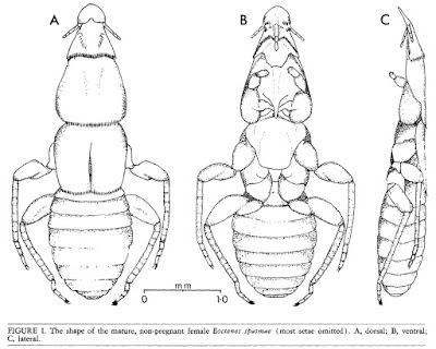
Dorsal, ventral and lateral views of Eoctenes spasmae, from Marshall (1982).


If you ever feel inclined to scan through host records for ectoparasites (and really, why wouldn't you?), you may be struck by the impression that bats seem to be peculiarly lousy animals. There seems to be an unexpected number of groups of ectoparasites that have their highest number of species on bats. One possible reason for this is that, with over 900 potential host species, bat-parasite diversity is high simply because bat diversity is high. Nevertheless, there are other features peculiar to bats that make them excellent parasite hosts. The modification of their fore-legs into wings means that their ability to groom themselves is curtailed. Because many bat species roost in dense colonies, transmission of parasites from one bat to another may happen freely. And because most bats will consistently return to the same roost, speciation is promoted by each colony becoming like an isolated island.

At the same time, referring to bats as 'lousy' is misleading because one ectoparasite group that is curiously absent from bats is the true lice (why this should be I have no idea). Instead, bats are often host to a number of parasite groups all of their own. One such group is the Polyctenidae, flightless true bugs that are found only on bats in tropical and subtropical parts of the world. Polyctenids are closely related to the bed bugs of the Cimicidae and are not dissimilar in appearance. Noticeable differences are their relatively shorter antennae and absence of eyes. They also possess a number of bristle combs at various places on the body, roughly similar in appearance to those on fleas. Their front legs are short and have sucker-like structures on the tarsi instead of claws; the hind two pairs of legs are longer and clawed. The manner of movement of the legs is specialised for crawling among the hair of their host; if removed from the host, the bug is unable to move on a flat surface. Transmission of bugs from one host to another presumably happens only through direct physical contact. Polyctenids share with bed bugs the notorious practice of traumatic insemination with each male injecting sperm directly into the female's body cavity via sharpened genitalia. However, unlike bed bugs they are viviparous, producing live nymphs instead of eggs. The developing embryos are nourished by a 'pseudoplacenta' with a single female potentially containing several developing embryos in a conveyor arrangement at different stages of development. The most mature of these embryos protrudes from the female's genital opening for some time prior to birth and may be a third of its mother's size when born (Marshall 1982).

Type specimen of Hesperoctenes giganteus, from here.


Five genera of polyctenids are generally recognised, with four genera found in the Old World and only a single genus, Hesperoctenes, in the New World (Maa 1964; Ueshima 1972). A second New World genus, Parahesperoctenes, was described in 1947 from a single female, but as the features supposedly distinguishing it from Hesperoctenes related to the consistent duplication of combs, etc., it is thought likely that this was an ordinary individual of Hesperoctenes on the cusp of moulting from a nymph to an adult (so the features of the adult cuticle were visible through the translucent nymphal cuticle). Most of the polyctenid species have a restricted host range, being found on only a single bat species or a small number of closely related species. Some species of Hesperoctenes are more flexible, being found on a range of host species. Hesperoctenes and the Old World genus Hypoctenes are found on free-tailed bats of the Molossidae. Of the other Old World genera, Adroctenes is found on horseshoe bats and leaf-nosed bats of the Rhinolophidae and Hipposideridae, Polyctenes is found on ghost bats of the Megadermatidae, and Eoctenes is found on Megadermatidae, Nycterididae and Emballonuridae. Records of polyctenids from other bat families are currently regarded as suspicious, due to either mislabelling or cross-contamination. Ueshima (1972) suggested that records of Hesperoctenes fumarius from the bulldog bat Noctilio labialis might result from bugs being transferred while the bulldog bats were sharing a roost with their more usual molossid hosts.

Relationships between the genera were discussed by Maa (1964) who divided the family between two subfamilies on the basis of comparative features; a formal phylogenetic analysis of the family appears to still be wanting. On the basis of Hesperoctenes being the 'most specialised' genus and its shared host family with Adroctenes, Maa suggested an Old World origin for Polyctenidae. Eoctenes, with its broad host family range, was regarded as 'least specialised' and likely to be evolutionarily older than other genera. Many of the features distinguishing the polyctenid genera relate to the arrangement of combs: which combs are present where and how they are developed. Prior to Maa's revision, Hesperoctenes had been regarded as likely to be primitive within the Polyctenidae due to its relatively low number of combs. The mid- and hind legs of Adroctenes are fairly short compared to those of other genera.

REFERENCES

Maa, T. C. 1964. A review of the Old World Polyctenidae (Hemiptera: Cimicoidea). Pacific Insects 6 (3): 494–516.

Marshall, A. G. 1982. The ecology of the bat ectoparasite Eoctenes spasmae (Hemiptera: Polyctenidae) in Malaysia. Biotropica 14 (1): 50–55.

Ueshima, N. 1972. New World Polyctenidae (Hemiptera), with special reference to Venezuelan species. Brigham Young University Science Bulletin, Biological Series 17 (1): 13–21.

In a Pichia

Culture of Pichia membranifaciens, from Tomas Linder.


In my previous post, I alluded to the revolutionary effect that DNA analysis had on the classification of bacteria. A similar thing happened for the study of yeasts. Previously, the taxonomy of yeasts (i.e. unicellular fungi) had suffered for the same reasons as bacterial taxonomy: a dearth of usable morphological features combined with uncertainty about the significance or otherwise of metabolic variations. With the availability of genetic information, the relations between yeast taxa became far easier to ascertain.

Needless to say, this lead to a significant shake-up in our understanding of individual yeast taxa. One of the harder-hit taxa was the genus Pichia, previously recognised as a large genus of close to 100 species. Molecular phylogenetic analyses showed that the various species of Pichia were widely scattered within the Saccharomycotina, a fungal clade that includes a large number of yeast species (including such familiar taxa as the brewer's or baker's yeast Saccharomyces cerevisiae). This probably did not come as a huge shock: part of the reason for Pichia's size was that it had not been very stringently defined. Members of this genus were characterised by multilateral budding (that is, buds could develop anywhere along the side of the yeast cell) on a narrow base. They could produce hyphae and/or pseudohyphae (except when they didn't), they might ferment sugars (except when they couldn't), and nitrate might be used as their sole source of nitrogen (except when it wasn't). Pichia spores might be hat-shaped, hemispheroidal or spherical, and they might or might not have a ledge or rim around the equator (Kurtzman 2011).

All of which adds up to a genus that probably tended to be defined as 'the genus that includes any yeast not belonging to these other genera'. In other words, the classic concept of a wastebasket taxon. As a result, the genus has been progressively pared down to a smaller array of species concentrated around the type, Pichia membranifaciens. This is a yeast commonly found as a spoilage organism on foods such as fruit or cheese. Among its other sins, it may grow as a film in the surface of wine, giving the wine an off taste. However, it's not all bad news: recently, P. membranifaciens has been studied as a potential biocontrol agent as it may produce a toxin that has an inhibitory effect on other contaminating fungi (Santos et al. 2009).

A growing culture of Komagataella pastoris, from here.


Somewhat unfortunately, one of the species to be expelled from Pichia is perhaps the best-studied: the yeast formerly known as Pichia pastoris (now supposed to be referred to as Komagataella pastoris though a quick Google Scholar search suggests that a great many authors are pretending that hasn't happened). This species can be grown using methanol as a sole carbon source, and protocols were developed in the 1970s for growing it in high densities at an industrial scale. The original plan was for it to be used for high-protein stock-feed using methanol produced as a by-product of oil refining (the modern agricultural industry has been described as the process of turning oil into food; this would have been a somewhat literal example). Rising oil prices rendered this proposal economically inviable but the P. pastoris industry was to have a reprieve, as the culture method was adopted as a means of producing active proteins (Cereghino & Cregg 2000). Procedures were developed for inserting foreign genes into the yeast, with the resulting pure methanol-based culture allowing the target protein to be generated at a greater rate and higher purity than might be possibly with a culture of the original source organism. Enzymes for laboratory studies, vaccines, medical products such as insulin: whatsitsname pastoris has been used in the production of them all.

REFERENCES

Cereghino, J. L., & J. M. Cregg. 2000. Heterologous protein expression in the methylotrophic yeast Pichia pastoris. FEMS Microbiology Reviews 24: 45–66.

Kurtzman, C. P. 2011. Phylogeny of the ascomycetous yeasts and the renaming of Pichia anomala to Wickerhamomyces anomalus. Antonie van Leeuwenhoek 99: 13–23.

Santos, A., M. San Mauro, E. Bravo & D. Marquina. 2009. PMKT2, a new killer toxin from Pichia membranifaciens, and its promising biotechnological properties for control of the spoilage yeast Brettanomyces bruxellensis. Microbiology 155: 624–634.

Alpha Bacteria

Two budding individuals of Caulobacter crescentus, from the US Dept of Energy.


From about the 1980s onwards, the increasing application of molecular data (particularly the sequences of ribosomal RNA genes) to bacterial phylogeny meant that what had previously been an intractable mass of diversity began to emerge into some sort of order. One of the major new groups of bacteria to be recognised in this way was the Proteobacteria, a hyperdiverse array that includes many of those bacteria of direct significance to ourselves. Within this bacterial supergroup, the phylogeneticists also resolved five major subgroups that, in the absence of any more obvious markers, they labelled alphabetically: the alpha, beta, gamma, delta and epsilon Proteobacteria. Eventually these convenient labels would become formalised, and it is with the group known as the Alphaproteobacteria that I am concerned today.

Like the other proteobacterial lineages, the Alphaproteobacteria are diverse in features and habits. To the best of my knowledge, no uniting characteristic has yet been identified for members of this group other than their shared ribosomal heritage. Many of the Alphaproteobacteria are associated with anoxic habitats. Many are at least facultatively photosynthetic, obtaining energy from sunlight by means of bacteriochlorophyll a and/or carotenoids; these factors give such bacteria a purple coloration. Other Alphaproteobacteria are intracellular parasites of eukaryotes, including a number that are of medical significance to humans. Earlier posts on this site have covered particular subgroups of the Alphaproteobacteria: the nitrogen fixers and plant pathogens of the Rhizobiales, and the diverse order Rhodospirillales. Another group of Proteobacteria, the Epsilonproteobacteria, was the subject of another post.

Culture of vinegar bacteria Acetobacter aceti, copyright Эрг.


A recent study of Alphaproteobacteria ribosomal phylogeny by Ferla et al. (2013) recognised three major lineages within the group which they dubbed the Magnetococcidae, Rickettsidae and Caulobacteridae. The Caulobacteridae include the greater number of the named free-living Alphaproteobacteria: both of the orders covered in earlier posts, for instance, belong to this lineage. Detailed coverage of the various members of Caulobacteridae would fill a book, so I'll just mention some highlights. The earlier post on Rhodospirillales mentioned the family Acetobacteraceae, but one important detail I neglected to mention was that many members of this family obtain their energy by oxidising ethanol to acetic acid: these are the bacteria responsible for producing ethanol. Also potentially belonging to the Rhodospirillales is Sporospirillum, a candidate genus of enormous bacteria that have been found in the intestines of tadpoles. Individuals of Sporospirillum reach up to one-tenth of a millimetre in length, potentially large enough to be observed with a standard dissecting microscope, though they are only up to 5 µm in width. Because Sporospirillum have never been cultured or studied from a molecular perspective, their relationships remain uncertain: they may alternatively belong to the Spirillaceae, a family of the Betaproteobacteria (Brenner et al. 2005).

Also belonging to the Caulobacteridae are the Caulobacterales. As recognised by Ferla et al. (2013), this order contains two families, the Caulobacteraceae and Hyphomonadaceae. Many (but not all) of the members of these families have a distinctive life cycle, in which a previously motile individual loses its flagellum and grows an elongate stalk. This now-immotile individual then produces a motile offspring by budding at one end. The manner of budding differs between the two families: in the Caulobacteraceae, the stalk functions as an attachment to the substrate and the offspring buds from the unattached end of the cell, but in the Hyphomonadaceae the stalk is not an attachment organ and the offspring buds from the end of the stalk. Similar modes of growth and budding are found in other families of the Caulobacteridae, such as the Hyphomicrobiaceae in the Rhizobiales.

TEM view of Magnetococcus marinus, showing the line of magnetic particles (magnetosomes).


The other subclasses of the Alphaproteobacteria are smaller than the Caulobacteridae in terms of numbers of named species, but this may reflect our low appreciation of bacterial diversity more than environmental reality. The Magnetococcidae are represented by only a single named species, Magnetococcus marinus. This is an aquatic chemolithoautotroph, obtaining energy from sulphur compounds. Cells of Magnetococcus contain a row of magnetic particles that the bacterium uses to orient itself. Though only one species of magnetococcid has been named to date, environmental DNA samples indicate that many more await description (Bazylinski et al. 2013).

The ciliate Paramecium, infected with Holospora (in the swollen nucleus in the lower part of the photo). Photo from here, by this network's own Psi Wavefunction (wherever she may be...)


The named members of the Rickettsidae are mostly placed in the order Rickettsiales, an assemblage of intracellular parasites of eukaryotes. This order contains two families, the Rickettsiaceae and Anaplasmataceae; a third family, the Holosporaceae, that contains intracellular endosymbionts of large protozoans such as Paramecium and Acanthamoeba, was found by Ferla et al. (2013) to be potentially closer to the Caulobacteridae than the Rickettsidae. Members of the Rickettsiales of significance to humans include those causing such diseases as typhus or spotted fever. The Anaplasmataceae also includes the genus Wolbachia which has come under the spotlight in recent years for the significance that its effects on reproductive compatibility may have for the evolution of insects.

The only free-living bacterium associated with the Rickettsidae to date is the marine Pelagibacter ubique but, again, environmental DNA samples suggest that this is merely a representative of a larger undescribed group, commonly referred to as the 'SAR11' clade. Indeed, Pelagibacter and its relatives may be the most numerous organisms on the entire planet, making up about a third of the planktonic cells in the surface layers of the world's oceans (Morris et al. 2002). Even by bacterial standards, Pelagibacter cells are small, and it has one of the smallest known genomes for any free-living organisms.

Stained sample of Pelagibacter ubique, copyright Thomas Lankiewicz & Matthew Cottrell.


There is one final important subgroup of the alphaproteobacterial lineage that I haven't mentioned yet: us. At some point in the distant past, a member of the Alphaproteobacteria developed a close and personal relationship with another micro-organism, either a member of the Archaea or a close relative thereof. Phylogenetic studies indicate that this early alphaproteobacterium was probably a close relative of the Rickettsiales. Over time, this relationship became ever closer, until the one became inseparable from the other. Together, these two microbes were to give rise to the eukaryotes, with the alphaproteobacteria becoming transformed into the mitochondria of a eukaryote cell. From the perspective of descent, then, we are all Alphaproteobacteria.

REFERENCES

Bazylinski, D. A., T. J. Williams, C. T. Lefèvre, R. J. Berg, C. L. Zhang, S. S. Bowser, A. J. Dean & T. J. Beveridge. 2013. Magnetococcus marinus gen. nov., sp. nov., a marine, magnetotactic bacterium that represents a novel lineage (Magnetococcaceae fam. nov., Magnetococcales ord. nov.) at the base of the Alphaproteobacteria. International Journal of Systematic and Evolutionary Microbiology 63: 801–808.

Brenner, D. J., N. R. Krieg & J. T. Staley. 2005. Bergey's Manual of Systematic Bacteriology 2nd ed. vol. 2 pt C. The Alpha-, Beta-, Delta-, and Epsilonproteobacteria. Springer.

Ferla, M. P., J. C. Thrash, S. J. Giovannoni & W. M. Patrick. 2013. New rRNA gene-based phylogenies of the Alphaproteobacteria provide perspective on major groups, mitochondrial ancestry and phylogenetic instability. PLoS One 8 (12): e83383. doi:10.1371/journal.pone.0083383.

Morris, R. M., M. S. Rappé, S. A. Connon, K. L. Vergin, W. A. Siebold, C. A. Carlson & S. J. Giovannoni. 2002. SAR11 clade dominates ocean surface bacterioplankton communities. Nature 420: 806–810.

Cichlids are Not the Only Radiation

The Congo River catfish Chrysichthys brevibarbis, copyright John P. Sullivan.


With their long barbels around the mouth and lack of scales, the catfish of the Siluriformes are one of most instantly recognisable groups of fishes. They are also one of the more diverse, with close to 3000 species and including a third of the world's freshwater fishes (Diogo & Peng 2010). Within the catfish, the Claroteidae are a distinctly African group of thirteen genera divided between two subfamilies, the Claroteinae and Auchenoglanididae. They are characterised by a moderately elongate body with a distinct adipose fin, and strong spines in the dorsal and pectoral fins (Geerinckx et al. 2003). Distinctive features of the Claroteinae include the presence of a toothplate on the palate. The Auchenoglanidinae have a rounded caudal fin and the anterior nostrils moved to the anteroventral side of the upper lip (Geerinckx et al. 2004). For a long time, the claroteids were included in the catfish family Bagridae before being raised to the level of their own family in 1991. A molecular phylogenetic analysis of the Siluriformes by Sullivan et al. (2006) placed the claroteids within a clade of African catfish that they somewhat whimsically labelled as 'Big Africa'. The Bagridae, meanwhile, were placed within 'Big Asia' (though one true bagrid genus, Bagrus, does occur in Africa). Sullivan et al. (2006) questioned claroteid monophyly, finding Auchenoglanidinae to be sister to a clade grouping the Claroteinae with the family Schilbidae, but other morphological studies have found claroteids as a monophyletic unit (Diogo & Peng 2010).

Lake Tanganyika catfish Lophiobagrus brevispinis, from tanganyikacichlide.nl.


The Claroteinae are notable for having undergone something of an adaptive radiation in one of African Great Lakes, Tanganyika. Though not as dramatic as the famous radiation of cichlids in the same lake, the Tanganyikan claroteines comprise over a dozen species divided between four genera (Bailey & Stewart 1984; Hardman 2008). Seven of these are placed in the genus Chrysichthys which has a wide distribution around Africa; the other three genera are unique to the lake. Molecular phylogeny indicates that the majority of Tanganyikan claroteines represent a single colonisation of the lake; only Chrysichthys brachynema has colonised Lake Tanganyika independently (Peart et al 2014). This indicates that the genus Chrysichthys as currently defined is non-monophyletic (something that had previously been suggested on morphological grounds) but any consequent reclassification is yet to occur. The species of Chrysichthys are mostly larger than the endemic Tanganyikan genera, ranging from 19 to 77 cm within Tanganyika (species elsewhere in Africa may reach up to 1.5 m). Of the endemic genera, the monotypic Bathybagrus tetranema is about 15 cm in length but the other two genera Phyllonemus and Lophiobagrus are even smaller, less than 10 cm in length. Bathybagrus and Lophiobagrus also both have reduced subcutaneous eyes. In Bathybagrus, this possibly reflects their occurrence at greater depths than other Tanganyika fish, occurring down to 80 m (nowhere near the depths reached by Lake Baikal sculpins but still impressive enough in the low-oxygen depths of a tropical lake). Lophiobagrus species are specialised to live in the gaps between rocky rubble on the lake bottom. The species of this genus have also been observed secreting a toxic mucus that can be fatal to other fish; this mucus is believed to be secreted from enlarged glands behind the pectoral fins.

Subcutaneous eyes are also found in two claroteines outside Tanganyika: the species Amarginops platus and Rheoglanis dendrophorus, both found in the Upper Congo (Hardman 2008). These two species are specialised for life in river rapids.

REFERENCES

Bailey, R. M., & D. J. Stewart. 1984. Bagrid catfishes from Lake Tanganyika, with a key and descriptions of new taxa. Miscellaneous Publication, Museum of Zoology, University of Michigan 168: 1–41.

Diogo, R., & Z. Peng. 2009. State of the art of siluriform higher-level phylogeny. In: Grande, T., F. Poyato-Ariza & R. Diogo (eds) Gonorynchiformes and Ostariophysan Relationships: A Comprehensive Review pp. 465–515. Science Publishers.

Geerinckx, T., D. Adriaens, G. G. Teugels & W. Verraes. 2003. Taxonomic evaluation and redescription of Anaspidoglanis akiri (Risch, 1987) (Siluriformes: Claroteidae). Cybium 27 (1): 17–25.

Geerinckx, T., D. Adriaens, G. G. Teugels & W. Verraes. 2004. A systematic revision of the African catfish genus Parauchenoglanis (Siluriformes: Claroteidae). Journal of Natural History 38: 775–803.

Hardman, M. 2008. New species of catfish genus Chrysichthys from Lake Tanganyika (Siluriformes: Claroteidae). Copeia 2008 (1): 43–56.

Peart, C. R., R. Bills, M. Wilkinson & J. J. Day. 2014. Nocturnal claroteine catfishes reveal dual colonisation but a single radiation in Lake Tanganyika. Molecular Phylogenetics and Evolution 73: 119–128.

Sullivan, J. P., J. G. Lundberg & M. Hardman. 2006. A phylogenetic analysis of the major groups of catfishes (Teleostei: Siluriformes) using rag1 and rag2 nuclear gene sequences. Molecular Phylogenetics and Evolution 41: 636–662.