Field of Science

In a Pichia

Culture of Pichia membranifaciens, from Tomas Linder.


In my previous post, I alluded to the revolutionary effect that DNA analysis had on the classification of bacteria. A similar thing happened for the study of yeasts. Previously, the taxonomy of yeasts (i.e. unicellular fungi) had suffered for the same reasons as bacterial taxonomy: a dearth of usable morphological features combined with uncertainty about the significance or otherwise of metabolic variations. With the availability of genetic information, the relations between yeast taxa became far easier to ascertain.

Needless to say, this lead to a significant shake-up in our understanding of individual yeast taxa. One of the harder-hit taxa was the genus Pichia, previously recognised as a large genus of close to 100 species. Molecular phylogenetic analyses showed that the various species of Pichia were widely scattered within the Saccharomycotina, a fungal clade that includes a large number of yeast species (including such familiar taxa as the brewer's or baker's yeast Saccharomyces cerevisiae). This probably did not come as a huge shock: part of the reason for Pichia's size was that it had not been very stringently defined. Members of this genus were characterised by multilateral budding (that is, buds could develop anywhere along the side of the yeast cell) on a narrow base. They could produce hyphae and/or pseudohyphae (except when they didn't), they might ferment sugars (except when they couldn't), and nitrate might be used as their sole source of nitrogen (except when it wasn't). Pichia spores might be hat-shaped, hemispheroidal or spherical, and they might or might not have a ledge or rim around the equator (Kurtzman 2011).

All of which adds up to a genus that probably tended to be defined as 'the genus that includes any yeast not belonging to these other genera'. In other words, the classic concept of a wastebasket taxon. As a result, the genus has been progressively pared down to a smaller array of species concentrated around the type, Pichia membranifaciens. This is a yeast commonly found as a spoilage organism on foods such as fruit or cheese. Among its other sins, it may grow as a film in the surface of wine, giving the wine an off taste. However, it's not all bad news: recently, P. membranifaciens has been studied as a potential biocontrol agent as it may produce a toxin that has an inhibitory effect on other contaminating fungi (Santos et al. 2009).

A growing culture of Komagataella pastoris, from here.


Somewhat unfortunately, one of the species to be expelled from Pichia is perhaps the best-studied: the yeast formerly known as Pichia pastoris (now supposed to be referred to as Komagataella pastoris though a quick Google Scholar search suggests that a great many authors are pretending that hasn't happened). This species can be grown using methanol as a sole carbon source, and protocols were developed in the 1970s for growing it in high densities at an industrial scale. The original plan was for it to be used for high-protein stock-feed using methanol produced as a by-product of oil refining (the modern agricultural industry has been described as the process of turning oil into food; this would have been a somewhat literal example). Rising oil prices rendered this proposal economically inviable but the P. pastoris industry was to have a reprieve, as the culture method was adopted as a means of producing active proteins (Cereghino & Cregg 2000). Procedures were developed for inserting foreign genes into the yeast, with the resulting pure methanol-based culture allowing the target protein to be generated at a greater rate and higher purity than might be possibly with a culture of the original source organism. Enzymes for laboratory studies, vaccines, medical products such as insulin: whatsitsname pastoris has been used in the production of them all.

REFERENCES

Cereghino, J. L., & J. M. Cregg. 2000. Heterologous protein expression in the methylotrophic yeast Pichia pastoris. FEMS Microbiology Reviews 24: 45–66.

Kurtzman, C. P. 2011. Phylogeny of the ascomycetous yeasts and the renaming of Pichia anomala to Wickerhamomyces anomalus. Antonie van Leeuwenhoek 99: 13–23.

Santos, A., M. San Mauro, E. Bravo & D. Marquina. 2009. PMKT2, a new killer toxin from Pichia membranifaciens, and its promising biotechnological properties for control of the spoilage yeast Brettanomyces bruxellensis. Microbiology 155: 624–634.

Alpha Bacteria

Two budding individuals of Caulobacter crescentus, from the US Dept of Energy.


From about the 1980s onwards, the increasing application of molecular data (particularly the sequences of ribosomal RNA genes) to bacterial phylogeny meant that what had previously been an intractable mass of diversity began to emerge into some sort of order. One of the major new groups of bacteria to be recognised in this way was the Proteobacteria, a hyperdiverse array that includes many of those bacteria of direct significance to ourselves. Within this bacterial supergroup, the phylogeneticists also resolved five major subgroups that, in the absence of any more obvious markers, they labelled alphabetically: the alpha, beta, gamma, delta and epsilon Proteobacteria. Eventually these convenient labels would become formalised, and it is with the group known as the Alphaproteobacteria that I am concerned today.

Like the other proteobacterial lineages, the Alphaproteobacteria are diverse in features and habits. To the best of my knowledge, no uniting characteristic has yet been identified for members of this group other than their shared ribosomal heritage. Many of the Alphaproteobacteria are associated with anoxic habitats. Many are at least facultatively photosynthetic, obtaining energy from sunlight by means of bacteriochlorophyll a and/or carotenoids; these factors give such bacteria a purple coloration. Other Alphaproteobacteria are intracellular parasites of eukaryotes, including a number that are of medical significance to humans. Earlier posts on this site have covered particular subgroups of the Alphaproteobacteria: the nitrogen fixers and plant pathogens of the Rhizobiales, and the diverse order Rhodospirillales. Another group of Proteobacteria, the Epsilonproteobacteria, was the subject of another post.

Culture of vinegar bacteria Acetobacter aceti, copyright Эрг.


A recent study of Alphaproteobacteria ribosomal phylogeny by Ferla et al. (2013) recognised three major lineages within the group which they dubbed the Magnetococcidae, Rickettsidae and Caulobacteridae. The Caulobacteridae include the greater number of the named free-living Alphaproteobacteria: both of the orders covered in earlier posts, for instance, belong to this lineage. Detailed coverage of the various members of Caulobacteridae would fill a book, so I'll just mention some highlights. The earlier post on Rhodospirillales mentioned the family Acetobacteraceae, but one important detail I neglected to mention was that many members of this family obtain their energy by oxidising ethanol to acetic acid: these are the bacteria responsible for producing ethanol. Also potentially belonging to the Rhodospirillales is Sporospirillum, a candidate genus of enormous bacteria that have been found in the intestines of tadpoles. Individuals of Sporospirillum reach up to one-tenth of a millimetre in length, potentially large enough to be observed with a standard dissecting microscope, though they are only up to 5 µm in width. Because Sporospirillum have never been cultured or studied from a molecular perspective, their relationships remain uncertain: they may alternatively belong to the Spirillaceae, a family of the Betaproteobacteria (Brenner et al. 2005).

Also belonging to the Caulobacteridae are the Caulobacterales. As recognised by Ferla et al. (2013), this order contains two families, the Caulobacteraceae and Hyphomonadaceae. Many (but not all) of the members of these families have a distinctive life cycle, in which a previously motile individual loses its flagellum and grows an elongate stalk. This now-immotile individual then produces a motile offspring by budding at one end. The manner of budding differs between the two families: in the Caulobacteraceae, the stalk functions as an attachment to the substrate and the offspring buds from the unattached end of the cell, but in the Hyphomonadaceae the stalk is not an attachment organ and the offspring buds from the end of the stalk. Similar modes of growth and budding are found in other families of the Caulobacteridae, such as the Hyphomicrobiaceae in the Rhizobiales.

TEM view of Magnetococcus marinus, showing the line of magnetic particles (magnetosomes).


The other subclasses of the Alphaproteobacteria are smaller than the Caulobacteridae in terms of numbers of named species, but this may reflect our low appreciation of bacterial diversity more than environmental reality. The Magnetococcidae are represented by only a single named species, Magnetococcus marinus. This is an aquatic chemolithoautotroph, obtaining energy from sulphur compounds. Cells of Magnetococcus contain a row of magnetic particles that the bacterium uses to orient itself. Though only one species of magnetococcid has been named to date, environmental DNA samples indicate that many more await description (Bazylinski et al. 2013).

The ciliate Paramecium, infected with Holospora (in the swollen nucleus in the lower part of the photo). Photo from here, by this network's own Psi Wavefunction (wherever she may be...)


The named members of the Rickettsidae are mostly placed in the order Rickettsiales, an assemblage of intracellular parasites of eukaryotes. This order contains two families, the Rickettsiaceae and Anaplasmataceae; a third family, the Holosporaceae, that contains intracellular endosymbionts of large protozoans such as Paramecium and Acanthamoeba, was found by Ferla et al. (2013) to be potentially closer to the Caulobacteridae than the Rickettsidae. Members of the Rickettsiales of significance to humans include those causing such diseases as typhus or spotted fever. The Anaplasmataceae also includes the genus Wolbachia which has come under the spotlight in recent years for the significance that its effects on reproductive compatibility may have for the evolution of insects.

The only free-living bacterium associated with the Rickettsidae to date is the marine Pelagibacter ubique but, again, environmental DNA samples suggest that this is merely a representative of a larger undescribed group, commonly referred to as the 'SAR11' clade. Indeed, Pelagibacter and its relatives may be the most numerous organisms on the entire planet, making up about a third of the planktonic cells in the surface layers of the world's oceans (Morris et al. 2002). Even by bacterial standards, Pelagibacter cells are small, and it has one of the smallest known genomes for any free-living organisms.

Stained sample of Pelagibacter ubique, copyright Thomas Lankiewicz & Matthew Cottrell.


There is one final important subgroup of the alphaproteobacterial lineage that I haven't mentioned yet: us. At some point in the distant past, a member of the Alphaproteobacteria developed a close and personal relationship with another micro-organism, either a member of the Archaea or a close relative thereof. Phylogenetic studies indicate that this early alphaproteobacterium was probably a close relative of the Rickettsiales. Over time, this relationship became ever closer, until the one became inseparable from the other. Together, these two microbes were to give rise to the eukaryotes, with the alphaproteobacteria becoming transformed into the mitochondria of a eukaryote cell. From the perspective of descent, then, we are all Alphaproteobacteria.

REFERENCES

Bazylinski, D. A., T. J. Williams, C. T. Lefèvre, R. J. Berg, C. L. Zhang, S. S. Bowser, A. J. Dean & T. J. Beveridge. 2013. Magnetococcus marinus gen. nov., sp. nov., a marine, magnetotactic bacterium that represents a novel lineage (Magnetococcaceae fam. nov., Magnetococcales ord. nov.) at the base of the Alphaproteobacteria. International Journal of Systematic and Evolutionary Microbiology 63: 801–808.

Brenner, D. J., N. R. Krieg & J. T. Staley. 2005. Bergey's Manual of Systematic Bacteriology 2nd ed. vol. 2 pt C. The Alpha-, Beta-, Delta-, and Epsilonproteobacteria. Springer.

Ferla, M. P., J. C. Thrash, S. J. Giovannoni & W. M. Patrick. 2013. New rRNA gene-based phylogenies of the Alphaproteobacteria provide perspective on major groups, mitochondrial ancestry and phylogenetic instability. PLoS One 8 (12): e83383. doi:10.1371/journal.pone.0083383.

Morris, R. M., M. S. Rappé, S. A. Connon, K. L. Vergin, W. A. Siebold, C. A. Carlson & S. J. Giovannoni. 2002. SAR11 clade dominates ocean surface bacterioplankton communities. Nature 420: 806–810.

Cichlids are Not the Only Radiation

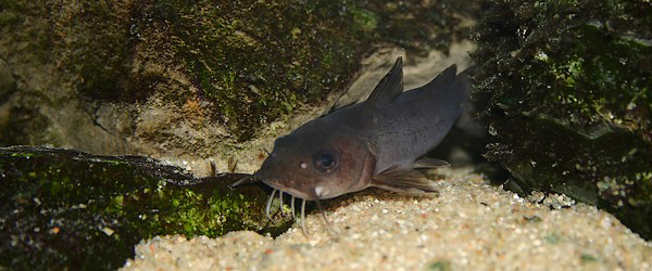
The Congo River catfish Chrysichthys brevibarbis, copyright John P. Sullivan.


With their long barbels around the mouth and lack of scales, the catfish of the Siluriformes are one of most instantly recognisable groups of fishes. They are also one of the more diverse, with close to 3000 species and including a third of the world's freshwater fishes (Diogo & Peng 2010). Within the catfish, the Claroteidae are a distinctly African group of thirteen genera divided between two subfamilies, the Claroteinae and Auchenoglanididae. They are characterised by a moderately elongate body with a distinct adipose fin, and strong spines in the dorsal and pectoral fins (Geerinckx et al. 2003). Distinctive features of the Claroteinae include the presence of a toothplate on the palate. The Auchenoglanidinae have a rounded caudal fin and the anterior nostrils moved to the anteroventral side of the upper lip (Geerinckx et al. 2004). For a long time, the claroteids were included in the catfish family Bagridae before being raised to the level of their own family in 1991. A molecular phylogenetic analysis of the Siluriformes by Sullivan et al. (2006) placed the claroteids within a clade of African catfish that they somewhat whimsically labelled as 'Big Africa'. The Bagridae, meanwhile, were placed within 'Big Asia' (though one true bagrid genus, Bagrus, does occur in Africa). Sullivan et al. (2006) questioned claroteid monophyly, finding Auchenoglanidinae to be sister to a clade grouping the Claroteinae with the family Schilbidae, but other morphological studies have found claroteids as a monophyletic unit (Diogo & Peng 2010).

Lake Tanganyika catfish Lophiobagrus brevispinis, from tanganyikacichlide.nl.


The Claroteinae are notable for having undergone something of an adaptive radiation in one of African Great Lakes, Tanganyika. Though not as dramatic as the famous radiation of cichlids in the same lake, the Tanganyikan claroteines comprise over a dozen species divided between four genera (Bailey & Stewart 1984; Hardman 2008). Seven of these are placed in the genus Chrysichthys which has a wide distribution around Africa; the other three genera are unique to the lake. Molecular phylogeny indicates that the majority of Tanganyikan claroteines represent a single colonisation of the lake; only Chrysichthys brachynema has colonised Lake Tanganyika independently (Peart et al 2014). This indicates that the genus Chrysichthys as currently defined is non-monophyletic (something that had previously been suggested on morphological grounds) but any consequent reclassification is yet to occur. The species of Chrysichthys are mostly larger than the endemic Tanganyikan genera, ranging from 19 to 77 cm within Tanganyika (species elsewhere in Africa may reach up to 1.5 m). Of the endemic genera, the monotypic Bathybagrus tetranema is about 15 cm in length but the other two genera Phyllonemus and Lophiobagrus are even smaller, less than 10 cm in length. Bathybagrus and Lophiobagrus also both have reduced subcutaneous eyes. In Bathybagrus, this possibly reflects their occurrence at greater depths than other Tanganyika fish, occurring down to 80 m (nowhere near the depths reached by Lake Baikal sculpins but still impressive enough in the low-oxygen depths of a tropical lake). Lophiobagrus species are specialised to live in the gaps between rocky rubble on the lake bottom. The species of this genus have also been observed secreting a toxic mucus that can be fatal to other fish; this mucus is believed to be secreted from enlarged glands behind the pectoral fins.

Subcutaneous eyes are also found in two claroteines outside Tanganyika: the species Amarginops platus and Rheoglanis dendrophorus, both found in the Upper Congo (Hardman 2008). These two species are specialised for life in river rapids.

REFERENCES

Bailey, R. M., & D. J. Stewart. 1984. Bagrid catfishes from Lake Tanganyika, with a key and descriptions of new taxa. Miscellaneous Publication, Museum of Zoology, University of Michigan 168: 1–41.

Diogo, R., & Z. Peng. 2009. State of the art of siluriform higher-level phylogeny. In: Grande, T., F. Poyato-Ariza & R. Diogo (eds) Gonorynchiformes and Ostariophysan Relationships: A Comprehensive Review pp. 465–515. Science Publishers.

Geerinckx, T., D. Adriaens, G. G. Teugels & W. Verraes. 2003. Taxonomic evaluation and redescription of Anaspidoglanis akiri (Risch, 1987) (Siluriformes: Claroteidae). Cybium 27 (1): 17–25.

Geerinckx, T., D. Adriaens, G. G. Teugels & W. Verraes. 2004. A systematic revision of the African catfish genus Parauchenoglanis (Siluriformes: Claroteidae). Journal of Natural History 38: 775–803.

Hardman, M. 2008. New species of catfish genus Chrysichthys from Lake Tanganyika (Siluriformes: Claroteidae). Copeia 2008 (1): 43–56.

Peart, C. R., R. Bills, M. Wilkinson & J. J. Day. 2014. Nocturnal claroteine catfishes reveal dual colonisation but a single radiation in Lake Tanganyika. Molecular Phylogenetics and Evolution 73: 119–128.

Sullivan, J. P., J. G. Lundberg & M. Hardman. 2006. A phylogenetic analysis of the major groups of catfishes (Teleostei: Siluriformes) using rag1 and rag2 nuclear gene sequences. Molecular Phylogenetics and Evolution 41: 636–662.

Amphiascus: Can a Copepod be a Friend of Mine?

Amphiascus sp., copyright Alexandra.


The animal shown in the image above is a member of Amphiascus, a cosmopolitan genus of about thirty known species of benthic harpacticoid copepods. Amphiascus is a genus of the family Miraciidae; in older texts, you will find it referred to the Diosaccidae, but this family is now regarded as a synonym of the former. Miraciids are somewhat elongate harpacticoids generally with a fusiform body shape and females with paired egg sacs; as with other copepod taxa, their specific characterisation depends on fairly fine characters of the appendage setation (Willen 2002). Wells et al. (1982) placed Amphiascus in association with a group of related genera in the miraciid family tree on the basis of its retention of a fairly extensive setation on the pereiopods, two inner setae on the endopod of pereiopod II in females, and two articulated claws on that segment in males. However, the proposed phylogeny of Wells et al. provides no apomorphies for Amphiascus itself, implying that it is characterised only by plesiomorphies relative to related genera.

The title of this post refers to the circumstances surrounding the discovery of a relatively recently described Amphiascus species, A. kawamurai Ueda & Nagai 2005. In the cultivation in Japan of nori, the edible alga used (among other things) in wrapping sushi rolls, the conchocelis phase of the life cycle is grown on oyster shells in outdoor tanks of seawater (like many algae, nori goes through an alternation of generations, with its life cycle including two very distinct forms; as well as the familiar large flat alga, the life cycle of nori includes a small filamentous shell-boring stage, initially mistaken for a distinct organism and called Conchocelis). Unfortunately, the oyster shells may also become overgrown with diatoms, retarding the growth of conchocelis. As a result, nori growers may be required to laboriously scrub the shells of diatoms several times over the conchocelis growth period. However, it was noticed in Ariake Bay in Kyushu that some form of copepod would sometimes appear in the nori tanks, presumably brought in with seawater from the bay. When this copepod was present, it would graze on the diatoms, reducing the need for other controls. Study of the nori-tank copepod revealed it to be a previously undescribed species, revealing once more that even the species we are not aware of have the potential to directly improve our lives.

REFERENCES

Ueda, H., & H. Nagai. 2005. Amphiascus kawamurai, a new harpacticoid copepod (Crustacea: Harpacticoida: Miraciidae) from nori cultivation tanks in Japan, with a redescription of the closely related A. parvus. Species Diversity 10: 249–258.

Wells, J. B. J., G. R. F. Hicks & B. C. Coull. 1982. Common harpacticoid copepods from New Zealand harbours and estuaries. New Zealand Journal of Zoology 9 (2): 151–184.

Willen, E. 2002. Notes on the systematic position of the Stenheliinae (Copepoda, Harpacticoida) within the Thalestridimorpha and description of two new species from Motupore Island, Papua New Guinea. Cah. Biol. Mar. 43: 27–42.

The Chromeurytominae: Australo-Asian Mystery Wasps

One of the most diverse groups of micro-wasps is the Chalcidoidea, a bewildering array of intricate little jewels. A number of chalcidoid taxa have been extensively studied due to their roles as parasitoids of insect pests, but there are also many groups of chalcidoids that remain little known. One such group is the Chromeurytominae.

Male Chromeurytoma sp., copyright John Heraty.


The Chromeurytominae are a small group of chalcidoids primarily known from Australia, where they are represented by two genera, fourteen species of Chromeurytoma and the monotypic Asaphoideus niger (Bouček 1988). A single species, Pitayana coccorum, has also been described from Bangladesh (Bouček & Bhuiya 1990). Characteristic features include a relatively large subrectangular pronotum (the first segment of the thorax) and an antenna with six segments between the pedicel and the clava (the club). They are more or less shiny, often with a blue or green metallic gloss, and the gaster is fairly robust and does not collapse in preserved specimens. The affinities of the Chromeurytominae have been rather uncertain and the subfamily was only established by Bouček in 1988. Chromeurytoma itself was originally described in the family Eurytomidae, with which it shares the large pronotum. Other features suggest a relationship with the family Torymidae, such as an occipital carina (a ridge around the back of the head) and prominent cerci. Currently the Chromeurytominae are treated as part of the family Pteromalidae, which is not really saying too much. As our understanding of chalcidoid phylogeny has improved in recent years, it has largely confirmed what many workers had long suspected: that once you account for the other families, the Pteromalidae is pretty much just what's left over. Nevertheless, the broad-scale analysis of chalcidoids by Heraty et al. (2013) places the Chromeurytominae within a cluster of 'pteromalid' subfamilies, closer to the type subfamily Pteromalinae than to either the Eurytomidae or Torymidae.

The chromeurytomines are a bit of a mixed bag in terms of host species, but there is the common thread that their hosts are immobile or semi-sedentary plant-feeding insects. Pitayana coccorum attacks mealybugs and other soft scales, with multiple larvae potentially developing on a single host. Asaphoideus niger attacks the citrus leaf-miner Phyllocnistis citrella. The Chromeurytoma species are associated with galls on trees such as Eucalyptus; presumably they are parasites of the insects forming the galls.

REFERENCES

Bouček, Z. 1988. Australasian Chalcidoidea (Hymenoptera): A biosystematic revision of genera of fourteen families, with a reclassification of species. CAB International: Wallingford (UK).

Bouček, Z., & B. A. Bhuiya. 1990. A new genus and species of Pteromalidae (Hym.) attacking mealybugs and soft scales (Hom., Coccoidea) on guava in Bangladesh. Entomologist's Monthly Magazine 126: 231–235.

Heraty, J. M., R. A. Burks, A. Cruaud, G. A. P. Gibson, J. Liljeblad, J. Munro, J.-Y. Rasplus, G. Delvare, P. Janšta, A. Gumovsky, J. Huber, J. B. Woolley, L. Krogmann, S. Heydon, A. Polaszek, S. Schmidt, D. C. Darling, M. W. Gates, J. Mottern, E. Murray, A. D. Molin, S. Triapitsyn, H. Baur, J. D. Pinto, S. van Noort, J. George & M. Yoder. 2013. A phylogenetic analysis of the megadiverse Chalcidoidea (Hymenoptera). Cladistics 29: 466–542.

Petrosia: The Sexual Life of the Sponges

It has to be admitted that sponges are not one of the best-publicised of animal groups. Even when they are given some grudging mention, there is little reference to the variety of sponges that can be found on our planet. But don't go thinking that all sponges are the same.

Stony sponge Petrosia ficiformis, copyright Véronique Lamare.


Petrosia is a genus of sponges found in tropical and subtropical oceans around the world. Members of this genus come in a variety of forms: branching, cylindrical, globular, lamellate or bowl-shaped. They may reach large sizes, with some species up to a metre or two in diameter, though others may be much smaller. Most species are dark colours such as red, brown or black, though the Sulawesi species Petrosia alfiani is a bright canary yellow (de Voogd & van Soest 2002). The reasons for classifying such superficially divergent forms in a single genus lie beneath the surface. However, it has a high proportion of skeletal spicules to soft tissue, giving Petrosia species a hard, brittle texture (hence they are sometimes known as 'stony sponges'). The spicules of Petrosia are mostly long, slightly curved rods that may be rounded or pointed at the ends; they may be large or smaller, with smaller spicules tending to be more common closer to the sponge's surface. Two subgenera are recognised within Petrosia on the basis of whether the spicules are mostly in a tangential (subgenus Petrosia) or reticulate (Strongylophora) arrangement. The subgenus Petrosia is known from the Atlantic and Pacific Oceans, whereas Strongylophora species are found in the Indian Ocean and the western Pacific (Desqueyroux-Faúndez & Valentine 2002).

Magnified view of surface of Petrosia ficiformis specimen, showing arrangement of spicules, from (Desqueyroux-Faúndez & Valentine (2002).


One of the best-studied species in this genus is the Mediterranean Petrosia ficiformis, which tends towards a cylindrical growth habit in sheltered spots. Like other sponges, P. ficiformis may provide an important habitat for other organisms. Smaller invertebrates live in and around the sponge, and molecular studies have shown that different sponge species tend to host their own distinct communities of bacteria. However, the niche provided by Petrosia in the Mediterranean can be vulnerable to damage: field observations have indicated that stony sponges grow exceedingly slowly. Maldonado & Riesgo (2009) found that in twenty years of diving off the Spanish coast, they saw almost no growth in individual sponges. When they took small (one by one-half centimetre) tissue samples from the sponges, it could take up to three months for the removed patch to regrow. Such a slow rate of growth definitely makes one wonder just how old some of the large Petrosia referred to above must be.

Bowl-shaped Petrosia lignosa, from de Voogd & van Soest (2002).


Maldonado & Riesgo (2009) were taking their samples to study how the sponges reproduced. Petrosia species are free spawners, releasing eggs and sperm directly into the water column. In the case of P. ficiformis, this happens in late autumn. Eggs develop at scattered locations through the sponge, but migrate within the body to form clusters before being released. The sexes are separate, with an individual sponge only producing either eggs or sperm. After fertilisation, the eggs develop into small ciliated larvae that may shift between a spherical and a multilobate form. Whereas the larvae of other sponges may be quite mobile, those of P. ficiformis are not active swimmers, presumably relying on the motion of water currents to carry them to a suitable resting spot. Maldonado & Riesgo (2009) noted that in the two years they observed Petrosia spawning, it occured at times when surge levels had risen immediately prior to the onset of stormy weather. Despite the regular associations of Petrosia with particular microbial populations, the larvae do not carry any sort of culture propagule from their parents, indicating that each individual sponge reacquires its associates from the surrounding waters. Larvae attach themselves to the substrate after two to four weeks of growth, and proceed to grow slowly (though, as is the way of sponges, if multiple larvae settle immediately adjacent to one another they may fuse into a single aggregate individual). Larvae grown in the lab took about one and a half months to develop distinct choanocyte chambers (the ciliated chambers in which a sponge filters water for food particles). They may share their environment with sea hares, but there is no question that Petrosia are sea tortoises.

REFERENCES

Desqueyroux-Faúndez, R., & C. Valentine. 2002. Family Petrosiidae van Soest, 1980. In: Hooper, J. N. A., & R. W. M. van Soest (eds) Systema Porifera: A guide to the classification of sponges pp. 906–917. Kluwer Academic/Plenum Publishers: New York.

Maldonado, M., & A. Riesgo. 2009. Gametogenesis, embryogenesis, and larval features of the oviparous sponge Petrosia ficiformis (Haplosclerida, Demospongiae). Marine Biology 156 (10): 2181–2197.

Voogd, N. J. de, & R. W. M. van Soest. 2002. Indonesian sponges of the genus Petrosia Vosmaer (Demospongiae: Haplosclerida). Zool. Med. Leiden 76 (16): 193–209.

Bizarre 'alien corpse' has idiots stumped

I know we've all become more familiar with 'mystery monster' corpse stories in the last few years, but I think they may have reached a new nadir. Fairfax featured a story today titled 'Bizarre 'alien corpse' discovered in Russia has experts stumped'. This is the corpse in question (image from linked article):


Supposedly, no-one (including a quoted 'biologist') has the slightest idea what this is, and the find has been sent off for extensive testing. Well, I don't know if I can claim to be any kind of expert myself, but even I can recognise the fricking Parrot of King Charles I when I see it.