Field of Science

The Sordariales: In the Soil and Under the Skin

Microfungi are a very important factor in our lives. They play a key role in assuring that we are not literally up to our armpits in shit. Their hungry little hyphae break down ordure, cleaning up the planet and unlocking nutrients that will then be made available to other organisms. And among the most significant lineages of these largely unseen decomposers are the members of the order Sordariales.

Lab culture of Sordaria fimicola, copyright BlueRidgeKitties.


Members of the Sordariales are, without exception, minute. Many species are coprophilous, growing on dung. Others may be found on rotting wood, or other decaying plant matter or soil. Fruiting bodies, when they appear, are flask-shaped perithecia protruding to a greater or lesser degree from the surface of their substrate. The walls of the perithecia are made up of large cells and have a membranous or coriaceous (leathery) texture. Within the fruiting body, the asci are single-walled and contain one- or two-celled ascospores that are often surrounded by a gelatinous sheath or bear various appendages. If the ascospores are two-celled, the cells are typically differentiated into an apical head and a basal tail (Kruys et al. 2015; Marin-Felix et al. 2020). Genera of Sordariales have historically been recognised on the basis of ascospore morphology but the advent of molecular data has indicated that such genera are highly polyphyletic. As a result, the Sordariales have seen (and are still seeing) a great deal of taxonomic reassessment. Miller & Huhndorf (2005) suggested that the structure of the fruiting body walls are more consistent with molecular phylogenies than ascospore morphology.

Cake of oncom-fermented beans, copyright Hariadhi.


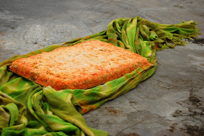
Apart from their significant role as decomposers, most Sordariales have little direct impact on human economics. The mould Neurospora intermedia is used to make oncom, a fermented food similar to tempeh. A number of species of Sordariales such as Neurospora crassa and Sordaria fimicola have been widely used in genetic research, to the extent that they have been labelled the 'fruit flies of the fungal world'. Seriously, it's one of those expressions almost every publication seems obliged to crow-bar in somewhere. The analogy is made even more apropos by the fact that one of the most widely used species, Triangularia née Podospora anserina, has been made the subject of debate whether taxonomic considerations should be allowed to shake up the name of a popular model organism.

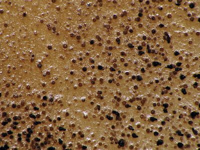
Molecular studies have also shown that the Sordariales encompass Madurella mycetomatis, a fungus causing subcutaneous inflammation in humans (van de Sande 2012). Seeing as sexual fruiting bodies are unknown in this species, and even asexual spore-producing structures are exceedingly rare, this organism would have previously been all but impossible to classify. Infection by M. mycetomatis is characterised by the production of granular swellings. It is most significant in central Africa but is also known from other tropical regions of the world. Madurella mycetomatis infects people via trauma such as animal bites and other wounds, and it has been isolated from soil and ant nests. In its normal state, M. mycetomatis is probably a quite innocent soil fungus. The trouble comes when it finds itself somewhere it shouldn't be.

REFERENCES

Kruys, Ã…., S. M. Huhndorf & A. N. Miller. 2015. Coprophilous contributions to the phylogeny of Lasiosphaeriaceae and allied taxa within Sordariales (Ascomycota, Fungi). Fungal Diversity 70: 101–113.

Marin-Felix, Y., A. N. Miller, J. F. Cano-Lira, J. Guarro, D. García, M. Stadler, S. M. Huhndorf & A. M. Stchigel. 2020. Re-evaluation of the order Sordariales: delimitation of Lasiosphaeriaceae s. str., and introduction of the new families Diplogelasinosporaceae, Naviculisporaceae, and Schizotheciaceae. Microorganisms 8: 1430.

Miller, A. N., & S. M. Huhndorf. 2005. Multi-gene phylogenies indicate ascomal wall morphology is a better predictor of phylogenetic relationships than ascospore morphology in the Sordariales (Ascomycota, Fungi). Molecular Phylogenetics and Evolution 35: 60–75.

van de Sande, W. W. J. 2012. Phylogenetic analysis of the complete mitochondrial genome of Madurella mycetomatis confirms its taxonomic position within the order Sordariales. PLoS One 7 (6): e38654.

The Age of the Perisphinctoid

During the Mesozoic era, the world's oceans were dominated by the ammonites. The coiled shells of these extinct cephalopods can be found preserved in rocks of this era around the planet, encompassing a bewildering array of species. During the latter half of the Jurassic, the most diverse ammonites were members of the superfamily Perisphinctoidea.

Likely Perisphinctes, copyright Spacebirdy.


Perisphinctoids first appear around the mid-point of the Jurassic, during what is known as the Bajocian epoch (Énay & Howarth 2019). As with other major ammonite groups, perisphinctoids are characterised by features of the folding around the edges of the septa that separate chambers of the shell. Perisphinctoids have basally five-lobed septa that differ from their ancestors in the Stephanoceratoidea in the loss of the UII lobe towards the outer edge of the whorl. The earliest perisphinctoids had more or less evolute shells (that is, later whorls did not significantly overlap the predecessors) with a rounded venter. Some later lineages would become more involute, with older whorls becoming partially hidden, and the venter might get sharper or flatter. Others would pretty much retain the original conformation to the end. The majority of perisphinctoids exhibited strong ribs on the outside of the shell, these ribs usually branching towards the outer rim of the whorl. Some forms developed further elaborations of the shells such as prominent nodules or spines.

Dimorphism was widespread in the perisphinctoids, if not universal. As with other dimorphic ammonites, populations included distinct microconches and macroconches (the majority interpretation is that macroconches were female and microconches male, but of course this is speculative). Macroconches usually had simple peristomes whereas microconches commonly had the mature shell aperture flanked by elongate lappets. The early Late Jurassic (Bathonian and Callovian) Tulitidae had a tendency in macroconches for the shell coiling to become eccentric in the outermost whorls, the peristome being distinctly skewed from the main plane of the shell.

Aspidoceras hirsutum, copyright Daderot.


Perisphinctoid faunas were often markedly provincial with many lineages being restricted to particular regions (such as the bipolar Perisphinctes or the western Eurasian Parkinsoniinae). They were mostly animals of shallower waters, perhaps foraging close to the bottom. This may go some way to explaining their high diversity but it can provide a challenge to their use in stratigraphy. Ammonites of the 'perisphinctoid' type would survive the end of the Jurassic but would fade from the fossil record not too long afterwards. Nevertheless, that would not be the end of their lineage: at the beginning of the Cretaceous, they would also spawn two derived descendants (Besnosov & Michailova 1991), the largely smooth-shelled Desmoceratoidea and the Ancyloceratoidea with four-lobed septa, that would continue to dominate the Mesozoic seas.

And while I'm on the subject of ammonites, I have another correction to make to an earlier post. However, while I was able to shift some of the blame for the correction in my last post onto my original source, in this case the blame is entirely mine. In a prior discussion of the live anatomy of ammonites, I discussed the evidence that the aptychus (a pair of calcified plates that probably functioned as an operculum) originated as a modification of the lower jaw. As such, I criticised reconstructions of ammonites that showed the aptychus articulating with the shell in the manner of a nautilus' hood. Unfortunately, I had overlooked a significant difference between ammonites and nautiluses. The coiled shell of a nautilus is exogastric—that is, when they evolved from their straight-shelled ancestors, the shell coiled upwards so the original lower edge corresponded to the outside of the whorl. However, the shell of ammonites was endogastric, with the shell coiled downwards so the original venter was on the inside (in the absence of preserved soft anatomy, we can infer this from the position of the siphuncle within the shell). This means that, even though the lower ammonite aptychus was anatomically on the opposite side of the animal from the upper nautilus hood, functionally they would have appeared in life to occupy much the same position. Entirely my mistake, and a reminder to me that describing orientation in coiled animals can be confusing.

REFERENCES

Besnosov, N. V., & I. A. Michailova. 1991. Higher taxa of Jurassic and Cretaceous Ammonitida. Paleontological Journal 25 (4): 1–19.

Énay, R., & M. K. Howarth. 2019. Part L, revised, volume 3B, chapter 7: Systematic descriptions of the Perisphinctoidea. Treatise Online 120: 1–184.

Ophiusini Corrections

Earlier this year, I presented a post on the noctuoid moth tribe Ophiusini. As it turns out, that post includes some notable errors. One of the main sources I used, Zahiri et al. (2012), stated that Ophiusini "have a strongly modified apex to the proboscis, with strong and enlarged spines and erectile, reversed hooks that are used in fruit-piercing or lachrymal-feeding behaviour". As reviewed by Zilli (2021), such hooks on the proboscis are unique to a separate subgroup of the family Erebidae, the Calpinae. Ophiusini have thin, nail-like spines on the proboscis but no erectile hooks. They are still fruit-piercers but no ophiusins have been observed to date engaging in lachrymal feeding.

Artena dotata, copyright Shipher Wu.


Zilli (2021) had further comments on the historically fraught concept of Ophiusini. As noted in my earlier post, 'Ophiusini' has historically been recognised as a cosmopolitan group of moths but molecular studies have lead to its restriction to the Old World, North American exemplars being transferred to the related tribe Poaphilini. However, though the two groups are each supported as monophyletic by molecular data, they are not well defined morphologically. Characters previously thought distinct to one or the other do not always hold true. Ophiusini have been described as having reduced coremata but some ophiusins have coremata larger than those of some poaphilins. Ophiusins have been supposed to lack the waxy bloom on the pupa found in other noctuoids but some species do indeed have such a bloom. Some have pointed to the use of Euphorbiaceae as host plants by Poaphilini but not Ophiusini, but not all poaphilins feed on Euphorbiaceae and their use of this plant family is generally correlated with species being more generalist feeders overall.

One character that may yet distinguish the two tribes is the location of the androteca, a groove along the top of one of the leg segments in the male that contains a long brush of dense hairs (I'm not sure just what the function of this structure is meant to be but I would suspect something to do with dispersing pheromones). In Ophiusini, this structure is found on the femur of the fore leg. In Poaphilini, it is on the tibia of the mid leg. Nevertheless, Zilli (2021) questions the reliability of this feature: both arrangments are found in other tribes and neither alone is diagnostic.

Conversely, molecular phylogenies support the two tribes as sister taxa, and they share a number of distinctive features of the terminalia. While he does not formalise the suggestion, Zilli (2021) seems to feel that we might be better served by a return to a broader Ophiusini uniting the two tribes as one. I commented in my previous post that noctuoid classification has been in a continuous flux for as long as it has been a thing. It would be presumptuous to believe that it has finally been settled.

REFERENCES

Zahiri, R., J. D. Holloway, I. J. Kitching, J. D. Lafontaine, M. Mutanen & N. Wahlberg. 2012. Molecular phylogenetics of Erebidae (Lepidoptera, Noctuoidea). Systematic Entomology 37: 102–124.

Zilli, A. 2021. Tabwecala robinsoni gen. nov., sp. nov., from Vanuatu and its systematic postion in the 'Ophiusini-Poaphilini' clade (Lepidoptera, Erebidae). Nota Lepidopterologica 44: 193–211.

Rasahus albomaculatus, the White-Spotted Corsair

Though the Hemiptera began their long evolutionary history as plant-feeders, many of their subgroups later switched to a predatory lifestyle, their suctorial mouthparts being just as suited for stabbing flesh as vegetation. Among the most successful of the predatory bugs where the assassin bugs of the family Reduviidae.

Image copyright Jacob Gorneau.


This is Rasahus albomaculatus, a widespread assassin of the Neotropical region, found from Mexico to Argentina (Coscaron 1983). Though not one of the largest members of its genus, R. albomaculatus is a decent-sized bug, growing close to an inch in length. Rasahus is a genus of the reduviid subfamily Peiratinae, commonly known as corsairs for their fearsome aspect. Features distinguishing Rasahus from other genera of corsairs include their large eyes, a deep grove across the head in front of the ocelli, long procoxae, and well-developed spongy pads on the fore- and mid-tibiae. Rasahus albomaculatus is distinguished from other species of the genus by its colour pattern. The body is mostly black with white patterning on the wings. Stripes along the top of the wing and across the mid-length form a crude H-shape when the wings are closed, with separate spots towards the base of the wing and towards the tip. Other noteworthy features include a lack of granulation on the pronotum, and a rounded apex to the scutellum (Swanson 2018).

Corsairs are mostly predators of other insects and not often dangerous to humans (though their bite is supposed to be very painful). Indeed, they may be beneficial to humans as among their prey are believed to be other reduviids of the subfamily Triatominae, the blood-sucking "kissing bugs" that spread Chagas disease (contrary to the Wikipedia page on the western corsair R. thoracicus, corsairs do not spread Chagas themselves). Rasahus albomaculatus may provide its vertebrate co-habitants with far more comfortable living conditions.

REFERENCES

Coscarón, M. del C. 1983. Revision del genero Rasahus (Insecta, Heteroptera, Reduviidae). Revista del Museo de La Plata (nueva serie) (Zoologia) 13: 75–138.

Swanson, D. R. 2018. Three new species of Rasahus, with clarification on the identities of three other Neotropical corsairs (Heteroptera: Reduviidae: Peiratinae). Zootaxa 4471 (3): 446–472.

The Dermacentor Ticks

Pacific Coast tick Dermacentor occidentalis, copyright Jerry Kirkhart.


Among the ticks of most concern to humans are species of the genus Dermacentor. This genus of about forty known species is widely distributed in Africa, Eurasia and the Americas. Examples include the meadow tick D. reticulatus in Europe, and the wood tick D. variabilis and Rocky Mountain wood tick D. andersoni in North America. They are parasites of mammals, including both generalist and more host-specific species; records of Dermacentor individuals from reptiles and even carpenter bees (Goddard & Bircham 2010) presumably represent incidental and/or accidental associations. Species of Dermacentor are responsible for the spread of bacteria causing diseases such as Rocky Mountain spotted fever (which, despite sounding like a 1950s dance craze, is presumably not much fun), Q fever and tularemia. The ticks can also be more directly hazardous, as their bites inject a toxin that can cause tick paralysis.

Distinguishing features of Dermacentor species relative to other ticks include a rectangular base to the capitulum, relatively short, broad palps, well-developed eyes and the presence of festoons (impressed divisions of the posterior margin of the body) (Keirans 2009). Most are ornate—that is, marked on the dorsum with contrasting pale patterns—with the notable exception of the tropical horse tick D. nitens of the Americas (until recently, often treated as forming its own genus Anocentor). The function of such markings is unknown though suggestions include environmental protection, warning predators of distastefulness, or sexual signalling.

Meadow tick Dermacentor reticulatus, copyright Ferran Turmo Gort.


The majority of Dermacentor species have a three-host life cycle, dropping off the host between each of the life stages of larva, nymph and adult, and seeking out a new host after moulting. However, at least two New World species, the aforementioned D. nitens and the winter tick D. albipictus (a parasite of deer), are one-host ticks that remain on their original host between instars. In general, Dermacentor species are more resilient to dry climates than many other tick species. Individual species can differ in their climate tolerance, however. In North America, the geographical divide between D. variabilis in the east of the continent and D. andersoni in the west seems to be driven by the need for the latter of drier conditions (Yoder et al. 2007). Older instars also tend to be hardier than younger. Females of the ornate sheep tick D. marginatus, a European species, leave their host after gorging at the beginning of winter and then wait for more amoenable spring conditions before laying their delicate eggs (Dörr & Gothe 2001).

Higher relationships within the genus do not appear to have been extensively studied. A preliminary molecular phylogeny of hard ticks has suggested the possibility of a basal division between Afrotropical, Eurasian and New World lineages (Barker & Murrell 2004). Comparison with related tick genera raises the possibility of an Afrotropical origin for Dermacentor, though the genus has only a relictual presence in that continent now. However, with only a handful of species subjected to broad phylogenetic analysis to date, further testing is demanded. Does the continental divide hold true? Do the one-host species form a single clade within the genus? Inquiring minds wish to know.

REFERENCES

Barker, S. C., & A. Murrell. 2004. Systematics and evolution of ticks with a list of valid genus and species names. Parasitology 129: S15–S36.

Dörr, B., & R. Gothe. 2001. Cold-hardiness of Dermacentor marginatus (Acari: Ixodidae). Experimental and Applied Acarology 25: 151–169.

Goddard, J., & L. Bircham. 2010. Parasitism of the carpenter bee, Xylocopa virginica (L.) (Hymenoptera: Apidae), by larval Dermacentor variabilis (Say) (Acari: Ixodidae). Systematic and Applied Acarology 15: 195–196.

Keirans, J. E. 2009. Order Ixodida. In: Krantz, G. W., & D. E. Walter (eds) A Manual of Acarology 3rd ed. pp. 111–123. Texas Tech University Press.

Yoder, J. A., D. R. Buchan, N. F. Ferrari & J. L. Tank. 2007. Dehydration tolerance of the Rocky Mountain wood tick, Dermacentor andersoni Stiles (Acari: Ixodidae), matches preference for a dry environment. International Journal of Acarology 33 (2): 173–180.

Shadow of the Palaeoniscoids

Palaeoniscum freieslebeni, copyright James St. John.


Depending how you cut it, the ray-finned fishes (Actinopterygii) are arguably the most diverse group of vertebrates in the modern fauna. They are the dominant vertebrates in all aquatic environments, they encompass an enormous array of species, and they have evolved a bewildering assemblage of morphologies. But despite their current pre-eminence, the early evolution of actinopterygians remains rather understudied. The earliest actinopterygians appear in the fossil record in the Late Silurian/Early Devonian but, until fairly recently, the majority of Palaeozoic ray-finned fishes have often been lumped into a catch-all holding tank, the 'Palaeonisciformes'. This was a vague assemblage of fishes united by plesiomorphic features such as ganoid scales (heavy, bony scales with an outer layer of enamel, also found in modern gars and sturgeons), a single dorsal fin and a heterocercal tail (with the upper arm of the tail fin longer than the lower). The key genus of the group, the Permian Palaeoniscum, had a fusiform (or torpedo-shaped) body; at first glance, it would not have looked dissimilar to a modern herring. However, it lacked the mobile jaw structure of modern teleost fishes, with the maxilla and preopercular bones being fixed together. As such, it would have lacked the modern fish's capacity for suction feeding (Lauder 1980). Prey capture by Palaeoniscum would have been a simple smash-and-grab affair. Palaeoniscoid fishes remained a component of both marine and freshwater faunas until the end of the Cretaceous before being entirely supplanted by modern teleost radiations such as the ostariophysans and percomorphs.

Reconstruction of Acrolepis gigas, copyright DiBgd.


The core concept of 'Palaeonisciformes' has united fishes with a fusiform body shape like Palaeoniscum; depending on the author, more divergent contemporary fishes such as the deep-body platysomoids might be combined in the same order or treated separately. By modern standards, former 'Palaeonisciformes' probably combine stem-actinopterygians, stem-chondrosteans, stem-holosteans and possibly even stem-teleosts. As such, the term Palaeonisciformes has tended to fall out of favour, though the less formal 'palaeoniscoid' remains a useful descriptor. Nevertheless, the exact phylogenetic position of many palaeoniscoid taxa remains unestablished. Part of this is due to a lack of observable detail: though those heavy ganoid scales preserve well, they effectively cover up internal skeletal features. Many palaeoniscoids are preserved as compression fossils, effectively not much more than intriguing silhouettes. However, part of the problem is simple neglect. Palaeoniscoids are not rare fossils; in some formations, they may be the dominant part of the fauna by a large margin. They certainly deserve a closer look.

REFERENCE

Lauder, G. V., Jr. 1980. Evolution of the feeding mechanism in primitive actinopterygian fishes: a functional anatomical analysis of Polypterus, Lepisosteus, and Amia. Journal of Morphology 163: 283–317.

Herbs of Dragons and Worms

Preparing for this post has inspired me to some low-key experimentation. When it came time to assign myself its topic, I landed on the plant genus Artemisia. This is the genus that, among others, includes the culinary herb tarragon, Artemisia dracunculus. Which got me thinking that I wasn't sure if I'd ever actually eaten tarragon. I asked Christopher if he was familiar with it; he responded that all he knew about tarragon was that you had to consume it in the 1970s. Without access to a functioning Delorean, I did the next best thing and prepared a dish of tarragon chicken myself. The verdict: very tasty, though I could appreciate why tarragon might have a reputation for being somewhat difficult as it had a light flavour that I could imagine being easily overwhelmed.

Tarragon Artemisia dracunculus, copyright Cillas.


Tarragon is not the only species of Artemisia of significance to humans. This genus of composite-flowered plants comprsises over five hundred species and subspecies of herbs and small shrubs. The greatest diversity is found in arid and semi-arid regions of the Northern Hemisphere temperate zone (Sanz et al. 2008). The genus is characterised by its distinctive pollen with surface spinules reduced or absent. This pollen type is associated with the wind pollination typical of the genus, though some species do exhibit features such as sticky pollen and colourful flower-heads associated with insect visitation (Hayat et al. 2009). The flower-heads or capitula (a reminder that the 'flowers' of composite plants such as daisies and thistles actually represent a fusion of multiple flowers) of Artemisia are either disciform, with an outer circle of reduced ray florets surrounding the inner disc florets, or discoid, with disc florets only. In disciform capitula, the outer limb of the ray florets is reduced to a membranous vestige, not readily visible without minute examination. The ray florets are female whereas the disc florets are ancestrally hermaphroditic (more on that shortly). In discoid capitula, where the ray florets have been lost, all florets are uniformly hermaphroditic.

Mugwort Artemisia vulgaris, copyright Christian Fischer.


Historically, there has been some variation in the classification of Artemisia but a popular system divides the genus between five subgenera. A phylogenetic analysis of Artemisia and related genera by Sanz et al. (2008) found that the genus as currently recognised is not monophyletic, with a handful of small related genera being embedded within the clade. Time will tell whether this inconsistency is resolved by subdividing Artemisia or simply rolling in these smaller segregates, but for the purposes of this post they can be simply set aside. The subgenus Dracunculus, including tarragon and related species, falls in the sister clade to all other Artemisia. As well as being united by molecular data, members of this clade are distinguished by disciform capitula in which the central disc florets have become functionally male (female organs have been rendered sterile).

Wormwoood Artemisia absinthium, copyright AfroBrazilian.


The second clade encompasses the subgenera Artemisia and Absinthium, with disciform capitula, and Seriphidium and Tridentatae, with discoid capitula. Not all authors have supported the distinction of Artemisia and Absinthium, and Sanz et al. identify both as non-monophyletic, both to each other and to the discoid subgenera. Because of their similar flower-heads, most authors have presumed a close relationship between the Eurasian Seriphidium and the North American Tridentatae (commonly known as sagebrushes). Some have even suggested the former to be ancestral to the latter. However, Sanz et al.'s results questioned such a relationship, instead placing the Tridentatae species in a clade that encompassed all the North American representatives of the Artemisia group.

As well as the aforementioned tarragon, economically significant representatives of Artemisia include wormwood A. absinthium, best known these days as the flavouring agent of absinthe (though historically it has also been used for more innocuous concoctions). Mugworts (A. vulgaris and related species) have also been used for culinary and medicinal purposes. Sagebrushes are a dominant component of the vegetation in much of the Great Basin region of North America, providing crucial habitat for much of the region's wildlife. Artemisia species have shaped the lives of many of their co-habitants, both animal and human.

REFERENCES

Hayat, M. Q., M. Ashraf, M. A. Khan, T. Mahmood, M. Ahmad & S. Jabeen. 2009. Phylogeny of Artemisia L.: recent developments. African Journal of Biotechnology 8 (11): 2423–2428.

Sanz, M., R. Vilatersana, O. Hidalgo, N. Garcia-Jacas, A. Susanna, G. M. Schneeweiss & J. Vallès. 2008. Molecular phylogeny and evolution of floral characters of Artemisia and allies (Anthemideae, Asteraceae): evidence from nrDNA ETS and ITS sequences. Taxon 57 (1): 66–78.